Paravalvular Leak Closure: Plugging a Leak Without Surgery
Paravalvular leak closure procedure repairs valve leaks without open-heart surgery.
Heart valve surgery or a transcatheter valve replacement can transform a person's life — but in a modest number of patients, a small gap opens between the new valve and the tissue around it. Blood then escapes backwards through that gap instead of flowing forward, a problem doctors call a paravalvular leak. When the leak is large enough to cause symptoms or strain the heart, the paravalvular leak closure procedure offers a way to plug it — usually through a thin catheter, without reopening the chest. This guide walks you through what a paravalvular leak is, how it is detected, what the closure procedure involves, and what recovery really looks like, so you can approach the decision with clear eyes and calm questions.
What Is a Paravalvular Leak?
When a surgeon replaces a damaged aortic or mitral valve — or when a valve is delivered through a catheter, as in TAVR (transcatheter aortic valve replacement) — the new valve must sit snugly inside the ring of tissue where the old valve lived. A paravalvular leak occurs when blood finds a channel around the valve's edge, between the artificial valve and the surrounding tissue, rather than through the valve itself. It is different from valve regurgitation inside the device, which is a problem with the valve's own leaflets.
These channels form at the sewing ring or the outer frame of the prosthesis. In surgical replacements, tiny gaps can remain where the sutures grip tissue; in TAVR valves, calcium in the native valve can prevent the new frame from sealing perfectly against the wall. Paravalvular Leak Closure was developed precisely for this situation: devices that can be delivered through the bloodstream to plug the channel at its source.
Small leaks versus significant leaks. Many small paravalvular leaks are found incidentally on routine scans and cause no symptoms at all. They are usually watched, not treated. A leak only becomes a treatment problem when it is large enough to make the heart work harder, when it damages red blood cells (causing anemia and dark urine), or when it produces symptoms such as breathlessness and fatigue. Size, location and symptoms together drive the decision.
How Serious Is a Leaking Heart Valve?
Seriousness depends on where the leak sits and how much blood it steals with each heartbeat. A leaking aortic valve — meaning blood flows backwards from the aorta into the left ventricle, the heart's main pumping chamber — forces that chamber to pump the normal amount plus the leaked volume, over and over. Over months or years this can enlarge the ventricle and weaken it. The same logic applies on the left side of the heart generally: significant heart valve regurgitation is never something to simply wait out if it is progressing.
With a paravalvular leak specifically, the concern is compounded by the fact that the valve has already been replaced. The heart has already been through one intervention. That is why doctors classify these leaks carefully — trivial, mild, moderate, severe — and why treatment is recommended when the leak is clinically significant, not merely because it exists.
When a leak becomes urgent. Warning signs that a paravalvular leak needs prompt attention include worsening shortness of breath, new or worsening heart failure symptoms, hemolytic anemia (destruction of red blood cells as they pass through the narrow leak), and endocarditis — infection around the prosthetic valve. Any of these changes the conversation from watchful waiting to active treatment, and the leak's severity is confirmed with imaging before anything is proposed.
What Causes a Paravalvular Leak After Valve Replacement?
The most common causes cluster around how the valve was implanted and what the anatomy looked like beforehand. Heavily calcified valve annuli, undersized prostheses, suture technique, and tissue fragility all play a role in surgical replacements. In TAVR valves, the degree and distribution of calcium in the native valve is one of the strongest predictors — a finding confirmed in studies such as the PREDICT PVL study of a self-expanding transcatheter valve, which examined exactly which factors predicted paravalvular leak after implantation (Open Heart).
Risk factors worth knowing. Several factors raise the likelihood that a leak will form:
- Heavy calcium in the native valve ring, which stops a new valve from seating evenly
- A prosthesis that ended up undersized relative to the patient's anatomy
- Tissue fragility or previous infection around the valve, weakening the suture line
- Very early leaks after surgery, which can reflect acute healing issues at the suture line
One study of early paravalvular leak after mitral valve replacement noted that percutaneous closure can be performed successfully even soon after the original operation, though careful timing decisions apply. The practical message for patients is this: a leak is usually a mechanical problem with a known mechanism, not a sign that something was done carelessly — and it has a defined, treatable cause.
What Are the First Symptoms of a Paravalvular Leak?
Initial symptoms are often subtle and easy to blame on aging or getting out of shape. The most common first complaints are breathlessness on activity that used to be easy, unusual fatigue, and a sensation of reduced exercise tolerance. Because a paravalvular leak forces the heart to recirculate the same blood, the body simply gets less forward flow than it should.
Anemia and dark urine. A distinctive symptom of some paravalvular leaks is mechanical hemolysis — red blood cells physically breaking apart as they squeeze through the narrow channel. This shows up as worsening anemia, jaundice or a yellowing of the eyes, and urine that turns dark or tea-colored. Patients are sometimes treated for anemia for months before anyone connects it to the valve. If you have a replaced valve and develop unexplained anemia, ask specifically about paravalvular leak.
Less common but important initial symptoms include a new heart murmur heard on examination, palpitations, swelling of the ankles, and in advanced cases symptoms of heart failure such as waking breathless at night. The pattern matters more than any single symptom: things that were fine after surgery slowly getting worse is the classic trajectory that should prompt a check.
Can a Paravalvular Leak Be Seen on an Echo?
Yes — echocardiography (an ultrasound of the heart) is the primary tool, and in most cases it is the only tool needed to make the diagnosis. On an echo, the leak appears as a jet of backward flow starting at the outer edge of the prosthetic valve rather than through the middle of it. Color Doppler mapping, which paints flowing blood in color, makes these jets visible and lets the cardiologist estimate how significant the leak is.
Why 3D imaging matters. Mitral paravalvular leaks in particular can be complex, with irregular crescent or slit-like shapes that are hard to measure on a standard 2D scan. Three-dimensional transesophageal echocardiography — a probe passed into the esophagus for a close-up view from behind the heart — has become the standard for mapping the leak's exact location, shape and size. Research has also explored 3D printing of the patient's valve anatomy from CT scans to plan closure and select the right occluder device ahead of time (Journal of Cardiothoracic Surgery).
In our team's experience, the planning scan is where the case is really won or lost: we review the transesophageal echo and CT images together as a multidisciplinary group before proposing any closure, because the leak's shape dictates which device will actually seal it. If your echo shows a suspicious jet, the next step is usually a transesophageal study — a routine outpatient procedure with throat numbing and light sedation, not an operation.
How Does the Paravalvular Leak Closure Procedure Work?
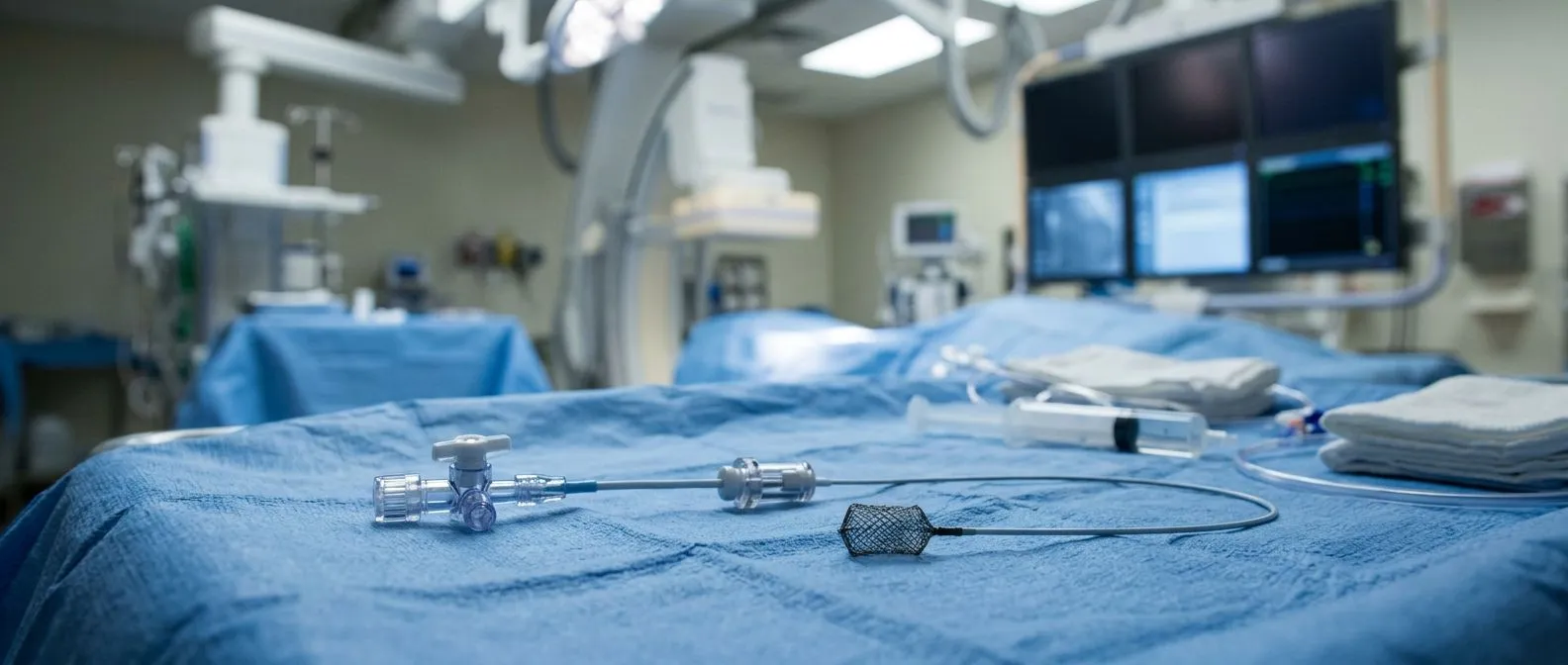
The transcatheter paravalvular leak closure procedure is done in a cardiac catheterization laboratory, the same setting used for angiograms and stent procedures. The patient receives local anesthetic at the access site (usually the groin) and light sedation. The cardiologist passes a long, thin tube — a catheter — up through a large vein or artery to the heart.
Reaching the leak. Getting the catheter to the leak is often the most delicate part. Aortic leaks can usually be reached through an artery from the groin, since they sit on the high-pressure side of the circulation. Mitral leaks, which sit between the left atrium and left ventricle, are usually approached through the vein system by crossing the wall between the right and left atria — a well-established maneuver called transseptal puncture — or occasionally by an advanced route looping from the aorta through the aortic valve backwards.
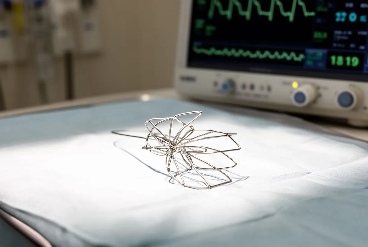
Plugging the channel. Once the catheter tip crosses the leak, the operator passes a soft guide wire through the channel and then slides the closure device — typically a self-expanding mesh occluder, similar in concept to the devices used for Septal Defect Closure — over the wire. The device is deployed so one disc sits on each side of the leak, clamping the channel shut. The team confirms the result with ultrasound and X-ray contrast before releasing the device. If the leak has several openings, more than one device may be placed.
The entire procedure usually takes one to three hours. Because the heart is not opened and no incision is made in the chest, the body's trauma is that of a catheter procedure, not an operation. The result is checked immediately: the echo shows whether the leak has been reduced, ideally to a trivial or mild residual flow, and the patient is watched overnight.
Paravalvular Leak Repair Without Surgery: Who Is a Candidate?
The catheter-based approach is now the preferred option for most clinically significant paravalvular leaks that need treatment, precisely because it avoids a second open-heart operation. Redo sternotomy — cutting through the breastbone a second time — carries substantially higher risk than the first operation, both because of scar tissue and because the heart is often adherent to the chest wall. For patients who are older, frail, or have had more than one previous operation, percutaneous paravalvular leak repair is often the only sensible route.
What makes a good candidate. The team looks for several things before recommending the procedure:
- A leak that is causing symptoms, hemolysis, or measurable heart strain — not a trivial incidental finding
- A leak shape and location that can be reached with a catheter and crossed with a wire
- Heart function that can tolerate the procedure, judged on echo and clinical review
- No active infection around the valve, which must be treated first
Not every leak qualifies. Very small leaks that cause no symptoms are usually observed. Some leaks sit in positions that are impossible to reach safely, or are so large or irregular that a device cannot seal them. In those cases, reoperation or careful medical management remains the alternative — and this is exactly the kind of trade-off that deserves an honest conversation rather than a push toward any single option.
Our team treats this decision as the patient's, not ours: every case is reviewed against international practice guidelines, the patient receives a clear proposal with the alternatives laid out, and the final call is always theirs. We encourage second opinions and consider them a normal part of deciding about heart surgery.
Paravalvular Leak Closure Recovery Time and What to Expect
Paravalvular leak closure recovery time is one of the procedure's main advantages. Most patients go home the next day, sometimes the same evening. Compare that with redo open-heart surgery, which typically means several days in hospital and weeks of healing. The catheter route trades a day in the cath lab and an overnight stay for what would otherwise be a major operation.
The first 48 hours. After the procedure, you lie flat for several hours while the access site in the groin seals. Nurses check your blood pressure, heart rhythm and the puncture site regularly. Some bruising and tenderness at the access site is normal. Most people are walking by the next morning and eating normally.
The first few weeks. You will be asked to avoid heavy lifting and strenuous exercise for roughly a week, to let the puncture site heal fully. Follow-up echo within a few weeks confirms the device is in place and the leak is closed. Symptoms such as breathlessness and fatigue often improve gradually over the following weeks as the heart stops wasting effort on recirculated blood. If hemolysis was present, blood counts typically recover over a similar period, though severe long-standing hemolysis can take longer to resolve.
We tell our patients to expect a catheter-procedure recovery, not an operation recovery: sore groin, tired for a few days, back to normal activities quickly. That said, we also set expectations honestly — some leaks are incompletely closed on the first attempt and may need a second device later, and we say so before the procedure, not after.
What Are the Risks of Transcatheter Paravalvular Leek Closure?
Every cardiac procedure carries risk, and honest discussion of risk is a mark of a good team, not a discouragement. The main risks of transcatheter paravalvular leak closure include vascular complications at the access site (bleeding, bruising, or damage to the vessel), interference with the prosthetic valve itself (a device can impinge on the valve leaflets and worsen its function), heart rhythm disturbances that occasionally require a pacemaker, stroke, and device embolization — the rare event of the device dislodging. The risk profile varies with leak location: mitral leaks are technically harder and carry somewhat higher procedural risk than aortic ones.
How risk is reduced. Risk is managed long before the procedure starts. Careful 3D imaging maps the leak's relationship to the valve leaflets. Device sizing is matched to the leak's measured dimensions. The procedure is done with continuous echo guidance. And the entire plan is reviewed by a multidisciplinary team — interventional cardiologist, cardiac surgeon, imaging specialist — before a recommendation is made, because a procedure that looks attractive on paper may not be right for a particular anatomy.
In our consultations we take the time to explain what the studies actually show, in plain words, and to answer every question before any decision. We also recommend the less invasive route only where the evidence supports it for that specific patient — if redo surgery is genuinely the safer option, we will say so.
How to Choose a Paravalvular Leak Closure Specialist
Because paravalvular leak closure is one of the more technically demanding structural heart procedures, the team performing it matters as much as the decision to do it. Look for a program with experience in structural heart interventions, full 3D imaging capability (transesophageal echo and cardiac CT on site), and a genuine multidisciplinary review process rather than a single-operator decision.
Questions worth asking any team. Before committing anywhere, ask:
- How many paravalvular leak closures does this team perform, and what is their success rate with leaks like mine?
- Who reviews my case — one doctor or a full multidisciplinary team?
- What are the specific risks in my anatomy, based on my imaging?
- What happens if the leak cannot be fully closed on the first attempt?
Dr Alejandro Molina trained in cardiothoracic surgery at the Universidad de Guadalajara, in cardiac, minimally invasive, robotic and structural surgery at the University of Montreal, and in structural cardiology and TAVR at CHU Bordeaux; the team also trained at Jena, Bad Berka and Buffalo, and Dr Molina performed the first robotic mitral valve repair in the Ángeles Health System and the first robotic heart surgery in Guadalajara. Our consulting rooms are in Guadalajara, Querétaro and Hermosillo, and the first contact is a free video consultation: we get to know the patient, talk through the condition, and identify which studies are still needed so no unnecessary ones are ordered — with a face-to-face visit following when the patient decides to go ahead.
The bottom line. A paravalvular leak that is causing symptoms, anemia or heart strain is a treatable problem, and for most patients the paravalvular leak closure procedure offers real repair without reopening the chest. The right first step is not self-diagnosis or waiting for things to worsen — it is a proper review of your echo and CT images by a qualified cardiac team, and a frank conversation about whether closure, reoperation, or careful observation is the lowest-risk path for your situation. If you have a replaced valve and any of the symptoms described here, bring them to a specialist consultation and let the imaging — not guesswork — decide the next step.