

Shock Wave Therapy for Calcified Arteries and Valves
Shock wave therapy arteries treatment breaks calcified plaque safely using intravascular lithotripsy.
For patients facing severe heart disease, learning that your blood vessels have become rigid with calcium can be a frightening and overwhelming experience. When standard treatments struggle to open these hardened arteries, modern cardiology offers a specialized, minimally invasive solution known as shock wave therapy arteries treatment. This advanced technique gently fractures stubborn calcium deposits, allowing vessels to expand properly and restoring healthy blood flow to the heart muscle. In this guide, we will explore how this technology works, what the procedure feels like, and how it compares to traditional surgical options, helping you feel prepared to discuss your treatment options with your cardiac care team.
What Is Shock Wave Therapy Arteries Treatment?
When plaque builds up in the arteries over many years, it often calcifies, turning from soft, waxy cholesterol into a hard, bone-like substance. How does calcification affect blood flow? This rigid atheromatous plaque acts like a concrete shell inside your artery, preventing the vessel from expanding when your heart needs more oxygen. It also creates a physical barrier that standard medical balloons cannot easily push aside, complicating traditional stent placements and increasing the risk of complications.
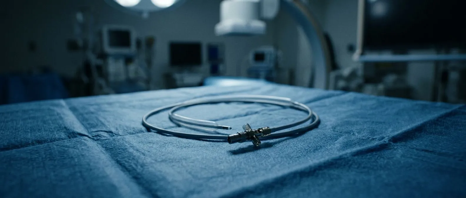
What makes this therapy different? Shock wave therapy arteries treatment, clinically known as intravascular lithotripsy, uses targeted acoustic pressure waves to safely break apart this hardened calcium. Instead of forcing a balloon against a rock-hard surface, the technology emits gentle sonic pulses that create micro-fractures in the calcium layers. This allows the artery wall to become compliant and flexible once again, making room for a stent to expand fully and securely.
This method represents a significant shift in how interventional cardiologists approach complex coronary artery disease. By focusing solely on the calcium and leaving the surrounding soft tissue unharmed, this therapy minimizes the trauma often associated with high-pressure balloon inflations. It provides a highly controlled way to prepare a heavily diseased vessel for definitive treatment, ultimately improving the long-term success rate of the procedure and the patient's overall quality of life.
How Intravascular Lithotripsy Works on Calcified Lesions
The technology behind intravascular lithotripsy is similar to the shock wave treatment used for decades to break apart kidney stones, but it has been precisely miniaturized for use inside the heart's delicate vessels. The mechanics of acoustic energy. A specialized, thin catheter is threaded through the groin or wrist directly to the site of the calcified blockage. Once positioned, the catheter emits focused sonic pressure waves that pass harmlessly through soft tissues like blood and vessel walls, but are absorbed by the dense calcium deposits.
Breaking the calcium safely. As the acoustic waves strike the calcium, they create micro-fractures throughout the rigid plaque. These tiny cracks weaken the structure of the calcification, transforming a stiff, unyielding barrier into a more flexible matrix. The cardiologist then inflates a standard balloon, which gently compresses the now-fractured calcium against the artery wall, creating a wider, smoother channel for blood to flow through without the risk of tearing the vessel.
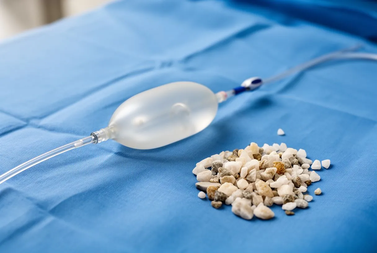
Understanding how this targeted energy works helps patients feel more comfortable with the procedure. The precision of the shock waves means that the risk of damaging the healthy portions of the artery is significantly reduced compared to older mechanical drilling methods. For a broader overview of how acoustic waves are utilized in medical treatments, you can read more from Healthline to understand the general safety and applications of these non-invasive energy waves in modern medicine.
When Do Cardiologists Recommend Shockwave Balloon Angioplasty?
Not every patient with coronary artery disease requires intravascular lithotripsy. Identifying heavy calcification. Cardiologists typically recommend this approach when imaging tests, such as intravascular ultrasound or optical coherence tomography, reveal that a blockage is heavily calcified. If a standard balloon fails to expand the vessel wall properly, or if the stent does not deploy uniformly due to calcium resistance, the medical team will often switch to a lithotripsy balloon to modify the plaque.
Why traditional methods fail. Pushing a standard balloon against severe calcium can cause the vessel to stretch unevenly, leading to a high risk of arterial dissection, a dangerous tear in the inner lining of the vessel. By using shockwave balloon angioplasty, the doctor can achieve full vessel expansion at much lower pressures, protecting the delicate endothelial cells and ensuring the stent fits flush against the artery wall to prevent future blood clots.
- Heavy calcification: Standard balloons cannot expand the vessel wall properly, leading to incomplete treatments.
- Stent deployment issues: Calcium prevents stents from fully opening and fitting flush against the artery, compromising blood flow.
- High complication risk: Forcing high-pressure balloons can cause vessel rupture or dissection, making a gentler approach necessary for patient safety.
During these complex catheterisations, maintaining the patient's physiological stability is paramount. Research on Interventions to prevent hypothermia in extremely preterm low-weight infants undergoing cardiac catheterisation highlights the critical importance of meticulous environmental and thermal management during vascular access procedures. While this study focuses on the most fragile pediatric patients, the core principle of protecting the body from environmental stress during any catheterisation remains a gold standard in modern interventional cardiology, ensuring the patient's body remains stable while the heart is being treated.
Treating Calcified Coronary Arteries with Lithotripsy
Coronary calcium removal therapy using lithotripsy is specifically designed for the arteries that supply oxygen directly to the heart muscle. The procedure step-by-step. After accessing the vascular system through a small puncture in the wrist or groin, the interventional cardiologist guides a guidewire across the narrowed, calcified segment. The specialized lithotripsy balloon catheter is then tracked over this wire and centered exactly within the hard plaque.
Delivering the pulses. Once the balloon is in position, the cardiologist inflates it slightly to ensure it makes contact with the artery walls, and then activates the shock wave generator. The device typically delivers a series of pulses, up to eighty or more, while the medical team monitors the vessel expansion under continuous X-ray imaging. This method is often used in tandem with traditional balloon angioplasty to achieve the optimal vessel diameter before a permanent stent is placed.
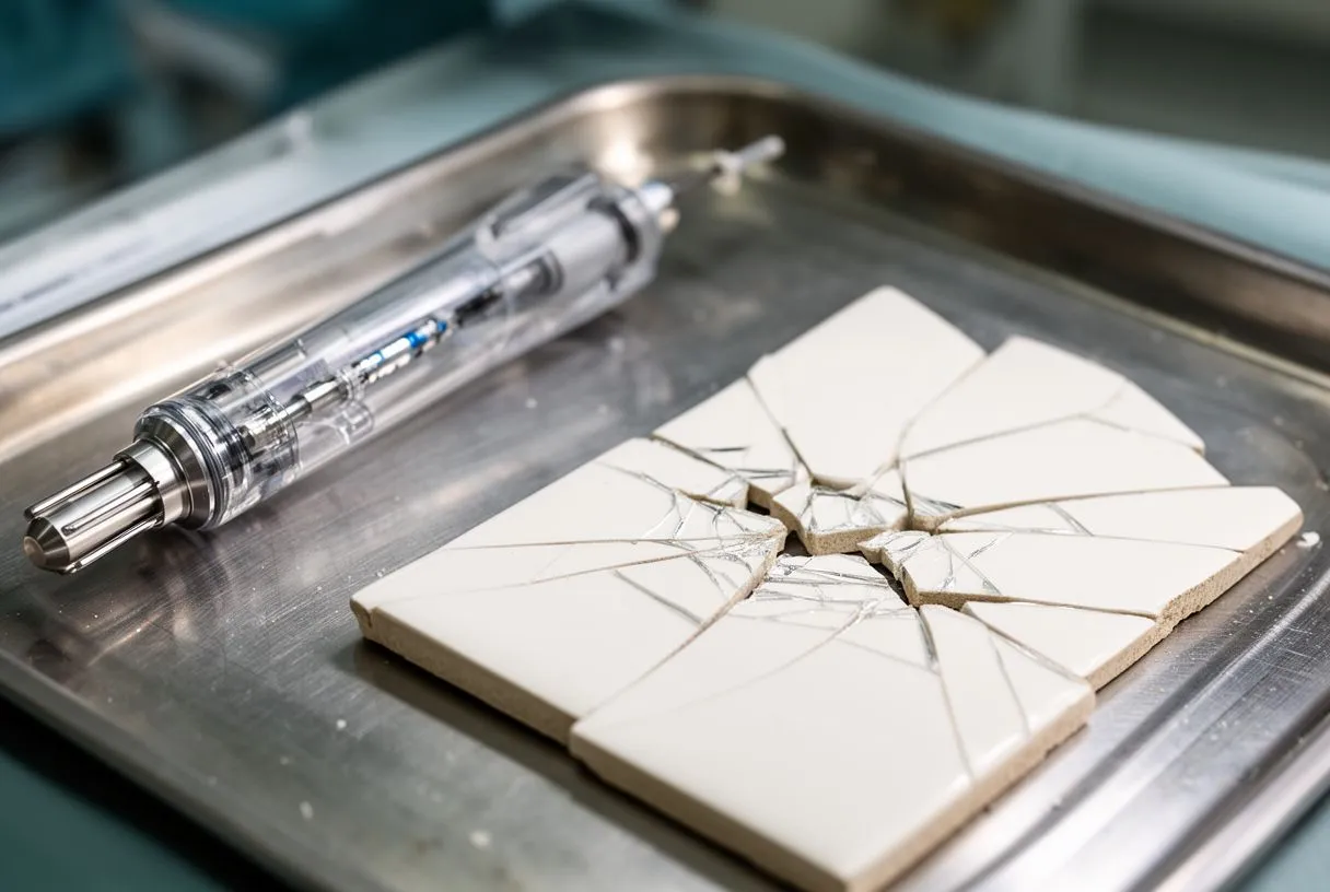
This precise approach allows cardiologists to tackle lesions that were once considered untreatable without open heart surgery. By fracturing the calcium from the inside out, the therapy restores the natural flexibility of the coronary artery, ensuring that the final stent achieves maximum apposition. This meticulous preparation is crucial for long-term outcomes, as a well-expanded stent is less likely to narrow again over time, reducing the need for repeat interventions.
Shockwave Therapy for Aortic Stenosis and Heart Valves
While intravascular lithotripsy is most commonly associated with coronary arteries, the principle of breaking calcium is increasingly being applied to heart valves. Valve calcification. The aortic valve, which controls blood flow from the left ventricle into the ascending aorta, can become severely calcified over time. This condition, known as aortic stenosis, restricts the valve's leaflets from opening fully, forcing the heart to work harder to pump blood to the rest of the body.
Valve preparation techniques. Before a transcatheter aortic valve replacement (TAVR) can be performed, the heavily calcified native valve often needs to be prepared to ensure the new artificial valve fits properly and functions without leaking. Doctors may first use balloon aortic valvuloplasty, a procedure where a balloon is inflated inside the valve to stretch the opening, combined with specialized techniques to crack the rigid calcium nodules that could otherwise prevent a balloon-expandable valve from seating correctly.
Adapting shock wave technology for valvular calcification represents an exciting frontier in structural heart disease treatment. By fracturing the large, obstructive calcium deposits on the valve leaflets beforehand, cardiologists can create a smoother, more circular landing zone for the transcatheter valve. This careful preparation minimizes the risk of paravalvular leaks, ensuring that the new valve opens and closes efficiently and dramatically improving the patient's cardiac output and daily energy levels.
What to Expect During a Coronary Lithotripsy Procedure
For patients, the experience of undergoing a coronary lithotripsy procedure is very similar to a standard cardiac catheterisation. Staying comfortable. You will typically be awake during the procedure, receiving a mild sedative through an intravenous line to help you relax, along with a local anesthetic at the puncture site. You should not feel the shock waves themselves, though you may feel a slight pressure or mild discomfort when the balloons are inflated inside your artery.
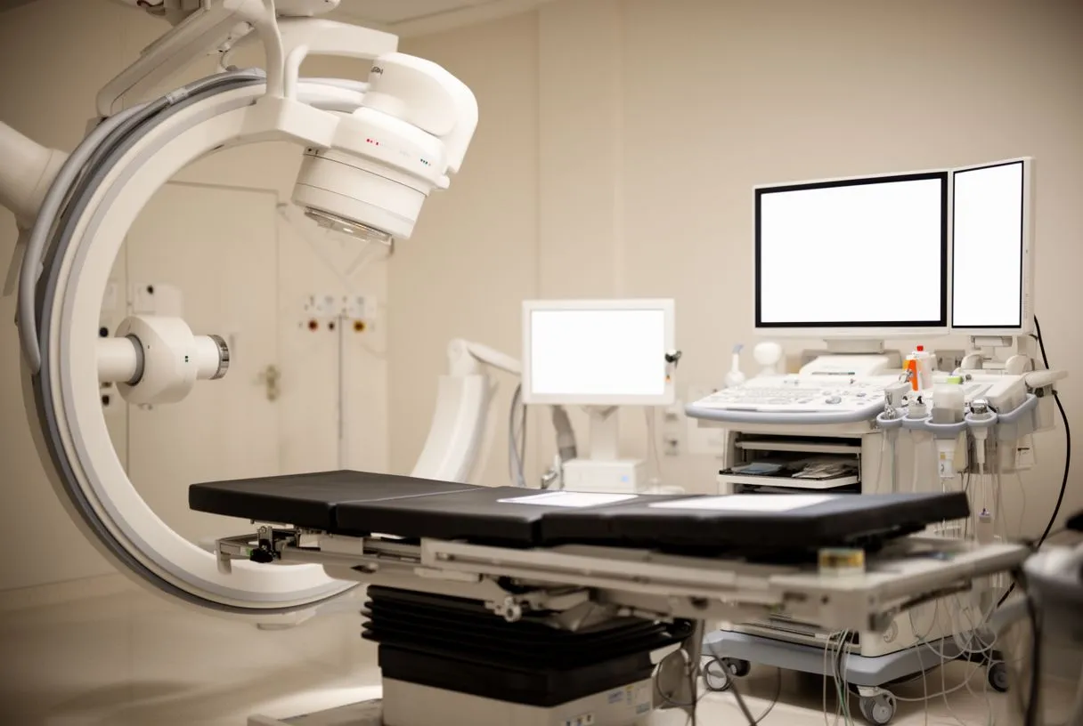
Monitoring and radiation safety. The medical team uses continuous X-ray imaging, or fluoroscopy, to guide the catheters and monitor the delivery of the acoustic waves. Radiation exposure is kept to an absolute minimum, and the clinical staff takes comprehensive precautions. The importance of managing this exposure is highlighted in ongoing medical discussions, such as those surrounding Lead Cap Use in Interventional Cardiology: Time to Protect Our Head in the Cardiac Catheterisation Laboratory?, reflecting the continuous efforts to improve safety for both patients and the medical professionals performing these intricate procedures.
Immediate results. The procedure usually takes between one to two hours, depending on the complexity and number of blockages being treated. Once the calcium has been fractured and the stent successfully deployed, the catheters are removed. Your cardiologist will review the final images to confirm that blood flow has been fully restored before you are transferred to the recovery area to begin your healing process.
Recovery and Outcomes After Minimally Invasive Decalcification
One of the primary advantages of this minimally invasive approach is the rapid recovery time compared to traditional bypass surgery. In the recovery room. Immediately after the procedure, you will rest in a specialized monitoring unit where nurses will check your heart rhythm and the access site in your wrist or groin. The doctor will use auscultation, listening to your heart and blood vessels with a stethoscope, to ensure your heart sounds are normal and that blood is flowing properly without any abnormal murmurs.
Going home. Most patients are able to go home within twenty-four hours of the procedure. You will be prescribed medications, often including aspirin and other blood thinners, to prevent clots from forming inside the newly placed stent. It is crucial to take these medications exactly as directed, as stopping them prematurely can lead to sudden blockage of the stent and serious cardiac complications.
Monitoring at home. While recovery is generally smooth, it is important to watch for any unusual symptoms. If the access site was in your wrist, you might experience mild bruising or temporary arm pain, which should gradually subside. You should avoid heavy lifting and strenuous exercise for a few days, but light walking is encouraged to promote healthy circulation. If you notice severe swelling, bleeding, or a sudden return of chest pain, you must contact your cardiac team immediately.
Are There Risks with Breaking Calcium in Arteries?
While intravascular lithotripsy is considered highly safe and effective, no medical procedure is entirely without risk. Understanding potential complications. Because the therapy involves fracturing hard calcium, there is a small chance that tiny pieces of plaque could break away and travel downstream, potentially causing a minor blockage in smaller vessels. However, the gentle, progressive nature of the shock waves makes this complication much less likely than with older mechanical drilling techniques.
- Vessel dissection: The artery wall could tear if the calcium fractures unevenly, often requiring an additional stent to repair the damage and seal the vessel.
- Slow flow: Calcium debris might temporarily slow blood flow, a condition cardiologists manage quickly with specialized medications to restore circulation.
- Access site bleeding: Bruising or minor bleeding at the catheter insertion point in the wrist or groin is possible, usually resolving with gentle pressure and rest.
It is important to have an open and honest conversation with your cardiologist about these risks before consenting to the procedure. Your medical team will evaluate your specific anatomy, overall health, and the severity of your calcification to determine if the benefits of breaking the calcium outweigh the potential downsides. For the vast majority of patients with severe calcification, this therapy provides the safest path to restoring blood flow without resorting to open chest surgery.
Alternatives to Shock Wave Therapy in Cardiology
If intravascular lithotripsy is not suitable for your specific anatomy, your cardiac team has several other tools at their disposal. Mechanical approaches. Rotational atherectomy, often called rotablation, uses a tiny, diamond-tipped burr spinning at high speeds to drill through the calcified plaque. While highly effective, this technique generates heat and fine debris, which can sometimes cause temporary spasms or slow blood flow in the artery during the procedure.
Surgical bypass. For patients with extensive disease that cannot be treated through a catheter, coronary artery bypass grafting remains the gold standard. In this open surgical procedure, a healthy arterial graft, often taken from your own chest or arm, is used to create a new pathway around the blocked artery. This provides excellent long-term results but requires a longer hospital stay and a significantly more intensive recovery period.
Choosing between these options depends heavily on the complexity of your coronary anatomy and your overall health status. Sometimes, a hybrid approach is used, combining minimally invasive catheter techniques with surgical grafting. Your multidisciplinary heart team will review your imaging and clinical history to recommend the approach that offers the lowest risk for your situation and the highest likelihood of a durable, successful outcome.
Is Shock Wave Therapy Right for Your Cardiac Care?
Shock wave therapy arteries treatment has truly revolutionized the way interventional cardiologists manage complex, heavily calcified blockages, offering a gentler, safer alternative to traditional high-pressure balloons and mechanical drills. By using targeted acoustic energy to fracture rigid plaque, this minimally invasive procedure allows stents to expand fully, restoring critical blood flow to the heart muscle with a remarkably short recovery time and a low complication profile.
Making the decision. Deciding whether this technology is right for you requires a comprehensive evaluation by a qualified cardiac team. They will carefully assess the location and severity of your calcium buildup, your overall heart function, and any other medical conditions you may have. Understanding the mechanism, risks, and recovery associated with this therapy empowers you to have a more meaningful conversation with your doctor about your treatment plan.
Navigating a diagnosis of severe coronary or valvular calcification can be daunting, but advanced therapies like intravascular lithotripsy provide new hope for patients who previously had limited options. We encourage you to discuss your specific symptoms and imaging results with a qualified cardiac team and to book a consultation to determine the best path forward for your heart health, rather than attempting to self-diagnose or rely solely on online research.






