

Sternal Closure After Heart Surgery: Wires, Plates and Healing
Learn about sternal closure methods after heart surgery—wires, plates, and healing timelines from Guadalajara specialists.
What Are Sternal Closure Methods After Heart Surgery?
When a cardiac surgeon needs to reach your heart, the most common pathway is through a median sternotomy—a vertical cut through the sternum, or breastbone. Once the operation is complete, that bone must be put back together securely. This is what we call sternal closure, and the method your surgeon chooses directly affects how you heal, how much pain you feel, and how quickly you can return to daily life. Every closure technique has trade-offs, and the decision is made based on your bone quality, body habitus, and the specific procedure performed.
Why the closure technique matters: The sternum is not just a protective shield; it anchors the ribs and supports every breath you take. If it heals poorly, the consequences range from chronic pain to life-threatening infection. Surgeons have used stainless-steel wires for decades, but newer rigid fixation systems using plates and screws are gaining ground for higher-risk patients. The goal of any sternal closure method is the same: hold the two halves of the bone in stable, close contact so that natural bone healing can occur without movement at the fracture line.
What factors influence the choice of closure: Your surgeon considers bone density, obesity, the presence of diabetes, and whether you had previous chest surgery. Patients who undergo Beating-heart Surgery may have slightly different closure dynamics, but the sternum is still divided the same way. The decision is always individualised, and you should feel comfortable asking your surgeon which method they plan to use and why.
How Sternal Wires Work to Hold the Breastbone Together
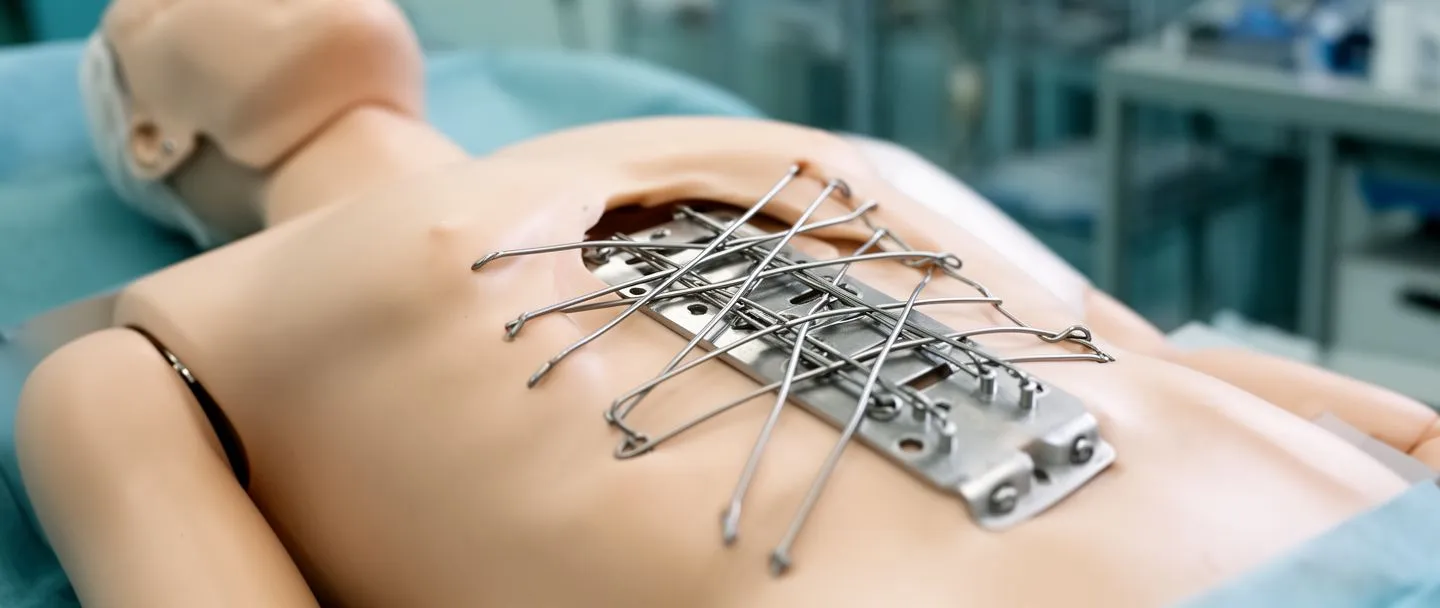
Sternal wiring is the oldest and most widely used closure technique in cardiac surgery. The surgeon passes heavy-gauge stainless-steel wires around or through the sternum at several levels, then twists them tight to draw the two bone halves together. The twisted ends are cut short and bent flat against the bone so they do not protrude through the skin. Most patients receive between six and eight wires, though the number can vary based on the patient's size and the surgeon's preference.
Why wires remain the default choice: They are fast to place, inexpensive, and have a decades-long track record of success in millions of patients. For a healthy adult with normal bone density undergoing a straightforward first-time sternotomy, wires provide more than enough stability. When bone healing proceeds normally, the sternum fuses within six to eight weeks, and the wires simply become inert passengers for the rest of the patient's life. Extensive medical databases like Europe PMC continuously catalogue research on these long-term hardware outcomes to ensure patient safety.
When wires are not enough: The limitation of wires is that they provide circumferential compression but no resistance to rotational or shearing forces. If you cough forcefully, twist your torso, or lift something heavy during the early healing phase, wires alone may allow microscopic movement at the fracture line, which delays bone union. This is why your team will give you strict precautions: no pushing, pulling, or lifting more than a few pounds for at least six weeks, and always splint your chest with a pillow when you cough.
Sternal Plates vs Wires: Which Closure Technique Is Better?
Sternal plating involves securing small titanium plates across the two halves of the sternum using screws that anchor into the bone on each side. Think of it like the plates an orthopaedic surgeon uses for a broken leg—the principle is the same: rigid fixation that prevents movement in every direction. Unlike wires, which only squeeze from front to back, plates resist separation, rotation, and sliding. This rigid stability can reduce postoperative pain and may allow earlier mobilisation, though the evidence is still evolving.
The trade-offs between plates and wires: Plates take longer to place and add cost to the procedure. They require precise drilling and measurement, and not every institution stocks the full range of plate sizes. However, for patients at elevated risk of sternal dehiscence—the separation of the two bone halves—plating offers measurable advantages. The decision is not about which is universally better; it is about which is the lowest-risk option for your individual anatomy and risk profile.
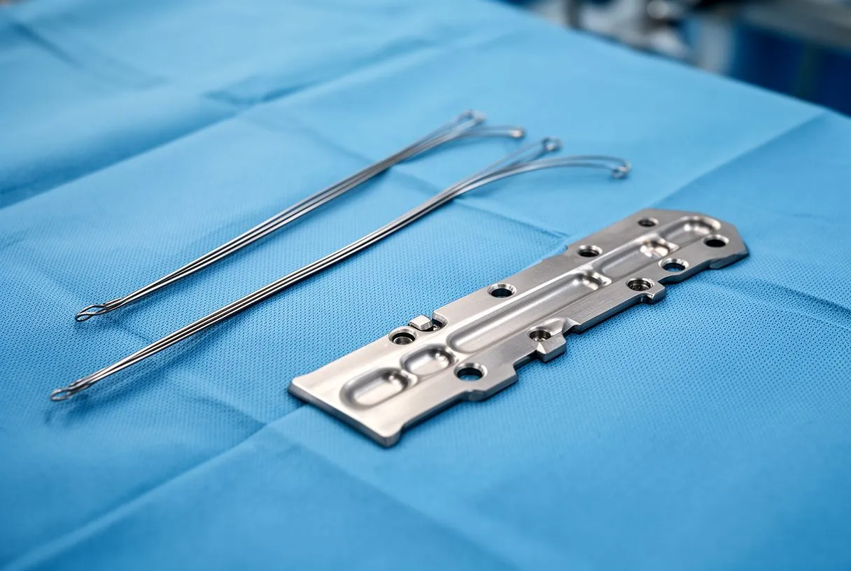
Can plates and wires be used together? Yes, and this hybrid approach is increasingly common. A surgeon may wire the central portion of the sternum and add plates at the manubrium or at the lower xiphoid process where bone is thinner. Some systems allow a single plate to span multiple segments, creating a bridge that distributes force more evenly. If you are concerned about your risk, ask your surgeon whether a hybrid closure is an option for your case.
Sternum Healing After Bypass: Stages and Timeline
Bone healing after a sternotomy follows the same biological stages as any other fracture, but in a location that is constantly stressed by breathing and movement. The first phase, the inflammatory phase, lasts about one to two weeks. During this time, a blood clot forms between the bone edges, and the body sends cells to clean up damaged tissue. You will feel the most pain during this stage, and the incision site may be swollen and tender.
Weeks two to six—the reparative phase: A soft callus of cartilage begins to bridge the gap between the two sternal halves. The callus is not yet strong enough to bear significant force, which is why your activity restrictions remain strict. Walking is encouraged, but you should avoid arm-heavy movements. Just as precise postoperative monitoring is essential for complex valve patients—highlighted in studies like EFFECT OF OXYGEN INHALATION ON MICROEMBOLIC SIGNALS IN PATIENTS WITH MECHANICAL AORTIC VALVE—your surgeon will monitor your sternal union closely to catch delayed healing early.
Weeks six to twelve—the remodelling phase: The soft callus is gradually replaced by hard, mineralised bone. By the six-week mark, most patients are cleared to resume light daily activities, though heavy lifting and contact sports remain off-limits. Full remodelling can take six months to a year, and the bone may continue to strengthen subtly beyond that. Smoking, poorly controlled diabetes, and malnutrition are the most common modifiable risk factors for delayed healing, and addressing these before surgery gives you the best chance of an uncomplicated recovery.
What Causes Sternal Closure Complications?
Sternal complications after cardiac surgery range from minor wound infections to catastrophic dehiscence and mediastinitis—infection of the central chest compartment that houses the heart. The overall rate of significant sternal complications is low in well-selected patients at experienced centres, but the risk rises sharply in certain groups. Understanding the risk factors helps you and your surgeon make decisions that reduce them.
- Obesity: Excess body weight places constant mechanical stress on the sternum and increases the risk of wound breakdown.
- Diabetes: Elevated blood sugar impairs wound healing and increases infection risk. Tight glucose control before and after surgery is essential.
- Smoking: Nicotine constricts the blood vessels that deliver oxygen and healing cells to the bone. Smoking cessation even four weeks before surgery measurably improves outcomes.
- Osteoporosis: Thin, brittle bone gives wires and screws less purchase, increasing the likelihood of hardware pulling through.
- Repeat sternotomy: A second or third chest operation means scar tissue, altered anatomy, and higher complication rates.
How COPD and chronic coughing affect sternal healing: Patients with chronic obstructive pulmonary disease face a unique challenge: forceful, repeated coughing generates significant shearing force across the healing sternum. Each cough can separate the bone edges by a fraction of a millimetre, and over thousands of coughs per day, this can prevent stable union. If you have COPD or a chronic cough, optimising your respiratory medications before surgery and using a splinting pillow after surgery is not optional—it is a critical part of protecting your closure.
Mitigating risk before surgery: Your surgical team will assess your risk factors well before the operation. If you need a Biological Valve or Bovine Pericardial Valve replacement, the team will coordinate your overall cardiac recovery plan to ensure your sternum heals alongside your new valve. Proper planning, including addressing malnutrition or uncontrolled blood sugar, is the most effective way to prevent sternal closure complications.
Signs of Sternal Dehiscence and When to Seek Help
Sternal dehiscence—the partial or complete separation of the breastbone halves—is one of the most feared complications of cardiac surgery. It can occur within days of the operation or weeks later if healing is interrupted. The most common early sign is a feeling of instability or movement in the chest bone, sometimes described as a "clicking" or "popping" that is new or worsening. You may also notice widening of the skin incision, drainage of clear or cloudy fluid, or increasing pain that does not improve with your prescribed pain medication.
Red flags that require immediate attention: If you develop fever, redness spreading from the incision, foul-smelling drainage, or severe chest pain with a visible step-off deformity along the sternum, go to the emergency department immediately. These may indicate mediastinitis, a deep infection of the chest cavity that can be life-threatening. Early detection is critical, and precise diagnostic evaluation is just as important here as it is in complex device management, such as the article Device console programming in patients undergoing TAVI with preexisting cardiac implantable electronic devices: a practical guide.

What happens if dehiscence is diagnosed: If the separation is partial and there is no infection, your surgeon may recommend a chest binder and strict activity restriction while monitoring healing. If the separation is complete or associated with infection, surgical revision is usually necessary. This involves reopening the incision, cleaning the wound, re-approximating the bone, and often upgrading the fixation from wires to plates. If infection is present, intravenous antibiotics will be required, sometimes for several weeks.
Sternal Nonunion Treatment Options for Failed Healing
Sternal nonunion is defined as failure of the bone to heal by six months after surgery. You may have persistent pain, clicking, or a visible gap. Nonunion is different from dehiscence—the bone edges may be stable but simply have not fused. The treatment approach depends on whether the nonunion is painful, whether hardware is intact, and whether there is any associated infection.
Conservative management for stable nonunion: If your sternum is stable and your main symptom is mild discomfort, your surgeon may recommend continued observation. Some nonunions are painless and do not require intervention. However, if wires have broken or migrated and are causing pain, sternal wire removal can provide significant relief. This is typically a short outpatient procedure—the surgeon makes a small incision over the wire end, removes the broken segment, and closes the skin.
Surgical revision for symptomatic nonunion: When nonunion causes persistent pain, instability, or cosmetic deformity, surgical reconstruction is the next step. The surgeon removes the old hardware, freshens the bone edges to stimulate new healing, and applies rigid plate fixation to hold the bone firmly while it fuses. The decision to proceed with revision surgery balances the severity of symptoms against the risks of a repeat operation. For patients who have already been through a major cardiac procedure, sternal repair is a much smaller operation than the original heart surgery.
Rigid Sternal Fixation Benefits for High-Risk Patients
Rigid sternal fixation using plates and screws was developed specifically for patients at high risk of sternal complications. The benefits extend beyond just holding the bone together—they can change the entire recovery experience. For patients with obesity, osteoporosis, COPD, or those undergoing repeat sternotomy, rigid fixation can be the difference between an uncomplicated recovery and a prolonged, painful one.
Reduced pain and earlier mobilisation: Because plates prevent micromotion at the fracture line, patients often report less pain with breathing and movement in the early postoperative period. Better pain control means patients can take deep breaths more easily, use their incentive spirometer more effectively, and ambulate sooner—all of which reduce the risk of pneumonia and blood clots. The faster a patient is up and moving, the shorter the hospital stay and the lower the complication rate.
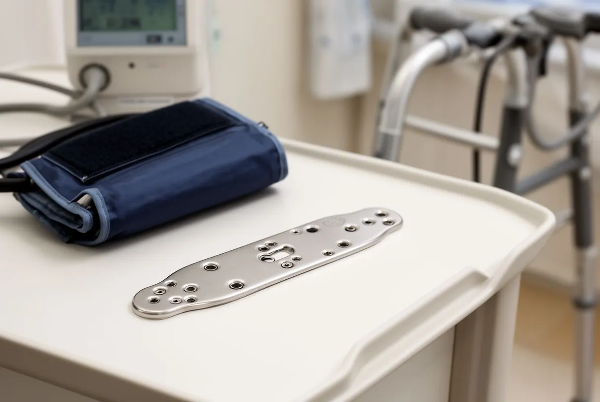
Lower dehiscence rates in selected populations: Multiple institutional studies have shown that rigid plating reduces the rate of sternal dehiscence compared to wire closure alone in high-risk groups. The hardware itself—titanium plates and screws—is biocompatible and rarely causes allergic reactions. Unlike stainless-steel wires, titanium does not interfere with future MRI scans, which is an important consideration for patients who may need cardiac imaging in the years following surgery.
Heart Surgery Recovery: Sternal Wound Care at Home
Caring for your sternal incision at home is one of the most important responsibilities you will have during the first month after discharge. The wound is healing from the inside out, and what you do—or avoid doing—during this period has a direct impact on whether your sternum heals without complications. Your surgical team will give you specific instructions, but the following principles apply to almost everyone.
- Keep the incision clean and dry: Shower with mild soap and let water run over the wound. Do not soak in a bath or pool until cleared by your surgeon.
- Sleep on your back: Side sleeping and stomach sleeping put pressure on the healing sternum. Use pillows for support.
- Splint when coughing: Hold a firm pillow against your chest every time you cough or sneeze to reduce stress on the bone.
- No driving for four to six weeks: Sudden braking engages chest muscles that can stress the healing bone.
- Lift nothing heavier than 5 kg: Avoid pushing, pulling, or carrying groceries, children, or pets until your surgeon clears you.
What to watch for during healing: Mild swelling, itching, and a pulling sensation along the incision are normal during the first few weeks. Small amounts of clear or slightly blood-tinged drainage from the edges can also be normal. However, spreading redness, increasing warmth, a foul odour, pus, or opening of the wound edges are not normal and should prompt a call to your surgical team. If you notice any change in the shape of your sternum, report it immediately.
When to call the surgeon: When in doubt, call your surgeon's office. No experienced cardiac team will be annoyed by a cautious phone call; they would much rather hear from you early than see you in the emergency department late. If you are on a Beta Blocker or other cardiac medications, your team will coordinate your general cardiac care with your sternal healing progress to ensure a holistic recovery.
Sternal Closure in Mexico: What International Patients Should Know
For patients considering cardiac surgery in Mexico—whether in Guadalajara, Querétaro, or Hermosillo—questions about sternal closure methods and postoperative care are common and important. Dr Alejandro Molina and his team follow the same evidence-based protocols used in leading international centres, and the choice of closure technique is always individualised. The surgical plan, including the method of sternal fixation, is discussed in detail during your preoperative consultation so there are no surprises on the day of surgery.
Follow-up and wound care after returning home: One of the most common concerns for international patients is what happens after discharge. You will receive detailed wound-care instructions, a schedule for follow-up imaging, and a clear plan for communication with the surgical team after you return to your home country. In most cases, a local physician can remove sutures or staples and monitor your incision, with any concerns relayed directly to Dr Molina's office. The team maintains continuity of care regardless of where you live.
Making an informed decision about your cardiac surgery: Every patient's situation is unique, and the information in this article is intended to help you ask better questions, not to replace a personal consultation. If you or a loved one are facing heart surgery and want to understand your options for sternal closure and recovery, we encourage you to book a consultation with a qualified cardiac team. Discuss your specific risk factors, ask which closure method the surgeon recommends, and make sure you feel confident in the plan before proceeding.






