Stroke Risk and Heart Surgery: How It Is Reduced
Stroke risk heart surgery teams reduce through screening, monitoring, and advanced techniques.
If you or someone you love is facing heart surgery, one of the quietest fears in the room is often the same: what happens to the brain while the heart is being operated on? Understanding stroke risk heart surgery involves is the first step toward reducing it. The good news is that stroke after cardiac surgery has become less common over the decades, not because of luck, but because surgical teams have learned exactly where the danger comes from and how to interrupt it. This guide walks you through what causes stroke during heart operations, how surgeons lower that risk before, during, and after the procedure, and what you can do as a patient to be part of that protection.
What Is Stroke Risk Heart Surgery?
A stroke happens when blood flow to part of the brain is suddenly blocked or when a vessel bleeds into brain tissue. During heart surgery, the concern is almost always the blocked type, called an ischemic stroke, caused by tiny fragments of clot, plaque, or air traveling upward to the brain.
Every operation on the heart carries some risk to the brain because the two are physically connected. The aorta, the body's largest artery, leaves the heart and carries blood directly to the arteries of the brain. Anything loosened from the heart chambers, the valves, or the wall of the aorta during surgery can ride that current upward. This is why teams talk about stroke risk as something to be managed, not eliminated.
The level of risk depends on several personal factors rather than on the operation alone. These include your age, whether you have disease in the carotid arteries of the neck, a history of a previous Stroke, atrial fibrillation (an irregular heart rhythm), diabetes, kidney disease, and the condition of the aorta itself. Surgeons weigh these together in a process called Risk Stratification, which helps predict who needs extra brain protection and which approach offers the lowest risk for your situation.
How Does a Stroke Happen During Cardiac Surgery?
The medical term for these traveling fragments is cerebral embolism — in everyday words, a small particle carried by the bloodstream that lodges in a brain artery. Most strokes connected to cardiac surgery are cerebral embolism cardiac surgery teams actively work to prevent.
There are three main sources of these particles. First, atherosclerosis — fatty plaque — inside the aorta can be disturbed when surgeons place clamps or cannulas (the tubes that connect the heart-lung machine). Fragile plaque crumbles easily, and even a particle smaller than a grain of sand can block a brain vessel. Second, the heart itself can hold clots, especially in atrial fibrillation or after a heart attack, and touching or opening the heart can release them. Third, air can enter the circulation during the operation and form bubbles that reach the brain.
Understanding the source matters because each one has a specific prevention strategy. This is also why a heart team looks at your imaging before deciding anything: the aorta's condition on a CT scan often shapes the entire surgical plan. In our consultation we take the time to explain what these studies actually show, in plain words, and to answer every question before any decision is made.
What Is the Stroke Risk During Cardiac Surgery for Each Procedure?
Different heart operations carry different levels of neurological risk, and knowing the general pattern helps you ask better questions.
- Coronary bypass (CABG): Bypass surgery on the beating heart, using the heart-lung machine or not, carries a low but real stroke risk, driven largely by the aorta and the carotid arteries.
- Valve surgery: Replacing or repairing the aortic or mitral valve means working inside the heart where calcium deposits live, so fragments are more likely to be released.
- Aortic surgery: Operating directly on a diseased aorta is where stroke prevention is most tested, because the surgical field is the very highway to the brain.
- Transcatheter procedures (TAVR): Catheter-based valve replacement navigates through the aorta, and embolic debris can be released as the new valve is placed.
No single number applies to everyone, and reputable surgeons avoid quoting percentages without seeing your specific anatomy. What matters is the combination of your health profile, the operation planned, and the protection measures the team uses. Every case we review is examined by our multidisciplinary team against international practice guidelines before a recommendation is given, so the trade-offs of each route — including the stroke question — are laid out openly.
Why Is Carotid Screening Before Heart Surgery Important?
The carotid arteries are the two large vessels on each side of the neck that carry blood to the brain. If they are narrowed by plaque, they become a ready-made source of stroke during any heart operation, independent of what happens inside the chest.
Carotid screening before heart surgery is now standard practice for patients over a certain age or with risk factors such as a neck bruit (a whooshing sound heard with a stethoscope), prior TIA, or peripheral artery disease. The screening itself is simple: a painless ultrasound, called a duplex scan, shows whether the arteries are open and how much plaque sits inside them. If a severe narrowing is found, the team may treat the carotid artery first, or during the same hospital stay, before the heart operation proceeds.
This is a good example of how modern cardiac care thinks upstream. Instead of reacting to a stroke after bypass surgery risk becomes reality, the team closes the side road before the main journey begins. Checking the carotid circulation is often part of the pre-surgical evaluation, and we recommend the less invasive route only where the evidence supports it for that specific patient.
In our first meeting with a patient — a free video consultation — we get to know their history, talk through the condition, and say exactly which studies are still needed so no unnecessary ones are ordered.
How Do Surgeons Minimize Stroke in Open Heart Surgery?
Surgeons have a whole toolkit of brain protection techniques used during open heart operations. Together they target the three sources of emboli described earlier: the aorta, the heart chambers, and air.
- Cooling the brain: Lowering body temperature during surgery slows the brain's metabolism, so brief reductions in blood flow are better tolerated — one of the established cooling techniques stroke prevention surgery relies on.
- Aortic scanning: The surgeon uses ultrasound directly on the aorta during the operation to find plaque-free spots before placing clamps or cannulas.
- Filtering and Cannula design: Modern heart-lung machine tubing includes filters, and specially shaped cannulas direct blood flow away from the brain's arteries, reducing the chance debris travels upward.
- De-airing the heart: Before the heart is closed, the team actively removes air from the chambers using ultrasound confirmation, because air bubbles reaching the brain are a preventable cause of injury.
Blood pressure management during the operation is another piece of the puzzle. The perfusionist — the specialist who runs the heart-lung machine — keeps pressure in a range that maintains steady flow to the brain, and this blood pressure management heart surgery stroke question is monitored minute by minute. None of these measures works alone; it is the combination, applied consistently, that makes the difference.
Dr Molina trained in cardiac, minimally invasive, robotic and structural surgery at the University of Montreal and in structural cardiology and TAVR at CHU Bordeaux, and he brings that training in brain-protection technique to every open operation our team performs.

Does Off-Pump Heart Surgery Lower Stroke Risk?
Off-pump coronary bypass means performing the bypass grafts on the beating heart, without connecting the patient to the heart-lung machine. The theory is appealing: no machine means no machine-related source of particles, and no clamping of the aorta in its diseased segments.
The evidence is mixed, and honesty matters here. Some studies suggest off-pump surgery may reduce certain complications in selected patients, while others find no clear advantage overall once all outcomes are considered. Non-randomized comparisons are particularly tricky to interpret, because patients chosen for off-pump surgery often differ from those chosen for the conventional route — a problem researchers address with tools like ROBINS-I, a structured method for assessing risk of bias in non-randomised studies of interventions (University of Manchester).
What this means for you as a patient is that off-pump is a genuine option, not a guaranteed shield. The right question to ask your surgeon is not "is off-pump better?" but "given my aorta and my arteries, which approach protects my brain best?" In our practice, that answer comes from the whole multidisciplinary team reviewing the case, not from a preference for one technique. The final decision is always the patient's, with the alternatives explained clearly.
What About Minimally Invasive Heart Surgery Stroke Risk?
Minimally invasive heart surgery — including robotic and small-incision approaches — avoids opening the chest fully, which shortens recovery. But any operation that works inside the heart still needs attention to minimizing stroke open heart surgery risk, because the sources of particles are the same.
In robotic and minimally invasive valve surgery, the surgeon works through small ports using long instruments and a camera. The heart-lung machine is still used, so the same principles apply: careful cannula placement, de-airing under ultrasound, and aortic management tailored to what the imaging shows. For some patients, a minimally invasive approach actually reduces aortic manipulation, which is a genuine advantage for the brain.
For patients who cannot tolerate open surgery at all, transcatheter options change the picture entirely. In TAVR, a replacement aortic valve is delivered through a catheter, usually from the groin, without opening the chest. This is where low-risk TAVR and Prohibitive Risk trials changed the field: patients at low surgical risk and patients at prohibitive risk were both studied, and TAVR became an established option across a range of risk levels. During these procedures, teams may use embolic protection devices — small filters designed to catch debris before it reaches the brain — and anticoagulation (blood-thinning medication) is managed precisely throughout.
Our team performs conventional, minimally invasive, robotic and transcatheter procedures, and we recommend the less invasive route only where the evidence supports it for that patient.
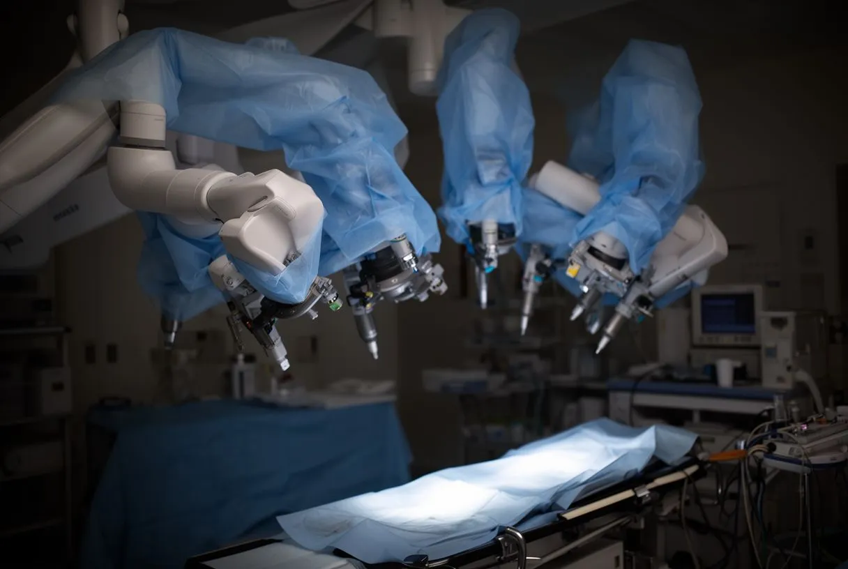
How Does Intraoperative Stroke Monitoring Protect the Brain?
Modern operating rooms do not wait for a problem to appear; they watch for it in real time. Neurological monitoring during surgery gives the team early signals so adjustments can be made before harm occurs.
Common techniques include transcranial Doppler, which uses sound waves to detect particles passing through the brain's arteries, and cerebral oximetry, small sensors on the forehead that measure how well the brain is being supplied with oxygen. If oxygen readings drop, the team can raise blood pressure, adjust the bypass circuit, or change cannula position immediately. Near-infrared spectroscopy and EEG monitoring serve similar purposes, catching changes invisible to the human eye.
Intraoperative stroke monitoring does not prevent every event, but it changes outcomes by shortening the gap between "something is wrong" and "something is done." For procedures on the aorta in particular, where the brain's blood supply is directly at stake, some teams also use selective perfusion — deliberately routing oxygenated blood to the brain's vessels during critical steps. I have found in my own work that a patient who arrives with a complete aortic and carotid picture before surgery is far easier to protect once we are operating, because nothing on the table is a surprise.
What Happens After Surgery: Postoperative Stroke Prevention After Cardiac Care?
The operation is not the only window of risk. The days following surgery carry their own challenges, and postoperative stroke prevention cardiac teams practice is a structured routine rather than an afterthought.
The most important late risk is atrial fibrillation, a rapid irregular rhythm that often appears in the first days after cardiac surgery. It matters because blood can pool and clot in the fibrillating atria, and those clots can travel to the brain. Teams monitor the rhythm continuously after surgery and treat new atrial fibrillation promptly, with medication to restore rhythm or to prevent clotting — this is anticoagulation heart surgery stroke protection in its most practical form.
Other pillars of prevention in the recovery unit are straightforward but powerful: keeping blood pressure in the target range, ensuring adequate oxygenation, getting the patient moving early, and watching for any new neurological sign. Nurses check for facial droop, arm weakness, or slurred speech as part of routine care, because a stroke caught within minutes can often be treated far more effectively. Recognizing the signs of a Stroke early — sudden face drooping, arm weakness, speech difficulty — is knowledge every family member should carry home.
- Bleeding Risk balance: Blood thinners protect against clots, but too much increases bleeding; teams adjust the dose daily to keep both risks low.
- Medication adherence: Following the prescribed anticoagulation and blood pressure plan after discharge is one of the strongest things a patient can do for their own brain.
We encourage second opinions and treat seeking one as a normal part of deciding about heart surgery — and we extend the same openness to questions about what happens in the recovery unit afterward.
Can You Make a Full Recovery From a Stroke After Heart Surgery?
This is one of the most common and most human questions patients ask, and the honest answer is: it depends on the type of stroke, how quickly it was treated, and the rehabilitation that follows. Small strokes, and especially those caused by air rather than clot, often recover well — sometimes completely. Larger strokes cause lasting deficits that rehabilitation works to improve, sometimes dramatically.
Early rehabilitation makes a real difference. Physical therapy, speech therapy, and occupational therapy begin within days in hospitals equipped to provide them, and the brain's capacity to reorganize — called neuroplasticity — means progress can continue for months. Recovery also depends on managing the underlying causes afterward: controlling blood pressure, treating atrial fibrillation, and keeping cholesterol and diabetes in check all reduce the chance of another event.
It is also worth knowing that not every brain symptom after heart surgery is a stroke. Some patients experience temporary confusion or memory fog that resolves on its own, sometimes called postoperative cognitive dysfunction, and there is an active research conversation about what causes it and how to prevent it. Research into specific anatomical causes continues — for example, the role of a patent foramen ovale, a small flap-like opening between the heart's upper chambers that many adults have without knowing, and whether it acts as a direct cause, a risk factor, or an incidental finding in unexplained stroke (Frontiers in Neurology). If you have questions like these about your own recovery, they belong in a conversation with your cardiac team — not in late-night internet searches.
Choosing a Heart Team That Protects Your Brain
Finally, the single most effective stroke prevention decision may be the choice of team itself. Ask direct questions before any operation: Will my carotid arteries be checked? What does my aorta look like on imaging, and how will that shape the approach? Will my brain be monitored during surgery? What is the plan if atrial fibrillation appears afterward? A confident team welcomes these questions and answers them in plain language.
Reducing the chance of stroke after bypass surgery risk, valve procedures, or aortic operations comes down to layered protection: careful screening beforehand, disciplined technique during the operation, vigilant monitoring in the recovery unit, and faithful follow-up after discharge. No layer alone is enough, but together they have transformed what was once one of surgery's greatest fears into a managed, measurable risk.
Our multidisciplinary team, led by Dr Alejandro Molina with consulting rooms in Guadalajara, Querétaro and Hermosillo, reviews every case against international practice guidelines before any recommendation, and the patient always receives a clear proposal with the alternatives — because an informed patient is the best protection we know.
If you are weighing a heart operation and want to understand the stroke risk heart surgery carries in your specific case, the next step is a conversation, not a search engine. Book a consultation with a qualified cardiac team, bring your questions, and make the decision with the full picture in front of you.