Structural Valve Deterioration in Tissue Valves
Structural valve deterioration tissue valves: learn warning signs, monitoring with echocardiograms, and when reoperation may be needed.
If you received a tissue valve to replace a diseased heart valve, you already know it is not metal and plastic. It is biological material, usually from a cow or a pig, and it can wear out over time. That gradual wearing process is called structural valve deterioration in tissue valves, and understanding it puts you in control. This guide explains what it is, why it happens, which symptoms matter, and what your options are if your valve starts to fail.
What Is Structural Valve Deterioration in Tissue Valves?
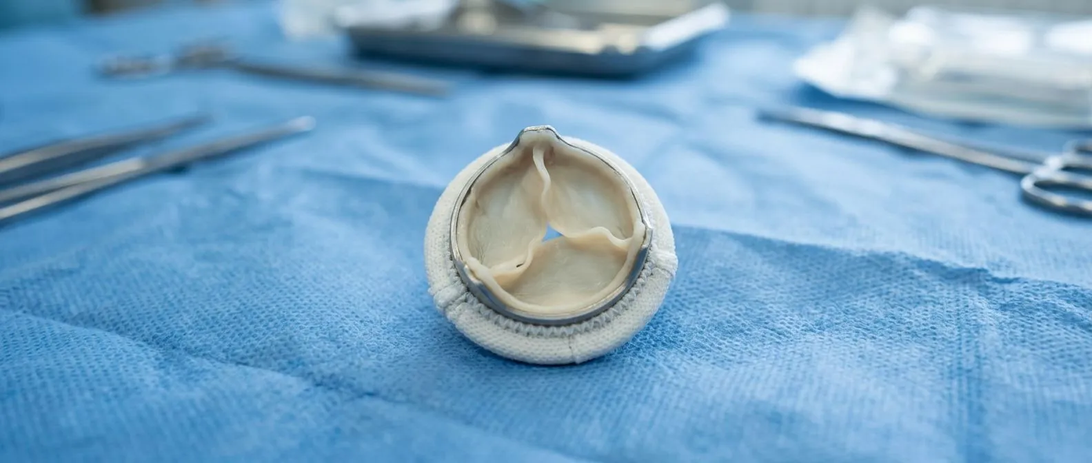
A tissue valve, also called a bioprosthetic valve, is built from animal tissue mounted on a frame or sewn into a ring. It opens and closes about 100,000 times a day. Over the years, that constant flexing, plus blood chemistry and the body's own healing response, slowly changes the valve's structure.
Specialists in valve disease and structural cardiology use the term structural valve deterioration to describe changes in the valve material itself: calcification, stiffening, tears, or fraying of the leaflets. It is different from other causes of valve failure, such as infection or a leak around the outside of the valve. Those have different treatments and different warnings.
When we review a patient with a replaced valve, our team looks specifically at whether the leaflets themselves are deteriorating or whether something else, like infection, explains the change — the distinction changes the whole treatment plan. Knowing the difference matters because deterioration usually develops slowly and can be monitored, while infection needs urgent care.
What Causes a Tissue Heart Valve to Deteriorate?
Calcification is the main driver. Calcium from your blood gradually deposits on the valve leaflets, the thin flaps that open and close with every heartbeat. As calcium builds up, the leaflets stiffen and stop opening fully. The valve can also start leaking if a leaflet tears or the tissue stretches.
Several factors speed this process up:
- Younger age: the body's immune and calcium processes are more active in younger patients, so deterioration tends to appear earlier.
- Kidney disease: impaired kidneys change calcium and phosphate balance, which accelerates calcification of biological tissue.
- Smoking and poorly controlled diabetes and high blood pressure: all of these promote the same vascular calcification process that stiffens the valve.
Where the valve sits matters too. An intra-annular valve sits within the natural ring of the native valve, while a supra-annular valve sits slightly above it; position can influence blood flow patterns and, in some designs, how the leaflets wear over time. In transcatheter valves, uneven expansion or a residual leak can also concentrate stress on parts of the leaflets. Research on specific devices, such as the PREDICT PVL study, has looked at which factors predict problems after implantation, helping teams choose and position valves more carefully.
Which Tissue Valve Failure Symptoms Should You Watch For?
Early deterioration is usually silent. That is why routine imaging matters more than symptoms. When symptoms do appear, they usually mean the valve has either narrowed (stenosis) or started leaking (regurgitation).
Breathlessness and reduced exercise tolerance. The most common early complaint is getting tired or short of breath with activities that used to be easy — climbing the same stairs, walking the same route. Fluid can also build up in the legs or abdomen.
Chest discomfort and dizziness. A narrowed valve makes the heart pump harder against resistance, which can cause chest pressure, lightheadedness, or fainting spells during exertion. Fainting with exertion always deserves prompt evaluation.
Palpitations or an irregular heartbeat. A leaking or narrowed valve changes pressures inside the heart, especially the left atrium behind the mitral position. That can trigger rhythm problems such as atrial fibrillation, which may be the first noticeable sign of valve trouble. If you notice any of these changes, tell your cardiologist rather than waiting for your next scheduled scan.
How Long Do Tissue Valves Last?
This is the question patients ask most, and the honest answer is a range, not a guarantee. Most tissue valves function well for 10 to 15 years, and many last longer. Some begin to show measurable deterioration earlier, especially in younger patients or those with kidney disease.
Durability depends on several things at once: your age at implantation, the type of tissue (a porcine valve comes from pig tissue, while a bovine pericardial valve is made from cow tissue lining), the position of the valve, and your overall health. Valves in the aortic position generally face less pressure than those in the mitral or tricuspid valve position, so they often last longer.
When a patient asks us how long their valve will last, we give them a realistic range and explain which of their own factors — age, kidney function, valve position — push that estimate up or down, because the average never tells the individual story. The goal of follow-up is to catch deterioration early, while you still feel well and all options remain open.
How Does Echocardiogram Monitoring Track Your Tissue Valve?
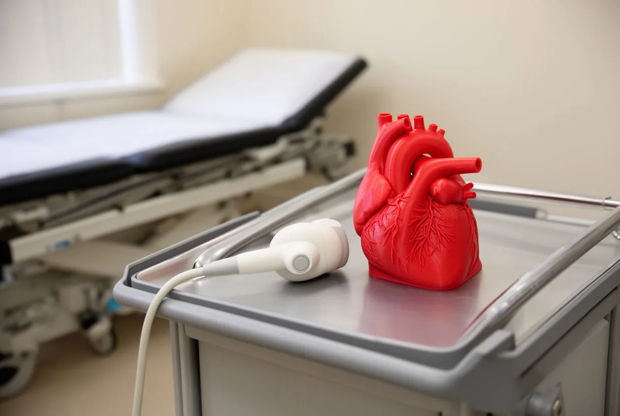
An echocardiogram is an ultrasound of the heart. It is painless, uses no radiation, and shows how well your valve opens and closes. It measures the valve's opening area, the pressure gradient across it, and whether any blood leaks backward. This is the primary tool for detecting tissue valve calcification and degeneration before symptoms begin.
Typical surveillance follows a rhythm: a baseline study soon after surgery, another around the first anniversary, then periodic checks that become more frequent as the valve ages — often yearly after the fifth or sixth year. Your own team will set the schedule based on your valve, your symptoms, and what each scan shows.
At each follow-up, comparing the new echocardiogram against the baseline study, rather than reading it alone, matters, because a gradual change in gradient or leaflet motion is the earliest signal that a valve is starting to deteriorate. Keep copies of your echocardiogram reports. Comparisons over time are far more informative than any single study.
Tissue Valve vs Mechanical Valve: Which Lasts Longer?
Mechanical valves are made of engineered materials and can last a lifetime, but they require lifelong blood-thinning medication with warfarin and carry a bleeding risk. Tissue valves usually do not require long-term warfarin, which many patients prefer, but they trade durability for that freedom.
The choice is not simply "longest lasting." It is about your life: your age, other conditions, whether you can take anticoagulation reliably, and whether you may need another procedure later. Women planning pregnancy, patients with bleeding risks, and older patients often favor tissue valves. Younger, active patients who tolerate warfarin well may favor mechanical ones — including some mechanical valve designs that permit somewhat lower anticoagulation targets in the aortic position.
Every valve choice in our practice is reviewed by the multidisciplinary team against international practice guidelines before we recommend anything, and we present the alternatives with their trade-offs so the final decision is always the patient's. A second opinion is a normal, sensible part of this decision, and we encourage patients to seek one whenever they want more certainty.
What Are the Signs Your Valve Replacement Is Failing?
Change in how your heart sounds. Your doctor may hear a new murmur on a routine examination before you feel anything at all. This is often the first clue and a reason for an echocardiogram.
Returning of old symptoms. If breathlessness, chest tightness, or swelling that improved after your original surgery slowly comes back, take it seriously. It does not always mean the valve is failing, but it always merits investigation.
Uneven decline rather than sudden collapse. Degenerative valve disease — the broader process of biological tissue wearing out — usually progresses over months to years. That gives you and your team time to plan rather than react. Sudden severe symptoms, however, need urgent assessment, because they can point to a torn leaflet, a blood clot, or infection. Persistent fever, chills, or night sweats in anyone with a prosthetic valve should be evaluated immediately, since prosthetic valve endocarditis — infection of the replaced valve — is an emergency that mimics ordinary illness at first.
How Durable Are Transcatheter Tissue Valves After TAVR?
Transcatheter aortic valve replacement, or TAVR, delivers a new valve through a catheter, usually from an artery in the groin, without opening the chest. The valves used in TAVR are tissue valves, so the same question of durability applies. Early TAVR valves were implanted mostly in older or frailer patients, so long-term data took years to accumulate.
Current evidence is reassuring for the short and middle term, and registries now follow patients well beyond five years. Concerns studied include leaflet thickening visible on imaging and, rarely, leaflet thrombosis — small clots on the valve leaflets — which is one reason some patients are prescribed anticoagulation or antiplatelet therapy for a period after the procedure.
Access-site management is part of what makes TAVR recovery possible. Closure devices seal the artery after the catheter is removed; the technique matters, as described in a published case report on achieving hemostasis after TAVR with a Perclose device. Our team performs both conventional and transcatheter procedures, and we recommend the less invasive route only where the evidence supports it for that specific patient — durability of the valve is one of the factors we weigh in that decision.
What Are Valve-in-Valve and Reoperation Options?
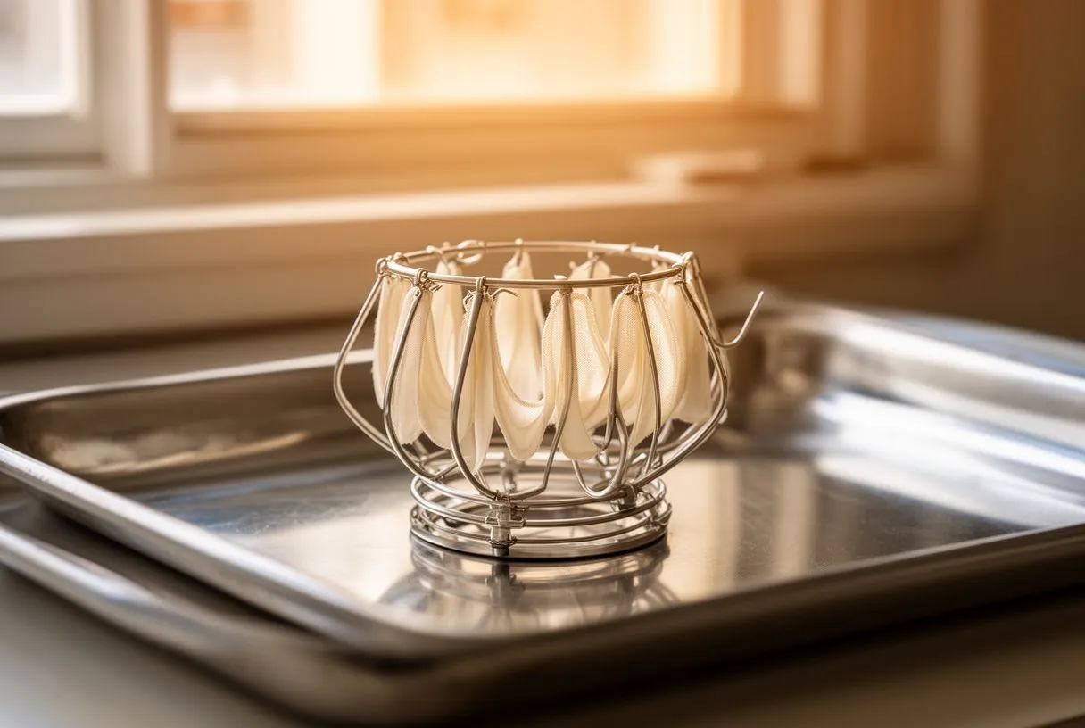
Valve-in-valve procedures. When a tissue valve fails, a new valve can often be placed inside the old one through a catheter, in a procedure called valve-in-valve implantation. This avoids removing the original valve and can be far less invasive than open surgery, though it depends on the size and type of the existing valve.
Surgical reoperation. Traditional valve replacement through open surgery remains a strong and well-established option, especially for younger patients, where a new surgical tissue valve or a mechanical valve may offer the best long-term result. The decision depends on your anatomy, your age, and how the first valve failed.
Newer valve designs. Options continue to evolve. A sutureless valve is designed to be seated quickly without extensive stitching, shortening surgery; a stentless valve omits the supporting frame and can behave more like a natural valve; a self-expanding valve opens to its final size gradually after placement, and some designs are intended to allow future valve-in-valve procedures. For failed mitral bioprostheses, transcatheter mitral valve replacement is an emerging route. Dr Molina trained in structural cardiology and TAVR at CHU Bordeaux, and our team reviews each failed-valve case together to determine which of these routes — surgical or transcatheter — offers the lowest risk for that patient's situation.
What Follow-Up Care Should You Expect, and How Do You Choose a Team?
Good follow-up is the single most controllable factor in managing a tissue valve. You should expect regular echocardiograms, a review of symptoms at every visit, dental hygiene guidance (since bacteria from the mouth can reach the valve), and clear instructions on which warning signs require urgent attention.
General guidance on valve disease, its symptoms, and testing is available from reliable patient resources such as MedlinePlus. But resources cannot replace a team that knows your valve, your scans, and your history.
At our practice, led by Dr Alejandro Molina with consulting rooms in Guadalajara, Querétaro and Hermosillo, the first contact is a free video consultation: we get to know you, talk through your condition and your valve, and tell you which studies are genuinely still needed so no unnecessary ones are ordered. If a face-to-face visit follows, we explain what every study shows in plain words before any decision is made. In conclusion: tissue valves are durable but not permanent, deterioration develops gradually, and regular monitoring is what keeps your options open. If your valve was placed years ago and you have new symptoms — or even if you simply have not had an echocardiogram in over a year — discuss your situation with a qualified cardiac team rather than waiting or self-diagnosing.