

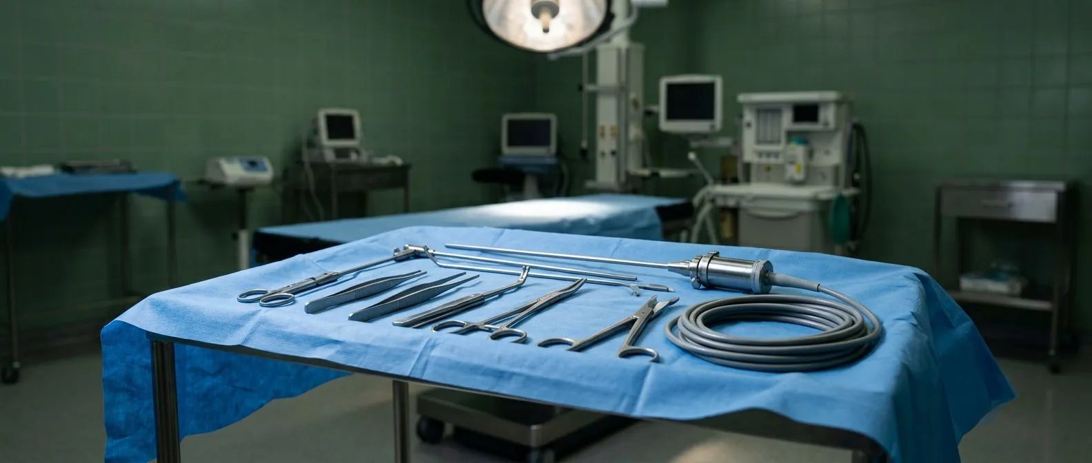
Surgical Exposure in Minimally Invasive Heart Surgery
Surgical exposure in minimally invasive heart surgery: how the surgeon reaches the heart through small incisions, explained by the cardiac team in Guadalajara.
When a surgeon opens this paragraph's central question — how does the heart become visible through a cut the length of a finger? — the answer lies in one concept: surgical exposure. In traditional open-heart surgery, the chest is divided down the middle through a full sternotomy, giving the surgical team a wide, direct view of the heart. In minimally invasive cardiac surgery, that wide window is replaced by a small incision, specialized instruments, and camera systems that work together to create an equally clear view of the surgical field. Understanding how this exposure is achieved helps patients and families make sense of what is actually happening during the operation — and why some patients are candidates for these approaches while others are not. At Cirugía de Corazón, we believe an informed patient makes calmer decisions, so this guide walks through the access techniques, the technology behind them, the safety evidence, and what recovery really looks like.
What Is Surgical Exposure in Minimally Invasive Heart Surgery?
Surgical exposure refers to how the surgeon gains visibility of and access to the heart during an operation. In conventional open-heart surgery, the breastbone (sternum) is divided completely, and a retractor holds the rib cage open. This full sternotomy provides unlimited direct vision — the surgeon sees the entire heart with the naked eye, can move instruments freely in any direction, and can convert to a larger operation without changing approach.
Minimally invasive cardiac surgery changes the geometry of the operation. Instead of opening the whole chest, the surgeon works through one limited incision — typically 5 to 8 centimeters — positioned to give the best possible angle on the specific valve or vessel being treated. The working space is smaller, so the surgeon relies on carefully planned instrument angles, lighting, and in many cases video magnification to see structures clearly.
The essential principle is that exposure is not sacrificed; it is redesigned. A right mini-thoracotomy for mitral valve surgery, for example, places the incision directly over the region of the heart where the mitral valve sits, so the surgeon's line of sight is actually more direct than it would be through a midline opening. Modern series of aortic valve replacement performed through limited access have described the technique as safe in experienced hands, with exposure adequate for the same steps as conventional surgery (see the center experience published in Cirugía y Cirujanos).
Why Surgical Exposure Matters for Your Safety
Exposure is not a cosmetic detail — it determines what the surgeon can see, how quickly problems can be handled, and how much tissue trauma the operation creates. Good exposure means the surgeon can identify delicate structures, control bleeding, and complete the repair with the same precision as in an open operation.
Direct vision vs video vision. In a full sternotomy, the surgeon looks directly at the heart. In video-assisted and robotic approaches, the surgeon views a high-definition, magnified image on a monitor. Magnification can actually reveal structures in greater detail than the naked eye, but it removes the natural depth perception of direct vision, which is why these techniques demand specific training and a consistent team.
The conversion question. Every minimally invasive heart operation carries a plan for what happens if exposure proves insufficient — for example, unexpected bleeding or difficult anatomy. Converting to a full sternotomy is not a failure; it is a built-in safety measure. When you discuss surgery with a cardiac team, it is fair and wise to ask how often conversion happens in their program and what triggers it.
What Are the Main Access Techniques for Surgical Exposure?
There is no single "minimally invasive" approach — several distinct techniques exist, each designed around the structure being treated. Knowing the vocabulary helps you follow your surgeon's explanation.
- Mini-sternotomy: a partial division of the breastbone, usually the upper or lower portion, most often used for aortic valve surgery while preserving most of the sternum.
- Right mini-thoracotomy: a small incision between the ribs on the right side of the chest, a common route for mitral and tricuspid valve surgery.
- Video-assisted (VATS) exposure: a thoracoscope — a thin camera — is inserted through a small port, transmitting a magnified view to monitors in the operating room.
- Robotic-assisted exposure: the surgeon sits at a console and controls wristed instruments inserted through small ports, with a 3-D endoscopic camera providing the field of view.
Each of these trades some of the "wide-open" familiarity of a sternotomy for smaller incisions and, in skilled hands, faster recovery. The right choice depends on the valve, the patient's anatomy, prior chest surgery, and lung function, because the lung on the operated side is typically temporarily deflated during thoracoscopic exposure to create the working space.

Mini-Sternotomy vs Right Thoracotomy: Which Exposure Is Used When?
The heart sits slightly rotated in the chest, and each valve has its own geography. This is why the incision is chosen to match the target.
Mini-sternotomy for aortic valve surgery. The aortic valve sits at the base of the aorta, near the top of the heart, close to the upper sternum. A partial upper sternotomy — often a "J" or inverted-T shape — gives direct exposure of the ascending aorta and aortic root while leaving the lower sternum intact. Studies of aortic valve replacement through this limited access have reported it as a safe technique in experienced centers, with operative steps essentially mirroring conventional surgery.
Right thoracotomy for mitral valve surgery. The mitral valve lies deeper and more to the left and back of the heart. Approaching it from the midline means working around the right side of the heart; approaching it through the right chest places the surgeon nearly in line with the valve itself. This is why the right mini-thoracotomy, often combined with video assistance, has become a standard minimally invasive exposure for mitral repair and replacement, as well as for many atrial procedures such as atrial septal defect closure and ablation for atrial fibrillation.
Coronary artery bypass exposure. Minimally invasive coronary artery bypass grafting (CABG) is among the more demanding applications, because multiple vessels on the beating or temporarily arrested heart must be reached. Exposure strategies include small left thoracotomies for grafting the front of the heart and hybrid strategies combining limited-access bypass with stents placed by cardiologists.
Thoracoscopic Surgical Exposure Methods Explained
Thoracoscopy means looking into the chest with a camera. In video-assisted cardiac surgery, the thoracoscope does more than assist — in many operations it becomes the primary means of visualization.
Port placement and lung isolation. Typically, a working incision of a few centimeters is made in the right chest, with one or two additional small ports for the camera and instruments. A specialized breathing tube isolates the right lung, letting it deflate gently so the chest cavity becomes a working space. Carbon dioxide is often gently insufflated to further lower the diaphragm and improve the field.
Peripheral cannulation. Because the surgeon cannot easily clamp the aorta through a tiny incision in every case, the heart-lung machine is often connected through cannulae placed in the groin vessels rather than directly into the heart and aorta. This is a defining feature of port-access cardiac surgery and is planned with ultrasound imaging of the vessels beforehand.
Guidance from within. A key adjunct to exposure in these operations is transesophageal echocardiography — an ultrasound probe positioned in the esophagus behind the heart. It confirms the placement of cannulae, guides the delivery of endoballoon clamps where used, and verifies the result of the valve repair before the chest is closed. Research on the early period after minimally invasive cardiac surgery has highlighted how intraoperative monitoring shapes the immediate postoperative course (see Reanimatology).
Robotic Heart Surgery Port Placement: How Does It Work?
Robotic systems take the video-assisted concept a step further. The surgeon does not stand over the patient holding instruments; instead, the surgeon operates from a console, and a machine translates hand movements into precise motions of wristed instruments inside the chest.
How the ports are arranged. Robotic mitral repair typically uses several small ports along the right chest side — one for the 3-D endoscope, two or three for instruments, and a small "service" incision through which suture material and the valve prosthesis, if needed, are passed into the chest. Port placement is individualized to the patient's chest shape and the valve's position, and it is rehearsed mentally by the surgical team before the first incision.
What the surgeon actually sees. The endoscope provides a three-dimensional, magnified view — often up to ten times life size — with articulation the human wrist could not achieve through a keyhole. This can make fine maneuvers such as placing tiny suture stitches in chordae or performing complex repairs feasible through very limited access.
Robotic surgery is not automatically better for every patient. It requires suitable anatomy, a team with substantial case volume, and the same readiness to convert to an open approach if needed. The robotic platform is a tool of exposure — not a substitute for surgical judgment.

Minimally Invasive Mitral Valve Exposure: What Should You Know?
The mitral valve has become the flagship operation for minimally invasive exposure, and for good reason: the right thoracotomy route aligns naturally with the valve's position in the heart.
Direct vision cardiac surgery still applies. Even through a small incision, portions of the operation are performed under direct vision — connecting the heart-lung machine, verifying instruments, handling unexpected findings. Video and robotic techniques supplement rather than replace the surgeon's ability to look at the field through the working incision. Experienced teams describe this blend as the essence of safe minimally invasive practice.
Repair versus replacement exposure. Mitral repair — reconstructing the patient's own valve — requires especially fine visualization, which the magnified camera view supports well. Exposure of the left atrium is achieved through the right chest, and the interatrial groove is opened to reach the valve. Degenerative mitral disease, the most common reason for surgery in this group, is generally favorable anatomy for these approaches.
When minimally invasive is not the right fit. Patients with severe lung disease, heavily calcified vessels in the groin, prior right-chest surgery, or complex multi-valve disease may be better served by a conventional approach. A frank evaluation — imaging, heart function tests, and a conversation about trade-offs — is what determines candidacy, not enthusiasm for a small incision.
Is Minimally Invasive Cardiac Surgery Safe?
For appropriately selected patients and experienced teams, minimally invasive heart surgery is regarded in established clinical understanding as a safe alternative to conventional surgery, with comparable outcomes for many standard operations such as isolated aortic or mitral valve procedures. Published center experiences and reviews — including those in The Lancet family of journals and the surgical literature cited above — consistently frame these techniques as safe when patient selection and team expertise are matched.
What the evidence emphasizes. Rather than promising universal superiority, the literature emphasizes trade-offs: less surgical trauma to the breastbone and chest wall, generally less pain and a faster return to daily activity in many patients, balanced against longer operative times in some settings, specialized equipment requirements, and a learning curve for the surgical team.
Risks are real, not eliminated. Bleeding, stroke, arrhythmias, infection, and the rare need for conversion to open surgery remain possible in any cardiac operation. The honest message is not "minimally invasive is risk-free" but "for your specific anatomy and diagnosis, one approach may offer the lowest risk profile." That determination requires a qualified cardiac team, imaging studies, and a risk assessment such as the EuroSCORE II calculation commonly used to estimate operative risk.
Recovery After Small Incision Heart Surgery
Recovery is where the differences between approaches become most tangible for patients and families.
The first days. Most patients wake in an intensive care unit or a closely monitored step-down unit. Breathing tubes come out within hours, and walking with assistance usually begins within the first day or two. Because the sternum may be intact or only partially divided in mini-sternotomy approaches, movement restrictions are often less strict than after a full sternotomy — though this varies by operation and surgeon's protocol.
The first weeks. Hospital stays for minimally invasive valve surgery are frequently shorter than for open surgery, and many patients describe returning to light activities within two to four weeks. Driving, lifting, and returning to work follow individualized timelines set by your surgical team. Chest-wall discomfort is common early and is managed with medication — we avoid promising a "painless" recovery, because no cardiac operation is painless.
Follow-up matters. Echocardiograms, wound checks, and medication reviews continue for weeks and months after surgery. Patients traveling internationally for care — common for our patients visiting Guadalajara, Querétaro, or Hermosillo — should plan adequate follow-up contact with the surgical team before returning home, and know exactly which local emergency resources to use if symptoms change.

Questions to Ask Your Surgeon About Surgical Exposure
You do not need medical training to have a productive surgical consultation — you need good questions. Bringing a family member and writing answers down helps enormously.
- Which exposure do you plan for my operation, and why? The answer should connect the incision to your specific valve or vessel.
- How many of these minimally invasive procedures does your team perform each year? Experience and volume are directly relevant to outcomes in these techniques.
- What would cause you to convert to an open approach? A clear answer signals preparedness and honesty.
- How will my heart-lung machine be connected, and what monitoring guides the operation? This reveals how exposure and cannulation are planned.
- What does recovery realistically look like for someone with my health profile? Ask for specifics, not generalities.
In summary, surgical exposure in minimally invasive heart surgery is the discipline of seeing the heart clearly through deliberately small doors — whether through a mini-sternotomy, a right thoracotomy, thoracoscopic cameras, or robotic ports. Each approach is chosen to match the target structure and the patient's anatomy, and each carries a well-defined plan for safety and, when needed, conversion. If you or a loved one has been told you need heart surgery, the most valuable next step is a consultation with a qualified cardiac team who can examine your imaging, discuss which exposure suits your situation, and answer your questions honestly. Rather than self-diagnosing from articles like this one, book a consultation with the team at Cirugía de Corazón and bring your questions — including the ones on this page.






