Surgical Wound Healing After Heart Surgery
Surgical wound healing heart surgery guide: stages, sternum care, and infection prevention from Guadalajara's cardiac specialists.
What Is Surgical Wound Healing After Heart Surgery?
Surgical wound healing after heart surgery is the biological process your body undertakes to repair the layers of tissue divided during a cardiac operation. When a surgeon opens the chest wall — typically through a median sternotomy, which means splitting the breastbone along its midline — multiple tissue layers are involved: skin, subcutaneous fat, muscle, and bone. Each layer closes on its own timetable, and understanding that timeline helps patients and families know what to expect and when something looks wrong. The focus keyword matters because the sternum is one of the largest bones in the body to be intentionally cut, and its reunion demands weeks of stability and careful management.
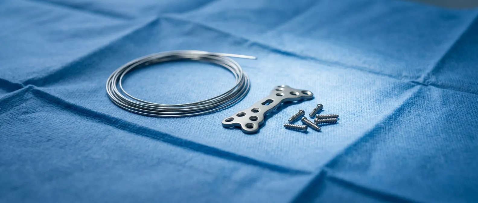
What tissues are involved in the closure? The outermost layer is the skin, which re-epithelializes within the first 48 to 72 hours, meaning new surface cells bridge the incision gap rapidly. Beneath the skin sits subcutaneous fat and connective tissue, which takes longer to regain structural integrity. The pectoralis fascia and the sternum itself form the deepest layers. Stainless steel wires hold the bone edges together, and these wires remain in place permanently in most patients — they do not typically need removal unless they cause pain or protrude. During this early phase, blood supply to the wound edges delivers oxygen, immune cells, and nutrients essential for collagen deposition.
Why does cardiac wound healing differ from other operations? Heart surgery wounds face unique mechanical stresses. Every breath expands the rib cage, placing tension across the sternum. Every cough or sneeze generates forceful pressure that can stress the closing bone. Patients may also experience Arm Pain from positioning during surgery or from harvesting an Arterial Graft, which adds to overall discomfort and can make protective postures difficult. Additionally, the heart and lungs lie directly beneath the sternum, so any deep infection — a condition called mediastinitis — can be life-threatening and demands immediate attention. These factors make surgical wound healing after heart surgery a distinct discipline within postoperative care.
How Does the Sternum Heal After Open Heart Surgery?
The sternum, or breastbone, is cut vertically down the center during a standard open-heart procedure to give the surgeon direct access to the heart and the Ascending Aorta. After the cardiac work is complete — whether that is a coronary bypass, a valve repair, or another intervention — the surgeon brings the two halves of the bone back together and wraps stainless steel or titanium wires around them. These wires act like internal splints, holding the bone edges in close approximation so that new bone can bridge the gap. Full bony union typically takes six to eight weeks, but complete remodeling — where the bone regains maximum strength — can take up to six months.
What happens at the cellular level? The body initiates a cascade of inflammatory, proliferative, and remodeling phases. Within the first several days, a blood clot forms between the bone edges, creating a scaffold called a callus. Osteoblasts — bone-building cells — migrate into this scaffold and begin depositing new mineralized tissue. Over weeks, this woven bone is replaced by stronger lamellar bone. Factors that slow this process include smoking, diabetes, poor nutritional status, and advanced age. Patients who received an Autologous Blood transfusion — their own blood collected before surgery — may have a slightly different inflammatory profile, but the fundamental healing sequence remains the same. According to Harvard Health, optimizing these modifiable risk factors before elective surgery measurably improves recovery outcomes.
What can patients do to protect the healing sternum? The most important external protection is avoiding lateral or twisting forces on the chest. Patients are taught to use a heart pillow or folded blanket to splint the chest when coughing, and to avoid pushing, pulling, or lifting anything heavier than a few kilograms for the first six to eight weeks. Driving is usually restricted until the sternum is sufficiently stable — often four to six weeks — because the steering wheel could strike the chest in a sudden stop. Sleeping on the back or slightly elevated side is recommended, and many patients find that getting out of bed by log-rolling — rolling as a unit without twisting — reduces sternal stress and discomfort.
What Are the Stages of Chest Incision Care?
Chest incision care evolves through clearly defined stages, each with its own priorities and red flags. In the first 48 hours, the incision is covered with a sterile dressing that the nursing team manages. Patients are typically still in the intensive care unit during this time, connected to an Arterial Line for continuous blood pressure monitoring and sometimes supported by Awake ECMO if the heart or lungs need temporary assistance. The wound is kept clean and dry, and the surgical team assesses it daily for signs of bleeding, excessive drainage, or early infection.

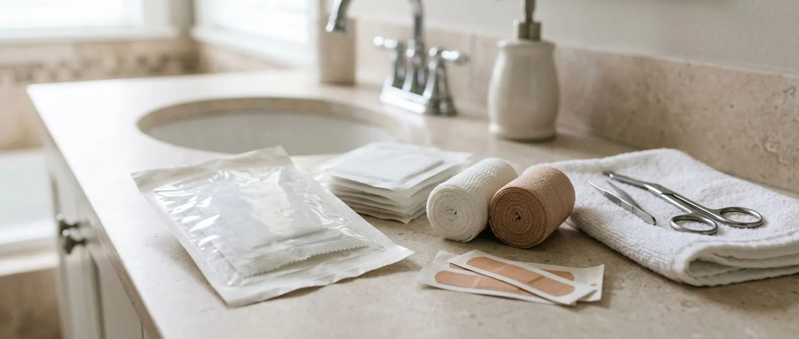
Days 3 to 14 — early outpatient care. Once the dressing is removed, patients or family members take over daily inspection. The incision should be gently washed with mild soap and water, patted dry, and left open to air unless otherwise instructed. Thin, clear or slightly pink drainage can be normal in the first few days, but thick, opaque, or foul-smelling drainage is not. Steri-strips — thin adhesive strips placed across the incision — will curl and fall off on their own within one to two weeks and should not be pulled off prematurely. Patients should avoid applying lotions, creams, or ointments unless their surgeon specifically recommends them, as these can trap moisture and bacteria against the healing skin.
Weeks 2 to 8 — maturation and strengthening. The incision will transition from bright red or pink to a paler, flatter color over several weeks. Some itching is common as nerve endings regenerate, but scratching should be avoided. The area around the wound may feel firm or slightly raised — this is normal scar tissue forming beneath the surface. Gentle movement of the arms and shoulders is encouraged to prevent stiffness, but overhead reaching and heavy lifting remain off-limits. Patients who underwent Auscultation — listening to the chest with a stethoscope — during follow-up visits will have their wound checked alongside heart and lung sounds to confirm healing is progressing as expected.
How Long Is the Heart Surgery Recovery Timeline?
The heart surgery recovery timeline spans weeks to months, and every patient's journey is somewhat individual. Hospital stays typically range from four to seven days for uncomplicated cases, though this can extend if complications arise. The first 24 to 48 hours are spent in intensive care, where continuous monitoring tracks heart rhythm, blood pressure, and oxygenation. Once stable, patients move to a step-down unit and begin walking short distances with assistance. Discharge happens when the medical team is satisfied that the wound is stable, the heart is functioning well, and the patient can manage basic activities safely.
Weeks 1 to 4 — the home recovery phase. Fatigue is the dominant sensation during this period. The body is expending enormous energy on surgical wound healing after heart surgery, and patients often need daytime naps alongside a full night's sleep. Walking is the primary exercise — starting with short distances and gradually increasing. Cardiac rehabilitation wound assessments may begin around week three, where a supervised program monitors exercise tolerance while keeping an eye on incision integrity. Patients on Aspirin or other blood thinners will have their dosing reviewed, and any signs of excessive bruising around the wound should be reported. According to research indexed at PubMed, structured early mobilization protocols correlate with shorter overall recovery times and fewer wound-related complications.
- Week 1: Hospital discharge, daily wound inspection, short walks, no lifting.
- Weeks 2–4: Gradually increased walking distance, light household activity, begin cardiac rehabilitation if cleared.
- Weeks 4–8: Sternal bones nearing union, may return to non-strenuous work, start supervised exercise progression.
- Months 2–6: Most patients feel significantly better, scar maturing, return to most normal activities with clearance.
Months 3 to 6 — return to full function. By three months, most patients feel a dramatic improvement in energy and breathlessness. The sternum has usually achieved clinical union — meaning the bone is solid enough to tolerate normal activity — though complete remodeling continues. Scars continue to fade and soften. Any persistent Arm Pain from positioning or graft harvesting should be evaluated and may respond to targeted physiotherapy. Follow-up appointments during this phase often include imaging or functional testing to confirm that the underlying cardiac repair is holding, and that the wound has healed without complications.
What Are Sternal Wound Infection Prevention Steps?
Sternal wound infection prevention begins before the operation and continues through every phase of recovery. Preoperatively, surgeons screen for risk factors such as diabetes, obesity, smoking, and chronic obstructive pulmonary disease. Blood sugar control is critical: elevated glucose impairs white blood cell function and slows collagen synthesis. Patients who smoke are strongly advised to stop well before surgery, as nicotine constricts the small blood vessels that deliver healing cells to the wound edges. Prophylactic antibiotics are given intravenously within one hour of the incision and continued briefly postoperatively.
What are the modifiable risk factors? Several patient-level factors can be optimized before surgery. Weight management reduces pressure on the wound and lowers infection risk. Nutritional status — particularly protein, vitamin C, and zinc levels — directly supports collagen production. Poor dental hygiene can seed bacteria into the bloodstream, so dental evaluation is sometimes recommended before elective surgery. For patients with an Atheromatous Plaque burden or other vascular disease, optimizing blood flow to the chest wall tissues improves oxygen delivery to the wound. The surgical team will also consider whether an Autograft or synthetic material is used in the repair, as this can influence the local healing environment.
What daily habits prevent infection at home? Hand hygiene is the single most effective infection prevention measure. Anyone touching the wound area — including the patient — must wash hands thoroughly with soap and water or use an alcohol-based sanitizer. The incision should be inspected daily for increasing redness, spreading warmth, new drainage, or separation of edges. Pets should be kept away from the wound area, and the incision should not be submerged in water — no baths, pools, or hot tubs — until the surgeon gives clearance, usually around four to six weeks. If a cough develops, splinting the chest with a pillow protects the wound from mechanical stress and reduces the risk of dehiscence, which is the separation of the sternal edges.
How to Care for Cardiac Surgery Scars at Home?
Cardiac surgery scar care at home is a gradual process that begins once the incision has closed and any surgical glue or adhesive strips have naturally separated. The scar will go through predictable changes: it may appear raised, red, and firm in the early weeks, then gradually flatten, soften, and fade over six to twelve months. Some patients develop thicker, wider scars called hypertrophic scars, particularly if they have a family history of abnormal scarring. Keloid scars — which extend beyond the original wound boundary — are more common in patients with darker skin tones and may require dermatologic treatment.
What promotes favorable scar appearance? Once the skin is fully closed and the surgeon approves, gentle massage with a fragrance-free moisturizer can help soften scar tissue and improve flexibility. Silicone gel sheets, applied overnight, have clinical evidence supporting their use for scar flattening. Sun protection is essential: UV exposure can permanently darken a healing scar, so the area should be covered or protected with sunscreen for at least the first year. Patients who had an Arterial Graft harvested — often from the internal mammary artery or radial artery — will have additional smaller incision sites to care for, and the same principles apply to each.
- Silicone sheets: Clinically shown to flatten and soften raised scars when used consistently for several months.
- Gentle massage: Circular pressure with moisturizer improves tissue flexibility and reduces tightness.
- Sun protection: UV exposure darkens new scars permanently; cover or use SPF 30+ for 12 months.
- Surgical review: Any spreading, thickening, or painful scar changes should be assessed by the cardiac team.
When should a scar concern you? A scar that becomes increasingly painful rather than less painful over time, or one that opens, drains, or develops surrounding redness, warrants prompt evaluation. Some patients experience a sensation of wires being prominent or sharp beneath the skin as swelling resolves — this is not always a problem, but if the wire causes pain or pokes through the skin, surgical removal may be discussed. Numbness around the scar is common and often permanent, as small cutaneous nerves are divided during the incision, but it does not affect function. The key principle is that scar care is part of surgical wound healing after heart surgery, not an afterthought.
What Are Common Heart Surgery Wound Complications?
Heart surgery wound complications range from minor, self-limiting issues to serious conditions requiring urgent intervention. The most common minor complication is a superficial infection — redness and drainage limited to the skin and subcutaneous layers, typically treated with oral antibiotics and local wound care. Seromas, which are collections of clear fluid beneath the skin, can also occur and may resolve on their own or require aspiration. Hematomas — pooled blood under the skin — can develop if a patient is on blood thinners and usually reabsorb over time without intervention.
What is mediastinitis and why is it dangerous? Mediastinitis is a deep sternal wound infection that reaches the mediastinum — the central compartment of the chest containing the heart and great vessels. It occurs in approximately one to two percent of cardiac surgery patients and is a severe complication. Symptoms include increasing chest pain, fever, wound breakdown with purulent drainage, and a feeling of instability or clicking in the sternum. Risk factors include diabetes, obesity, bilateral internal mammary artery harvesting, and prolonged operative time. Treatment requires surgical debridement, sometimes reconstructive flap surgery, and extended intravenous antibiotics. According to a clinical study tracked on ClinicalTrials.gov, optimizing intraoperative and postoperative protocols remains an active area of investigation to reduce these serious outcomes.
What is sternal dehiscence? Sternal dehiscence is the separation of the sternal halves, which can occur when the wires break, pull through the bone, or when infection weakens the bone edges. Patients may describe a popping or clicking sensation, visible separation of the incision, or a feeling that the chest is unstable. This is a surgical emergency because the underlying heart and vessels become vulnerable. Re-operation is usually required to re-wire the sternum, sometimes with additional reinforcement techniques such as plates or a muscle flap. Recognizing the signs early — through daily wound inspection and prompt reporting of any change — is the best protection a patient has against catastrophic progression.
When Are Sternal Wires Removed During Healing?
Sternal wires are designed to remain permanently in the vast majority of patients. These thin stainless steel or titanium loops hold the bone edges together while the callus forms and the bone reunites. Once the sternum has healed — typically by eight to twelve weeks — the wires serve no further mechanical purpose, but they are inert and generally cause no problems. Leaving them in place avoids the risks of a second surgical procedure, including anesthesia, infection, and additional wound healing.
When is wire removal actually necessary? A small percentage of patients develop symptoms that make removal appropriate. These include a wire that protrudes through the skin, causing pain or a visible bump; a wire that breaks and migrates, sometimes causing discomfort or palpable sharpness; or a localized infection around a wire that cannot be cleared without hardware removal. Patients with thin subcutaneous tissue — particularly those with low body weight — may be more likely to feel wires beneath the skin as postoperative swelling resolves. Some patients describe a clicking or popping sensation that, while not always pathological, can be distressing enough to warrant removal.
What does the removal procedure involve? Wire removal is typically a day-case procedure performed under local or light general anesthesia. The surgeon makes small incisions over the wire ends, cuts and extracts the loops, and closes the skin with minimal suturing. Recovery is far shorter than the original surgery — usually a few days of wound care and activity restriction. The sternum itself is already healed, so no additional bracing is needed. It is worth noting that wire removal does not undo the healed bone; the sternum remains solid. Patients who have had a Balloon Angioplasty or Balloon Aortic Valvuloplasty as part of their cardiac workup may have additional access sites to consider, but sternal wire management remains specific to the sternotomy itself.
How Does Robotic Heart Surgery Healing Compare?
Robotic and minimally invasive cardiac surgery approaches have transformed surgical wound healing after heart surgery for selected patients. Instead of a full sternotomy, these techniques use small incisions between the ribs — typically on the right side of the chest — through which instruments and a camera are inserted. The heart is accessed without splitting the breastbone, which fundamentally changes the wound healing profile. Recovery is often faster, pain is typically less, and the risk of sternal complications is eliminated because the sternum is not divided.
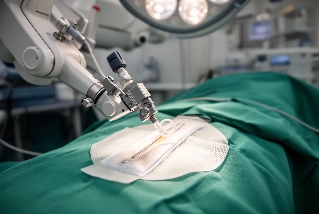
Who is a candidate for minimally invasive approaches? Not every cardiac procedure can be performed this way. Minimally invasive techniques are well established for certain valve repairs and replacements — including implantation of a Balloon-expandable Valve — and for some coronary bypass grafting strategies. Patient selection depends on anatomy, body habitus, the specific cardiac pathology, and the surgeon's experience. Patients with prior chest surgery, severe lung disease, or complex multi-vessel coronary disease may not be suitable candidates. An Arterial Cannula is still placed for cardiopulmonary bypass, but the chest wall trauma is substantially reduced. The decision is always individualized, weighing the benefit of faster wound healing against the technical feasibility of the approach.
What is the healing timeline difference? Without a sternotomy, the restrictions on lifting, pushing, and driving are typically shorter — often two to four weeks instead of six to eight. The incisions are small and heal quickly, though the thoracotomy approach can cause intercostal nerve irritation that produces localized discomfort for weeks to months. Hospital stays may be shorter, and return to work can be faster for appropriate patients. However, the internal cardiac repair is identical to open surgery, so the heart itself needs the same recovery time regardless of the access method. Patients should not confuse smaller external scars with a shorter internal recovery.
When to Seek Help During Post Heart Surgery Wound Care?
Knowing when to seek help during post heart surgery wound care can prevent minor issues from becoming serious complications. Patients receive detailed discharge instructions, but the volume of information can be overwhelming. The guiding principle is simple: any change from the expected healing pattern warrants a phone call to the surgical team. It is always better to ask and be reassured than to wait and risk escalation. Most cardiac surgery practices have a direct line or nurse coordinator for exactly these situations.
What symptoms require immediate attention? Fever above 38°C (100.4°F), particularly after the first 48 hours, should be reported. Increasing redness that spreads outward from the incision, warmth, swelling, or new drainage — especially if thick, yellow, green, or foul-smelling — are signs of possible infection. Separation of the incision edges, visible bone, or a wire protruding through the skin require urgent evaluation. A sensation of sternal clicking or instability that is new or worsening should not be ignored. Chest pain that feels different from the expected postoperative soreness — particularly if it is sharp, sudden, or associated with shortness of breath — may indicate a serious problem and warrants emergency assessment.
What symptoms can wait for a scheduled appointment? Mild itching as the incision heals, small amounts of dry flaking skin around the scar, slight bruising that is fading, and minor numbness around the wound are all expected parts of healing. Questions about scar appearance, return-to-work timing, and activity progression are appropriate for routine follow-up. Patients who notice a small seroma — a soft, fluid-filled lump beneath the skin — should mention it at their next visit unless it is growing or painful. The distinction between expected healing and warning signs becomes clearer as patients gain confidence, but in the early weeks, the surgical team expects and welcomes questions. Close with this: every patient's surgical wound healing after heart surgery is unique, and the best decision you can make is to discuss your specific situation with your qualified cardiac team and book a consultation rather than self-diagnosing from this or any article.