

The Arterial Cannula in Heart Surgery and ECMO
Arterial cannula heart surgery explained: placement, ECMO support, types, sizes, and recovery.
If you or a family member is facing open heart surgery or needs advanced life support, the word "cannula" will come up early. An arterial cannula in heart surgery is a short, specially designed tube that connects the patient's main artery to the heart-lung machine, or to an ECMO circuit in the most critical cases. It is the point where oxygen-rich blood, pumped by the machine, returns to the body. This guide explains what these tubes are, where they are placed, how they differ from the venous cannulas that drain blood away, what risks they carry, and what recovery involves — in plain language, from the patient's point of view.
What Is an Arterial Cannula?
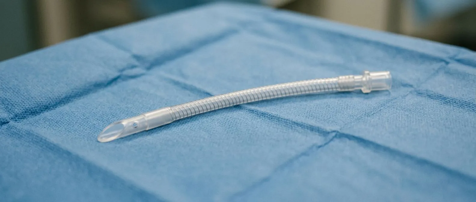
An arterial cannula is a sterile, plastic tube — usually made of medical-grade PVC or polyurethane — that is inserted into an artery so that blood can be delivered back into the circulation under pressure. In the operating room, its tip is often beveled and wire-reinforced so the tube stays open (does not kink) while blood flows through it at high speed. A thinner wire-reinforced design is called a thin-walled cannula: it gives the surgeon maximum flow through a smaller hole in the artery, which matters because a smaller hole means less damage to the vessel.
Why the design matters to you. Every detail of the tube — the angle of the tip, the number and size of side holes, the coating that reduces clotting — exists to move blood safely and gently. The tip is designed to direct the flow of blood toward the aorta's natural direction, away from the brain's main branches, which reduces the risk of dislodging deposits of calcium or plaque from the artery wall.
It is worth separating two very different things that share the word "arterial cannulation." In an intensive care unit, a tiny 20- or 24-gauge needle-like tube (a 24-gauge is the smallest common size, about the width of a pencil lead) is placed in the wrist to monitor blood pressure continuously. That is an arterial line, and it only measures — it does not pump blood. The large tube used in heart surgery and ECMO serves a completely different purpose: it carries the entire output of the heart-lung machine back to the patient. When we explain this to families in consultation, we make the distinction explicit, because the two procedures carry very different risks and are often confused in hospital conversations.
What Is the Purpose of Arterial Cannulation?
The purpose of arterial cannulation in heart surgery is simple to state and profound in effect: it creates the return path for oxygenated blood. During open heart surgery, the heart is stopped so the surgeon can work inside it. The heart-lung machine (the cardiopulmonary bypass circuit) takes over — it drains blood from the veins, adds oxygen, removes carbon dioxide, and pumps it back through the arterial cannula into the aorta, the body's largest artery. From there, the oxygenated blood reaches the brain, kidneys, and every other organ.
Where the tube goes. Most often the tip of the arterial cannula is placed in the ascending aorta — the first portion of the aorta, just above the heart. This is called ascending aorta cannulation, and it is the standard approach for most coronary bypass and valve operations. When the aorta itself is diseased, too fragile, or being replaced, the surgeon can instead use the femoral artery in the groin, the axillary artery under the collarbone, or the innominate artery at the base of the neck. Each site has trade-offs in flow, stroke risk, and the type of operation being done.
What it buys the surgical team. With a reliable arterial return path, the team can operate on a still, bloodless field while the patient's organs are continuously perfused. Surgeons monitor pressures, blood gases, and flow rates through the arterial line throughout the case.
Arterial Cannula vs Venous Cannula: What Is the Difference?
The arterial cannula vs venous cannula distinction is one of the clearest ways to understand the circuit. A venous cannula drains blood away from the patient; an arterial cannula returns it. The two are not interchangeable — they are built differently, sized differently, and inserted into different vessels.
- Direction of flow: the venous cannula siphons deoxygenated blood from the right atrium or the great veins (often helped by gravity or a vacuum); the arterial cannula pushes oxygenated blood back under pressure.
- Wall thickness: venous cannulas are thin-walled with large openings to let blood drain easily by gravity; arterial cannulas have reinforced walls and a shaped tip to withstand high pumping pressure.
- Size logic: venous sizes are chosen to maximize drainage without obstructing the vein; arterial sizes are chosen to deliver full flow without raising pressure dangerously in the artery.
- Insertion site: venous cannulas go into the right atrium, superior vena cava, or femoral vein; arterial cannulas go into the aorta, femoral artery, or axillary artery.
Why the vein-versus-artery question comes up. People sometimes ask why a cannula is "inserted in the vein but not in the artery" for routine IV access. The answer is that veins are low-pressure, easily compressed, and heal quickly — so for ordinary drips and medication, a vein is far safer. An artery is a high-pressure vessel with thick muscular walls; puncturing it needs care, and bleeding from an artery can be serious. That is exactly why arterial punctures in the ICU are reserved for arterial lines and arterial cannulation is reserved for situations where the benefit is essential: bypass surgery, ECMO, and continuous blood pressure monitoring in critically ill patients.
What happens if an artery is accidentally cannulated? In practice, if an IV line intended for a vein ends up in an artery, it is recognized quickly — bright red, pulsing blood, or a monitor reading arterial pressure — and the line is removed with firm pressure applied. Serious injury is rare, but the episode is treated with respect and the arm is watched closely for signs of compromised circulation. Deliberate arterial cannulation is different: it is done with sterile technique, ultrasound guidance, and the correct equipment, which is what makes it safe.
How Is Arterial Cannula Placement Done in Cardiac Surgery?
Arterial cannula placement in cardiac surgery follows a sequence that patients rarely see but should understand. After the patient is asleep and the chest is opened, the surgeon inspects the aorta. Using the fingers and, increasingly, ultrasound and epiaortic scanning, the team assesses the aortic wall for calcified plaque — hard chalky deposits that could be broken loose by the cannula. The insertion site is chosen to avoid these areas.
The aortic approach. For ascending aorta cannulation, the surgeon places two purse-string sutures (circular stitches that tighten around the tube like a drawstring) on the aorta, makes a small opening, and slides the cannula in. The sutures are snugged and tied, sealing the tube in place. Heparin — a blood thinner — is given before cannulation so clots do not form on the circuit. The cannula is then connected to the heart-lung machine's arterial line, and bypass begins gradually.
Alternative sites for a diseased aorta. When the ascending aorta is heavily calcified or aneurysmal, direct cannulation of it is dangerous. In that case, the femoral artery in the groin or the axillary artery under the collarbone is used. Axillary cannulation is particularly valued because it provides antegrade flow — blood moving in the body's natural direction — which also protects the brain during aortic arch surgery. In our team's practice, every case is reviewed by the multidisciplinary team against international guidelines before any decision about cannulation strategy, and the reasoning is explained to the patient in plain words before surgery.
Femoral arterial cannula insertion is also the usual route for minimally invasive and robotic operations, where the chest is not opened widely. In those procedures, the surgeon works through small incisions while the heart-lung machine runs through groin-based cannulation. Dr Molina performs robotic and minimally invasive procedures regularly, including the first robotic mitral valve repair in the Ángeles Health System, and cannulation strategy is tailored to each patient's anatomy in those cases.
How Does an Arterial Cannula Provide ECMO Support?
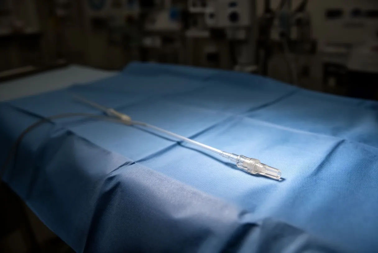
ECMO — extracorporeal membrane oxygenation — is a portable heart-lung machine that can support a patient for days or weeks. An arterial cannula is central to one of its two main configurations. In veno-arterial ECMO, blood is drained from a large vein and returned, via the arterial cannula, into an artery — usually the femoral artery or the axillary artery. The machine then does the work of both the heart and the lungs, which is why veno-arterial ECMO is chosen when the heart itself is failing: after massive heart attack, in severe cardiogenic shock, or during cardiotomy emergencies.
Veno-arterial versus veno-venous. In veno-venous ECMO, blood is both drawn from and returned to veins, and the machine supports only the lungs — the heart keeps doing its own pumping. Veno-venous ECMO is used for severe lung failure, such as devastating pneumonia or COVID-related respiratory failure. The choice between the two configurations determines where the arterial cannula goes — or whether one is needed at all.
Awake ECMO and other extensions. In some patients, ECMO support can be delivered without sedation, allowing the patient to breathe, eat, and participate in rehabilitation — a strategy sometimes called awake ECMO. Where arterial cannulation is involved, the insertion site is monitored daily for the pulse distal to the cannula (the pulse beyond it), because the large tube in the femoral artery partially blocks blood flow to the leg.
A landmark randomized trial of intraarterial treatment for acute ischemic stroke published in Maastricht University's research repository demonstrated how directly delivering therapy into the arterial tree can change outcomes — a principle that echoes the broader idea behind arterial cannulation: when the artery is the right access point, the path to the patient's organs is shorter and more effective. ECMO carried to the operating room, the catheterization lab, or even the ambulance has made this principle portable in emergency care.
What Types of Arterial Cannulas Are Used for Heart Surgery?
There is no single arterial cannula. Manufacturers design several families of cannula, each shaped for a specific insertion site and flow requirement. Knowing the main types helps patients ask informed questions about their own operation.
- Straight aortic cannulae: the classic workhorse for ascending aorta cannulation; simple, rigid, with a beveled or angled tip.
- Right-angled aortic cannulae: bent at a right angle so the tube exits the surgical field at a low profile, useful in minimally invasive operations where the chest opening is small.
- Wire-reinforced cannulae: built with a spiral metal mesh inside the wall so the tube resists kinking even when bent sharply; essential for femoral and axillary cannulation, where the tube takes a turn.
- Pediatric cannulae: much smaller versions, sized in fractions of a millimeter, designed for infants and children whose aortas are tiny — pediatric arterial cannula heart surgery demands precision because the entire circulation of a small child flows through it.
- Safety cannulae: designs with integrated features — such as a tapered tip or an introducer sheath — that reduce the risk of aortic injury during insertion.
The heart-lung machine connection. Every arterial cannula connects to the pump's arterial line through a standardized connector. The tubing leading to the pump is then monitored continuously for pressure, clots, and air. On the circuit, this arterial limb is the final common path back to the patient; any problem there is an emergency, which is why multiple alarm systems watch it throughout the case.
Which cannula is 24 gauge? In critical care, a 24-gauge arterial line catheter is the smallest common size, used for fragile or small arteries — for example, in low-weight infants or in adults with severe peripheral vascular disease. In cardiac surgery and ECMO, cannulae are sized in French units (Fr) — a measure of outer diameter — and typical adult arterial cannula sizes range from about 16 Fr to 24 Fr, chosen to deliver full cardiac output. The gauge system and the French system are different scales for different contexts.
How Is Arterial Cannula Size Selected?
Arterial cannula size selection is a genuine engineering decision, made before the skin is cut. The cannula must be large enough to carry the patient's full cardiac output without forcing the pump to generate dangerous pressure, and small enough not to damage the artery wall or make closure difficult. Surgeons use published flow charts that map cannula size to expected flow, combined with the patient's body surface area and the target blood flow for the operation.
Too small, and the circuit strains. An undersized cannula restricts flow. The heart-lung machine compensates by raising pressure, which creates turbulence, increases the risk of red blood cell destruction (hemolysis), and can make it impossible to deliver adequate perfusion to the organs. The perfusionist will report rising pressures and reduced flow margins.
Too large, and the artery suffers. An oversized cannula can tear the aortic wall, make it impossible to close the entry site cleanly, or partially obstruct the vessel around it. The pressure gradient across the cannula is checked during bypass; if it is too high, the team may need to reposition or change the cannula — a rare but planned-for eventuality. In our consultations, we take the time to explain in plain words what the imaging studies show about the patient's aorta, because the aortic diameter and condition drive the choice of cannulation site and size.
What Are the Risks and Complications of Arterial Cannulation?
Arterial cannula complications are uncommon but real, and an honest discussion of them is part of any informed decision. The most feared is aortic dissection — a tear in the layers of the aortic wall — which can occur during insertion if the vessel is fragile or calcified. Modern techniques, including ultrasound assessment of the aorta before cannulation and the use of soft-tipped cannulae, have reduced this risk substantially, but it is never zero.
Stroke and debris. Because the arterial cannula sits in the aorta and receives flow from a pump, any calcium or plaque dislodged during insertion can travel toward the brain. Strategies to lower this risk include choosing a cannulation site away from plaque, using filters or special cannula designs, and in selected patients avoiding the aorta entirely by using axillary or femoral access. Research on the circulation of cerebrospinal fluid has shown that arterial pulsations drive flow through the brain's fluid channels and that this flow is reduced in hypertension — one of many reasons surgeons pay close attention to blood pressure management during bypass as well as to the mechanics of cannulation itself.
Limb ischemia with femoral cannulation. When the arterial cannula is placed in the femoral artery, the tube partially blocks the artery, and the leg beyond it may receive reduced blood. Teams handle this in two ways: a smaller cannula with a side arm that perfuses the leg, or a second small catheter (an antegrade perfusion catheter) inserted into the femoral artery beyond the cannula to restore leg flow. The foot is monitored continuously for color, warmth, and Doppler signals.
Other risks include bleeding from the cannulation site, infection (rare with sterile technique), hematoma in the groin, and — rarely — injury to the artery wall requiring repair. Each risk is weighed against the alternative: without a functioning arterial return, the operation cannot proceed. The relevant question is not whether risk exists, but whether the team has planned for it — and whether the less invasive route, where the evidence supports it, has been considered for that specific patient. We recommend the less invasive approach only where the evidence supports it for that patient, and every patient receives a clear proposal with the alternatives laid out.
What Does Arterial Cannula Removal and Recovery Look Like?
Arterial cannula removal is one of the most carefully choreographed moments of the operation. When the heart-lung machine is stopped and the heart has taken over, the surgeon removes the aortic cannula while the purse-string sutures are tightened and tied. The arterial pressure inside the aorta is high, so the closure must be secure. The surgeon watches the site, checks the pulse, and confirms hemostasis — the complete stop of bleeding.
For aortic cannulation sites. Recovery from an aortic cannulation site is usually uneventful — it is inside the chest, closed with the rest of the operation, and patients rarely think about it again. The other chest tubes and incisions dominate the early recovery experience.
For femoral cannulation sites. A groin cannulation site is more visible and more sensitive. Patients may notice a small incision, bruising, or temporary swelling. The leg is monitored for the first hours after surgery: warmth, color, and the pulse in the foot. In rare cases where the leg was perfused with a separate catheter, that catheter is removed once the circulation is confirmed. Walking usually resumes within the first days of mobilization; the groin site heals like any other small incision.
For ECMO cannulation sites. When ECMO is decannulated — the term for removing the cannulas — the femoral artery is usually repaired with a small patch or closed with a purse-string technique, and sometimes the vessel is reconstructed. The limb is observed closely for the following days. The nursing team checks for pulses, temperature, and any change in the leg's appearance, and rehabilitation begins with careful attention to the cannulation site. Most patients, once decannulated, describe the memory of the cannula as less frightening than the anticipation of it — the tubes themselves are not painful, and the sedation or recovery period carries them past the worst of it.

When Should You Discuss Arterial Cannulation with a Cardiac Team?
If you have been told you need open heart surgery, valve intervention, or TAVR, arterial cannulation will be part of your operation — and it is a legitimate topic to bring up with your surgical team. The questions worth asking are simple: where will the arterial cannula be placed, why that site, and what does the imaging of my aorta show? A good team will answer these plainly. We encourage second opinions and treat seeking one as a normal part of deciding about heart surgery, and the first contact with our practice is a free video consultation in which we talk through the condition, the studies already done, and which studies are still needed — so no unnecessary ones are ordered.
Specific situations where the cannulation strategy matters more. Patients with a heavily calcified or aneurysmal aorta, previous aortic surgery, severe peripheral vascular disease, or a history of stroke benefit from a team that has a full range of cannulation strategies — not only the standard approach. In these cases, the choice of cannulation site can meaningfully influence the risk of the operation. Dr Molina trained in cardiothoracic surgery at the Universidad de Guadalajara, in cardiac, minimally invasive, robotic and structural surgery at the University of Montreal, and in structural cardiology and TAVR at CHU Bordeaux, and the team also trained at Jena, Bad Berka and Buffalo — this breadth of training is precisely what allows cannulation strategy to be matched to the patient rather than to the surgeon's habit.
Choosing the right team. Look for a multidisciplinary team that reviews every case against international practice guidelines before recommending anything, and that involves you in the decision. Our team reviews each case in this way, and the final decision is always the patient's. If you are facing surgery in Guadalajara, Querétaro, or Hermosillo, or would like to travel for care, the first step is a consultation — a conversation in which the condition is explained in plain language and every question is answered before anything is scheduled.
The arterial cannula is a small piece of equipment that sits at the center of some of the most consequential moments in medicine: the moment the heart-lung machine takes over, and the moment life support bridges a failing heart to recovery. Understanding what it is, where it goes, and what risks it carries will not make you a surgeon — but it will make you a better-informed partner in your own care. The decision that follows from this understanding is the one worth making with a qualified cardiac team by your side.






