The Arterial Media: The Muscle Layer Behind Aneurysms
Learn how the arterial media layer weakens and causes aneurysms.
What Is the Media in Your Artery Walls?
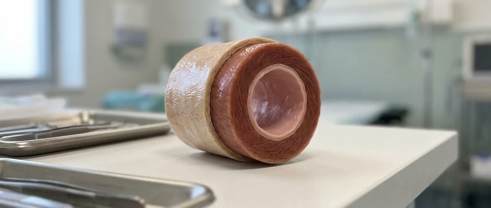
The media is the middle layer of your artery wall, and it sits at the heart of this article. Every artery has three Arterial Wall Layers: the intima, a thin inner lining; the media, a muscular middle layer; and the adventitia, a tough outer sheath. The media is the layer that gives your arteries their strength and their ability to stretch and recoil. When this layer weakens, the artery can slowly balloon outward — and that is the beginning of an aneurysm.
Inside the media are smooth muscle cells and elastic fibers woven in careful patterns. In the aorta, the body's largest artery, elastic fibers dominate so the vessel can absorb the surge of every heartbeat. In smaller arteries, muscle cells take over, letting the vessel narrow or widen as your body needs. This design keeps blood moving smoothly through a system that endures thousands of pressure waves every day.
Why the media matters to patients: If a doctor has told you that you have an aortic aneurysm, the underlying problem is almost always in this middle layer. Understanding it helps you understand why blood pressure control matters, why imaging is repeated over years, and why surgery sometimes becomes the safest path. It also explains why the same condition can behave very differently from one person to the next.
How Does the Media Contribute to Aneurysm Formation?
An aneurysm begins when the media can no longer hold back the pressure inside the vessel. Its muscle cells degrade or die, and its elastic fibers fragment. Each heartbeat then pushes the weakened wall outward a little more, creating a slowly growing bulge. This process is usually silent and can take years before anything shows on an imaging test.
Pressure and the arterial wall: High blood pressure places constant mechanical stress on the media, accelerating the breakdown of elastic fibers and damaging muscle cells. Over years, the wall thins and the risk of aneurysm formation rises. The aorta, which carries the highest pressure in the body, is the most common site for this damage.
What the research shows: Recent work on abdominal aneurysm remodeling describes how aging-related oxidative stress and DNA damage in the vessel wall reprogram its cells and drive aortic dilation — a mechanism documented in a study published in the Journal of the American Heart Association. In plain terms, the older the vessel, the harder it becomes for its cells to repair themselves. The same forces that thicken the wall in one place can thin it in another, which is why aneurysms and hardened arteries often appear in the same patient.
Why the ascending aorta is vulnerable: The segment of the aorta that rises directly from the heart absorbs the full force of every heartbeat. Its media is rich in elastic fibers, which makes it effective — but also fragile. People with inherited conditions may have a weak media from birth, which changes how early and how often they should be monitored.
What Are the Types of Aneurysms Caused by Media Weakness?
Aneurysms are classified by location, shape, and cause. The most common types linked to media weakness occur in the aorta. A Thoracic Aortic Aneurysm develops in the chest, while an Abdominal Aortic Aneurysm forms lower down, below the arteries that feed the kidneys. Each location reflects a slightly different pattern of wall damage and follows its own timeline for monitoring and intervention.
True aneurysms versus pseudoaneurysms: A true aneurysm involves all three layers of the wall bulging together — the media has weakened but not ruptured. A pseudoaneurysm occurs when the inner and middle layers tear and blood leaks out, contained only by the outer sheath. That distinction matters because a pseudoaneurysm carries a higher rupture risk and usually needs more urgent treatment.
Saccular and fusiform shapes: A fusiform aneurysm widens the whole circumference of the vessel, forming a spindle-shaped bulge. A saccular aneurysm bulges from only one side, forming a pouch. Both arise from media weakness, but saccular aneurysms concentrate stress on a smaller area of tissue, so they tend to grow faster and rupture more easily. Imaging determines the shape and how quickly it changes.
Why Does the Aortic Media Weaken Over Time?
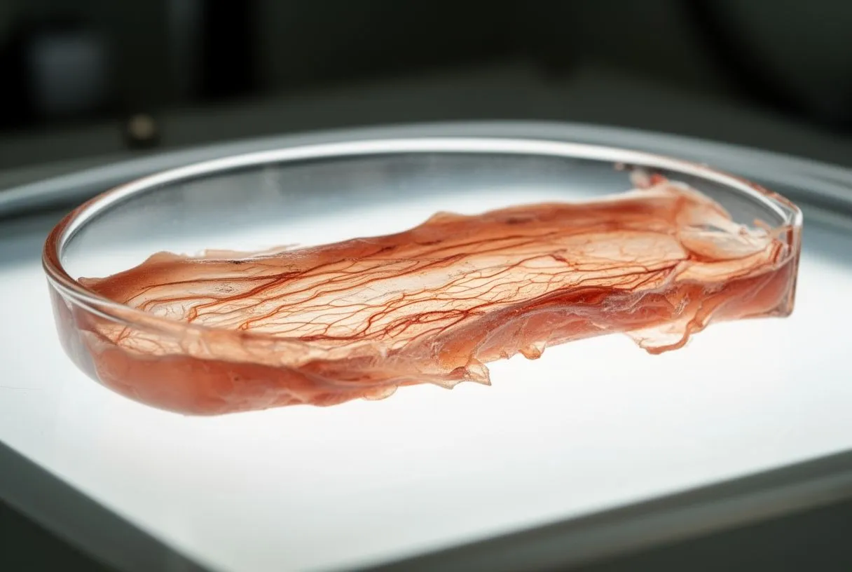
Several forces weaken the aortic media, and most patients have more than one at work. Age is the most universal. Elastic fibers degrade naturally as we grow older, and muscle cells gradually lose their ability to maintain the wall. Some stiffening is nearly universal by the sixties, although it does not always lead to an Aortic Aneurysm.
High blood pressure and plaque: Sustained hypertension breaks down elastic fibers faster than the body can repair them. Cholesterol plaque inside the wall displaces and damages the muscle cells of the media. Together they create a hostile environment for the vessel, raising the likelihood that an aneurysm will develop in the aorta or its major branches.
Genetic and inflammatory causes: Connective tissue disorders such as Marfan syndrome and vascular Ehlers-Danlos syndrome produce abnormal collagen and elastin, the proteins that give the media its strength. A family history of aortic aneurysm or dissection can signal similar vulnerabilities without a formal diagnosis. Inflammatory conditions and certain infections can also damage the media, though these are less common today.
What Are the Early Signs of Media Damage?
Most aneurysms cause no symptoms until they become large or rupture. This is what makes media damage so dangerous: the weakening happens silently, and the first sign may be an emergency. Small aneurysms are usually found by accident, on a chest X-ray or abdominal ultrasound done for something else entirely. That is why screening matters so much for patients with known risk factors.
When symptoms do appear: As an aneurysm grows, it may press on nearby structures. A thoracic aneurysm can cause chest pain, back pain, or a persistent cough if it compresses the airway or nerves. An abdominal aneurysm may produce a pulsing sensation in the belly, deep back pain, or discomfort radiating to the side. None of these symptoms are specific to aneurysms, which is why imaging is essential.
Warning signs of rupture: If an aneurysm begins to tear or burst, the symptoms are sudden and severe: a tearing or ripping sensation in the chest or back, a rapid heartbeat, dizziness, or fainting. This is a medical emergency requiring immediate care at a hospital with cardiac surgical capability. The goal of every screening and monitoring program is to intervene before the wall ever reaches this point.
How Do Doctors Diagnose an Aortic Aneurysm?
Diagnosis begins with a history and physical examination, but imaging is the cornerstone. A doctor may listen over the abdomen or chest for abnormal sounds suggesting turbulent flow, though many aneurysms are silent. The interview focuses on risk factors: blood pressure history, smoking, family history of aortic disease, and any connective tissue conditions. Occasionally a pulsatile mass in the abdomen can be felt, but not in every patient.
Imaging tests that show the wall: Echocardiography, which uses sound waves, is often the first test when an aneurysm is suspected, and it shows the heart and the ascending aorta well. CT scans give detailed cross-sectional images of size, shape, and location. MRI offers similar detail without radiation, which is useful for patients who need repeated scans over many years.
Monitoring: Once an aneurysm is identified, your doctors set a surveillance schedule based on its size and growth rate. Our team reviews every case together against international practice guidelines before recommending anything, and we explain what the studies actually show in plain words so you can decide with clear alternatives in front of you. If surgery becomes likely, additional tests check heart, kidney, and overall surgical risk. During an operation, an Arterial Line — a thin catheter placed directly into an artery — gives the team continuous blood pressure readings, moment by moment.
What Treatment Options Exist for a Media Aneurysm?
Treatment depends on the aneurysm's size, location, and the patient's overall health. When the aneurysm is small and stable, the usual approach is watchful waiting plus blood pressure control. Medications such as beta-blockers and angiotensin receptor blockers lower the mechanical stress on the media by reducing pressure and the force of each heartbeat. The goal in this phase is to slow progression and keep the aneurysm stable.
When surgery becomes the recommendation: Guidelines generally point to surgery when the ascending aorta reaches 5.5 centimetres in diameter, with lower thresholds for people with genetic conditions or a family history of rupture. An aneurysm growing more than half a centimetre per year may also warrant earlier intervention. These thresholds are not arbitrary; they mark the point where the risk of the wall failing outweighs the risk of repairing it.
Open repair versus a stent graft: Traditional open surgery replaces the weakened segment with an Arterial Graft — a synthetic tube that permanently takes over the damaged media's job. It requires a larger incision and a longer recovery, but results are durable. Endovascular Aneurysm Repair is a less invasive alternative in which a stent graft is threaded through a small groin incision and positioned inside the aneurysm to reinforce the wall from within. Not every anatomy supports a stent graft, so the decision is individual.
- Size drives the decision: A diameter above 5.5 cm generally triggers surgical evaluation, with lower thresholds for inherited conditions.
- Growth rate matters too: More than 0.5 cm of growth in a year may move the plan forward regardless of absolute size.
- Overall health shapes the approach: Heart, lung, or kidney disease may point toward a less invasive route or extended monitoring instead.
We offer conventional, minimally invasive, robotic and transcatheter procedures, and we recommend the less invasive route only where the evidence supports it for that specific patient — the final decision is always yours.
What Is Recovery Like After Aortic Aneurysm Repair?
Recovery after Aortic Aneurysm Repair depends on whether the repair was open or endovascular. After open surgery, patients typically spend several days in the intensive care unit followed by a longer hospital stay. The early days focus on stabilizing blood pressure, managing pain, and watching for complications. Movement is reintroduced gradually as the body heals around the new graft.
How your starting condition affects recovery: A patient's preoperative physical condition strongly influences the recovery trajectory. Older patients, or those with limited mobility, poor nutrition, or multiple chronic conditions, may face longer recoveries and higher complication rates. Cardiac teams increasingly assess frailty before surgery to decide who benefits most from an operation and who might do better with a less invasive approach. This does not mean frail patients cannot have surgery — it means the plan is tailored to give the lowest risk for their situation.
Returning to daily life: Most patients resume light activities within six to eight weeks after open surgery, with full recovery taking three to six months. Endovascular repair usually allows a faster return, often within two to four weeks. Lifelong follow-up imaging is essential in both cases, and blood pressure control remains a lifelong priority, because the same forces that weakened the original wall can affect other segments of the aorta over time. Before you leave the hospital, ask for your follow-up plan, so you know which scan comes next and why.
Can Lifestyle Changes Protect the Arterial Media?
You cannot reverse genetics or undo decades of wear on the media, but you can slow the progression of wall damage. The single most important step is controlling blood pressure. Every reduction in pressure means less mechanical stress on the middle layer, giving the remaining elastic fibers and muscle cells a better chance of holding the vessel together. Regular monitoring, medication adherence, and diet work together toward that goal.
Smoking and the artery wall: Tobacco smoke injures the cells lining the artery, triggers inflammation inside the wall, and accelerates elastic fiber breakdown. Quitting does not restore the media to its original state, but it halts the acceleration of damage. Former smokers have a lower risk of aneurysm growth and rupture than current smokers, and every year tobacco-free measurably reduces that risk.
Exercise and nutrition: Moderate, regular exercise supports healthy blood pressure and overall cardiovascular function. Patients with known aneurysms should discuss exercise intensity with their cardiac team, since very heavy lifting or straining can transiently spike pressure and stress the wall. A diet low in sodium and rich in vegetables, fruits, and whole grains supports blood pressure management. If you take aspirin or other medications as part of your plan, keep taking them as prescribed and discuss any change with your team first.
- Control blood pressure: Keep readings at or below the target your cardiologist sets, often under 140/90 mmHg, adjusted to your risk profile.
- Stop smoking: Each tobacco-free year reduces aneurysm growth and rupture risk.
- Stay active moderately: Brisk walking and swimming are excellent; clear heavy lifting with your cardiac team first.
When Should You Seek Help for an Aneurysm Concern?
If you have a family history of aortic aneurysm, a known connective tissue condition, or long-standing high blood pressure, discuss screening with a cardiac specialist even if you feel completely well. Screening is typically recommended for men over 65 who have ever smoked, and for anyone with a first-degree relative who has had an aneurysm or dissection. The goal is to detect media weakening before it becomes dangerous, when monitoring and medication can keep the aneurysm stable for years.
Red flag symptoms: Seek emergency care immediately for sudden, severe chest or back pain that feels like tearing, especially with a rapid heartbeat, dizziness, or fainting. These may signal that the aneurysm has begun to rupture or dissect — a separation of the wall layers that splits the media from within. Time is critical, and the outcome depends on how quickly you reach a hospital with cardiac surgical capability. Do not wait to see whether the pain resolves on its own.
Living with a known aneurysm: If you are under surveillance, keep every imaging appointment and stay in open communication with your cardiac team between visits. Discuss any change in symptoms, medication side effects, or new diagnoses promptly rather than waiting for the next scheduled scan. The media may be hidden inside your body, but its health shapes your entire cardiovascular future. Our team, led by Dr Alejandro Molina, offers a free first video consultation in which we get to know you, talk through your condition, and tell you which studies are still needed so no unnecessary ones are ordered — and we welcome second opinions as a normal part of deciding about heart surgery. If you have questions about your own risk, the next step is a conversation with a qualified cardiac team rather than self-diagnosis.