The Recovery Room After Heart Surgery: Your First Hours
Recovery room after heart surgery: what happens in your first hours, from monitors and tubes to waking up safely.
What is the recovery room after heart surgery
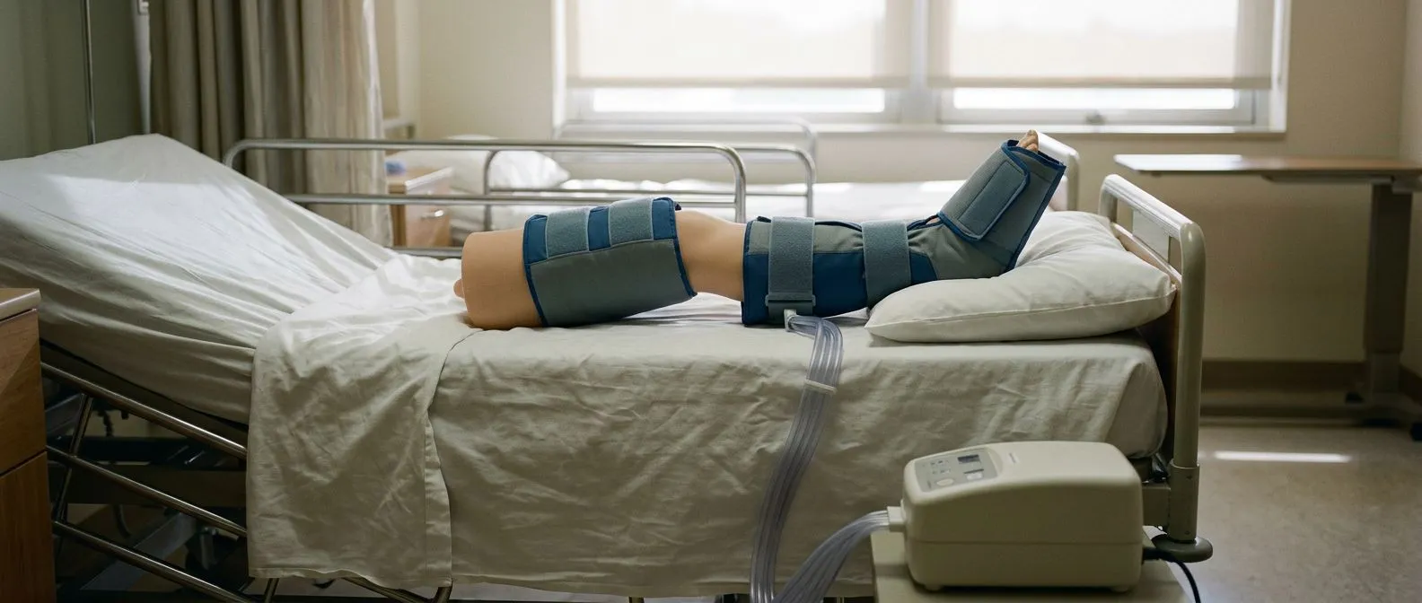
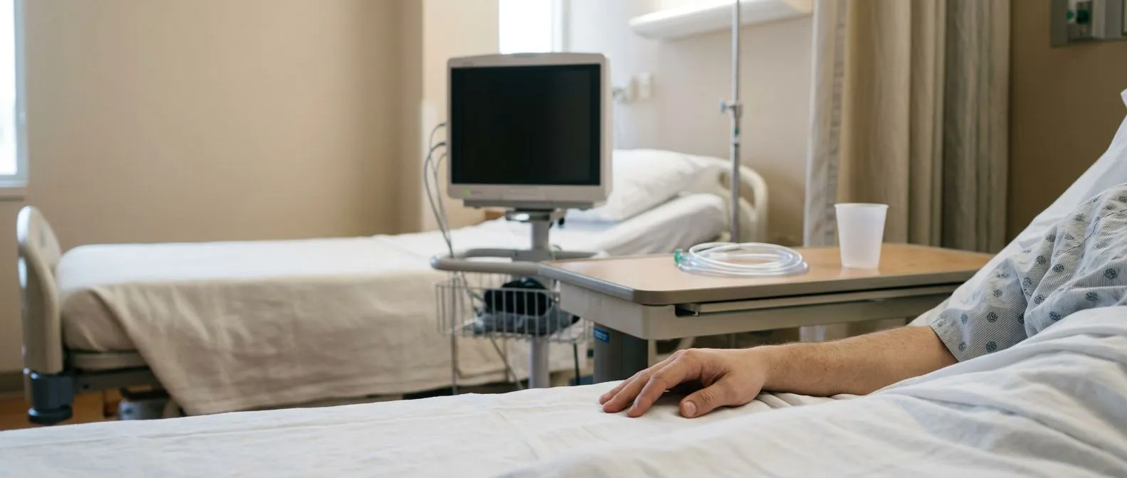
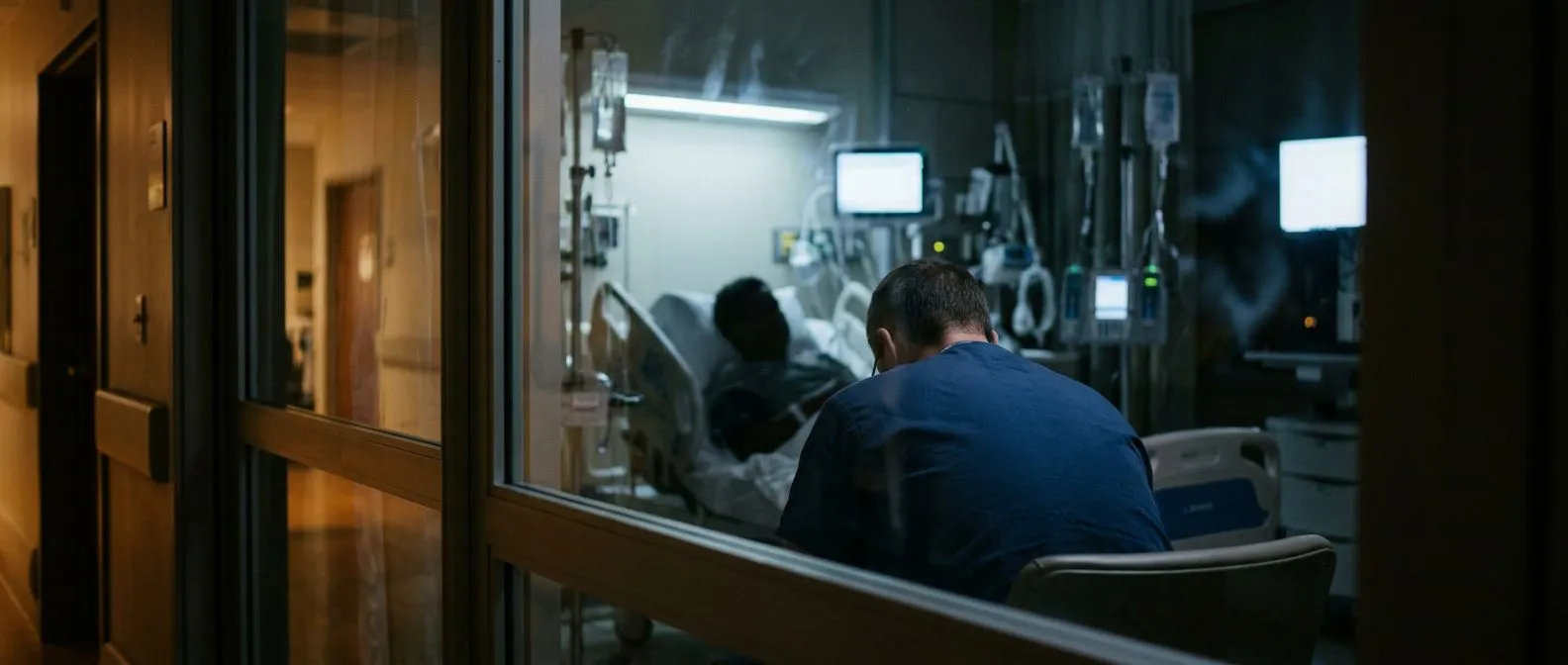
The recovery room after heart surgery is a highly specialized intensive care unit where the surgical team closely monitors your first critical hours. Transitioning from the operating theatre to this controlled environment marks the beginning of your active recovery. Many families feel anxious about this space, but it is specifically engineered to keep you safe, warm, and physiologically stable while your body wakes from anesthesia.
During your time in this environment, specially trained nurses and cardiologists use advanced monitors to track every beat of your vital signs. This intensive observation is essential because your body is adapting to the immediate changes in blood flow following a valve repair or bypass. The atrium and the ascending aorta begin their healing process here, and you will be kept deeply sedated initially to ensure no unnecessary stress is placed on your newly repaired cardiovascular structures.
As you slowly wake from the anesthesia, you might feel cold, slightly confused, or notice the bright lights and rhythmic beeping of the room. These sensory inputs are a normal and expected part of the intensive care environment. Your clinical team is continuously assessing your condition, utilizing techniques such as auscultation to carefully listen to your healing heart and lung sounds. Remember that these first few hours are about keeping you completely stable rather than providing immediate comfort; your care team will manage every discomfort as you need it.
How the recovery room team monitors your heart
Continuous electrocardiographic observation: Your care team will attach small pads to your chest to constantly observe your heart rhythm. This continuous electrical tracking helps immediately detect any irregular heartbeats or rhythm changes the moment they occur. As noted by extensive publications from the New England Journal of Medicine, postoperative cardiac monitoring remains essential for preventing complications during the vulnerable early hours.
Arterial line precision: A specialized arterial cannula, typically placed in your wrist, provides beat-by-beat measurements of your blood pressure. An arterial line is vastly more accurate than a standard blood pressure cuff, giving your doctors exact readings while your heart recovers from the stress of cardiopulmonary bypass. Your surgeon relies on this precise data to carefully adjust medications and ensure your circulatory system remains completely stable.
Your oxygen levels will be closely tracked via a small sensor clipped to your finger, while a temperature probe monitors your core warmth. If your surgical procedure required autologous blood collection, the team will meticulously manage your fluid volume throughout your stay. The combined data from these various monitoring tools gives your care team a real-time, comprehensive picture of your overall hemodynamics, ensuring your heart is pumping efficiently and returning to its baseline function as quickly as possible.
Waking up after cardiac surgery and what to expect
Emerging from anesthesia is a gradual process rather than an instant switch. You will slowly drift in and out of awareness, and it is completely normal to experience a sense of disorientation or mild confusion as the medications wear off. The nursing staff is specifically trained to orient you, often speaking calmly to let you know the surgery is finished and that you are in a safe, protected environment.
During these initial moments of wakefulness, you will likely become aware of the breathing tube in your throat. This endotracheal tube is connected to a mechanical ventilator that is temporarily breathing for you. Because you cannot speak while the tube is in place, the nurses will ask simple yes or no questions and encourage you to nod, ensuring you feel understood and supported despite the temporary loss of your voice.
Your hands may be gently secured to prevent you from accidentally pulling at your lines or tubes while you are still groggy. This practice can feel alarming to waking patients, but it is a standard safety protocol. As the anesthesia clears your system and your breathing stabilizes, the team will slowly reduce the sedation. They will keep you comfortable with pain medications, ensuring your transition from deep sedation to wakefulness is as smooth and stress-free as your clinical condition allows.

Common tubes and lines in the cardiac ICU
Navigating the intensive care unit means adjusting to several different medical tubes attached to your body. While the sheer number of lines can appear overwhelming to family members, each one serves a very specific, temporary purpose to keep you safe. Knowing what to expect can reduce the fear of the unknown and help you focus entirely on your physical recovery.
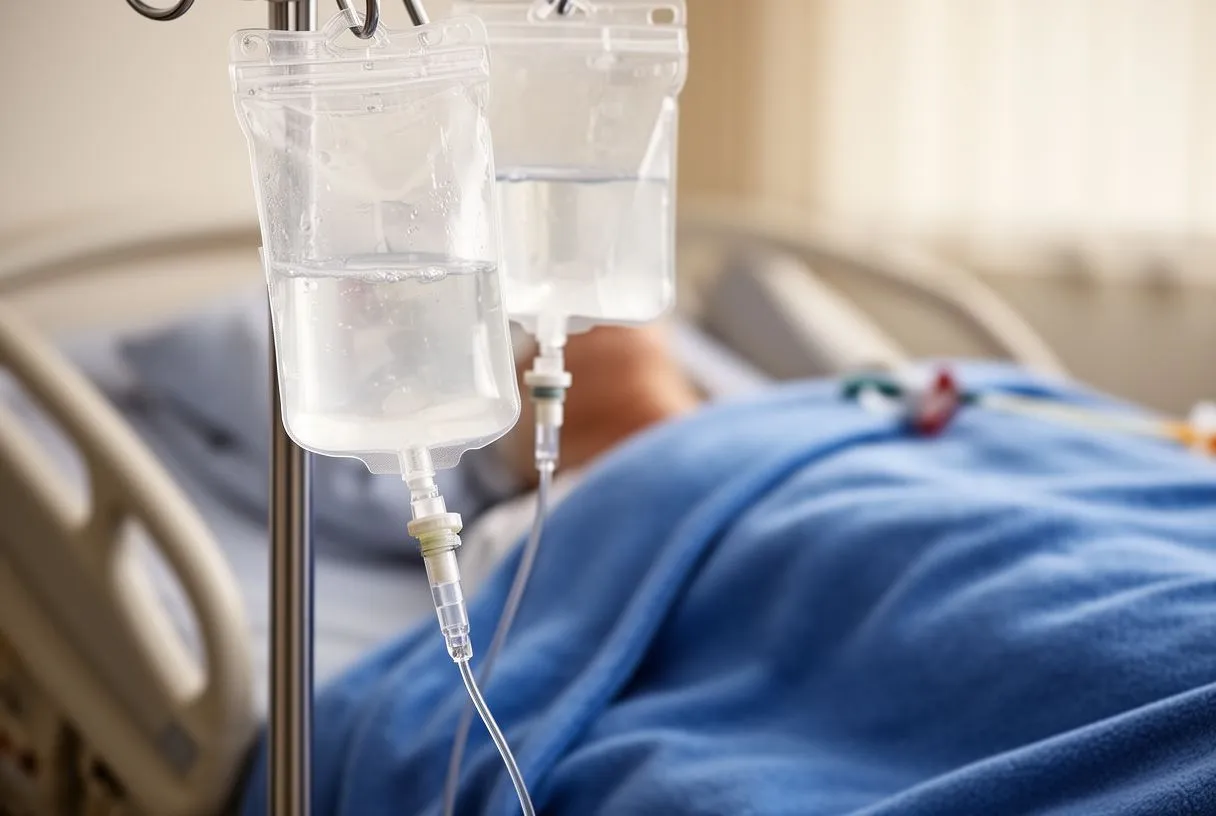
You will have intravenous lines in your neck and arms to deliver fluids, nutrition, and essential medications directly into your bloodstream. A urinary catheter, inserted while you are asleep, continuously drains your bladder so the team can precisely measure your kidney function and fluid output. Additionally, temporary pacing wires are often threaded just beneath the skin near your chest; these act as an electronic backup to keep your heart rate steady if the natural rhythm temporarily slows down.
As your condition stabilizes over the first day, your medical team will systematically begin removing these tubes when they are no longer clinically necessary. This de-escalation of care is a positive milestone, signaling that your body is taking over its own regulation. Here is a quick reference for the most common attachments you will encounter:
- Chest tubes: Drain excess blood and fluid from around the heart and lungs to prevent accumulation.
- Urinary catheter: Tracks exact hourly kidney output to guide fluid management.
- Pacing wires: Provide temporary electrical support to stabilize a slow heart rate.
Managing chest tube drainage after open heart surgery
Preventing fluid accumulation: Chest tubes are flexible plastic drains placed near your ribs to evacuate seeping fluid, preventing it from pooling around your healing tissues. When a surgeon removes an atheromatous plaque or repairs a valve, some degree of internal weeping is a normal and expected physiological response. These drains are generally removed once the fluid output drops to a safe, minimal level, usually within a day or two.
Managing drainage expectations: Seeing blood collect in these drainage containers can be startling for visiting families, but it is a predictable anatomical part of the postoperative process. Your nurses will carefully measure and record the color and volume of the drainage every hour. If your surgery involved placing an arterial graft, your surgical team will watch these tubes closely to ensure adequate blood flow is reaching your newly created bypasses without any internal obstruction.
Medications play a crucial role in keeping your blood flowing smoothly during this delicate healing phase. Aspirin is typically administered early in your recovery to prevent dangerous clotting at the freshly repaired surgical sites. Your team may also use autologous blood to replenish volume lost during the operation, minimizing the need for external donor transfusions. To help manage your long-term cardiovascular risk, your doctor may also check Apolipoprotein B levels, and medications like those studied in the ARISTOTLE trial help guide the blood-thinning protocols required for your specific recovery.
Understanding post-operative care heart surgery pain management
Setting realistic pain expectations: Having zero pain is an unrealistic expectation, but your nursing team will proactively manage any discomfort to help you breathe deeply and move gently. You will likely feel a deep, aching soreness from the chest incision and experience radiating shoulder or arm pain, which is usually caused by your surgical positioning and the stretching of tissues. A mild pulling sensation in your chest when taking a deep breath is normal, but sharp, severe pain is addressed immediately.
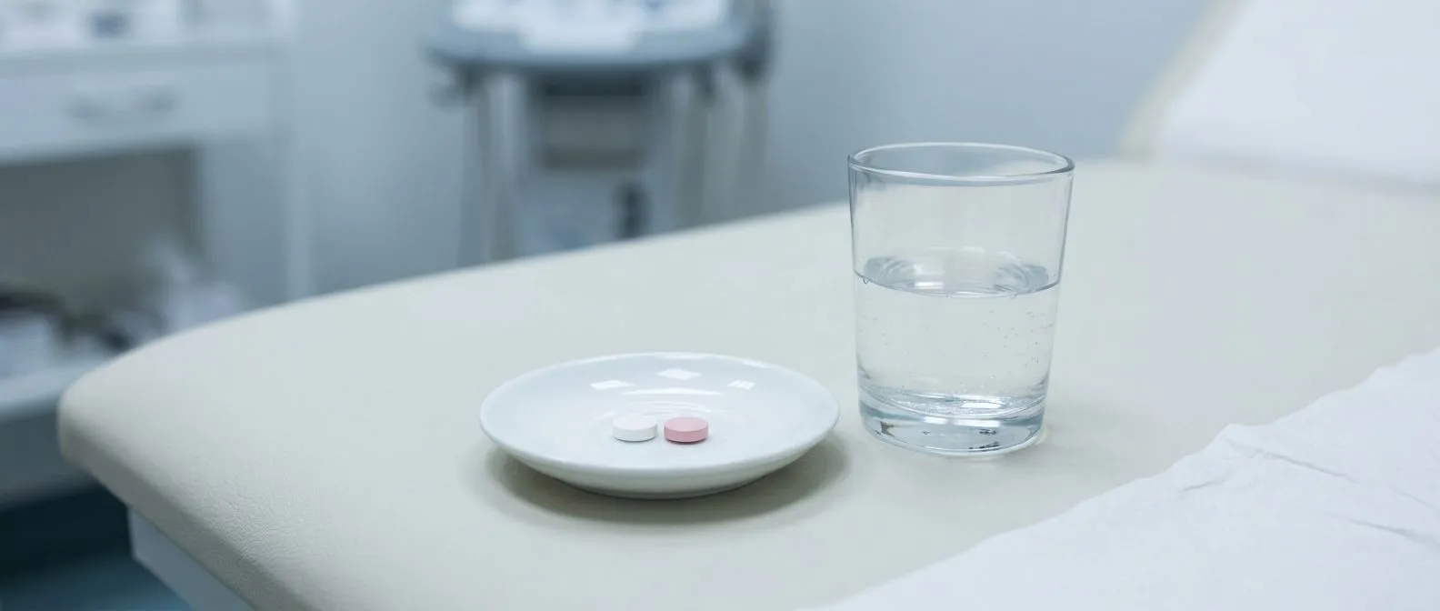
Medication strategies: Pain control begins through your intravenous lines and transitions to oral tablets as you wake up and your digestion improves. Combining mild opioids with simple analgesics like acetametaphin provides layered relief, managing the pain at multiple nerve receptors while minimizing side effects. Effectively controlling your pain is a critical clinical goal because unmanaged pain prevents you from taking the deep breaths needed to clear your lungs, which drastically increases the risk of postoperative pneumonia.
Distinguishing between surgical soreness and the warning signs of a cardiac issue is a critical clinical skill your care team constantly monitors. An atypical infarction can sometimes present as vague chest discomfort rather than the classic crushing pain, making continuous assessment vital. By actively communicating your pain levels using the hospital's numeric scale, you help your team safely titrate your medications and quickly identify any abnormal symptoms that might require immediate medical intervention.
First hours after heart surgery breathing and extubation
The breathing tube timeline: At the conclusion of your operation, you will have a breathing tube resting in your trachea, connected to a ventilator that temporarily takes over the mechanical work of breathing. This tube usually remains in place until the anesthesia completely clears and your respiratory muscles regain their normal strength. The ventilator is entirely controlled by your respiratory therapist, and you will be weaned from it as soon as you can independently maintain safe oxygen levels in your blood.
The extubation process: Extubation is the quick, controlled removal of the breathing tube once you are sufficiently awake and strong. You may experience a brief sore throat or a temporary cough, which is a normal response to having a foreign object in your airway. While extremely rare and complex cases might require advanced support like Awake ECMO, the vast majority of patients are successfully extubated within the first few hours after their heart surgery concludes.
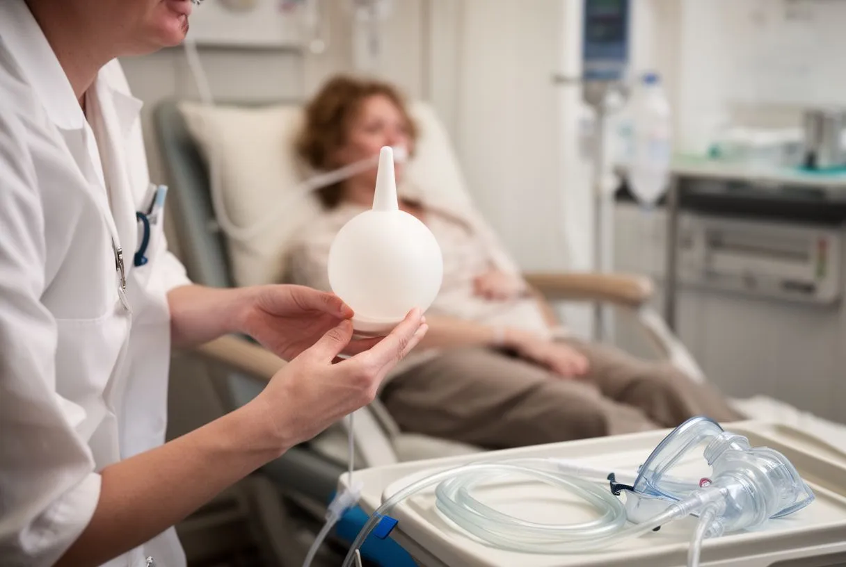
Once the tube is removed, you will be given an incentive spirometer, a simple plastic device used to practice and measure your deep breathing. These deep breathing exercises expand the tiny airways in your lungs, preventing fluid from settling and keeping your lung tissue fully inflated. While advanced safety equipment like an Automated External Defibrillator is always present at the bedside, the actual focus remains on gentle, persistent respiratory therapy to safely restore your full lung capacity.
Cardiac recovery room protocols for blood pressure and rhythm
Hemodynamic stabilization: Fluctuations in blood pressure are very common during the early postoperative period as your heart adjusts to its new workload. Your blood pressure will be carefully regulated through continuous intravenous medications to prevent excessive strain on your delicate surgical sutures. Whether you previously had a balloon angioplasty or received a balloon-expandable valve, your vascular system now requires a careful balance of medication and fluid to heal securely.
Addressing rhythm changes: You may experience a fast or irregular heartbeat, such as atrial flutter, during this phase as the heart muscle reacts to surgical manipulation and fluid shifts. If this occurs, your team will use medications or a brief, controlled electrical treatment to guide the heart back into a normal rhythm. Understanding these electrical impulses requires looking at advanced models of cellular behavior, such as those outlined in the Simulation of the Undiseased Human Cardiac Ventricular Action Potential: Model Formulation and Experimental Validation, which explains how heart cells repolarize after each pump.
The cellular mechanisms behind these electrical pulses rely heavily on complex calcium channels, as detailed in research concerning Ryanodine Receptors: Structure, Expression, Molecular Details, and Function in Calcium Release. When these microscopic receptors coordinate properly, your heart contracts effectively and maintains a steady rhythm. Your medical team continuously monitors this electrical stability, ensuring your heart muscle moves smoothly through the acute recovery phase without dangerous arrhythmias.
When can family visit the recovery room after surgery
Family visiting hours in the intensive care unit are usually staggered and scheduled to align with nursing shift changes and critical care routines. Because the recovery room requires strict infection control and continuous monitoring, visitation is often limited to short windows and restricted to a very small number of immediate family members. The nursing staff will keep your family updated frequently, explaining your status and letting them know exactly when it is safe to enter.
When your family finally comes to your bedside, they should be prepared to see you surrounded by monitors, IV poles, and the various tubes we have discussed. This highly clinical setting can be emotionally jarring, especially if they expected to see you sitting up and smiling. It is important for your loved ones to remember that even if you appear tired or restricted, you are receiving world-class, minute-by-minute observation designed to protect your healing heart.
Interacting with you during these first visits should be calm and gentle to help conserve your precious energy. Holding your hand or speaking in a soft, reassuring voice is far more beneficial than engaging in long, exhausting conversations. Here is how families can best support you during these early visits:
- Staggered visiting: Visits are timed around critical nursing care to ensure your monitors are never disrupted.
- Calm presence: A gentle touch and a familiar voice provide immense emotional comfort without draining your energy.
- Ask the nurse: Direct any questions about your tubes or monitors to the bedside nurse for clear, clinical explanations.

Transitioning from the recovery room to the cardiac ward
Meeting discharge criteria: You will only transition out of the recovery room after heart surgery to a standard cardiac ward once all your vital signs are stable and intensive monitoring is no longer required. This means your breathing tube has been removed, most of your invasive lines have been safely taken out, and your heart rhythm has remained steady and predictable. Leaving the ICU is a significant, positive milestone indicating that your heart has healed enough to begin regulating itself.
Shifting care to the ward: Once transferred to the regular ward, the frequency of your monitoring decreases, and you will be encouraged to begin gentle, assisted movements. Whether you underwent a structural intervention like a balloon aortic valvuloplasty or a complex bypass using an autograft, the focus of your recovery shifts entirely toward physical mobility. The ward nurses will continue to help you sit up, stand, and eventually walk short distances to rebuild your physical strength and independence.
Leaving the intensive care environment can sometimes make families feel nervous, but it clearly signals that the surgical team is confident in your heart's ability to sustain itself. Your care team will guide you through the rehabilitation process; ask them any questions you have about your upcoming discharge and home care. We strongly encourage you to discuss your specific recovery timeline with our qualified cardiac team and book a consultation to ensure you have personalized, expert guidance rather than relying on self-diagnosis.