

The Venous Cannula in Heart Surgery and ECMO
The venous cannula in heart surgery and ECMO: what it does, the types used, how it is placed, and what patient care involves.
What Is a Venous Cannula in Heart Surgery?
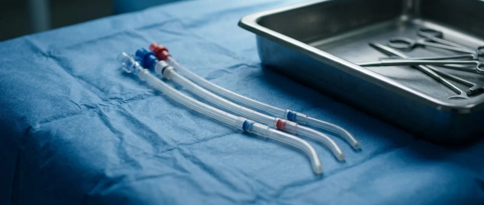
A venous cannula is a specialized sterile tube that cardiac surgeons insert into a patient's major veins to drain blood away from the body and route it into a heart-lung machine. This drainage step is essential during procedures where the heart must be temporarily stopped and emptied so the surgeon can operate on it safely. Without reliable venous drainage, the surgical field would be flooded with blood and the heart-lung machine could not maintain circulation to the rest of the body. The venous cannula therefore acts as the first critical link in the cardiopulmonary bypass circuit.
When you or a loved one is scheduled for CABG — coronary artery bypass grafting — or a valve repair, the surgical team will plan cannula placement long before the first incision. They choose the size, type, and insertion site based on the specific anatomy of the heart, the planned procedure, and the patient's body size. For instance, a patient with a Bicuspid aortic valve may need a different approach than someone undergoing a standard bypass, because the altered valve anatomy can influence how blood returns from the upper body.
Understanding the role of the venous cannula helps demystify what happens behind the operating room doors. The tube is not simply a passive pipe — it is a carefully engineered device with flow characteristics, wall stiffness, and tip design all matched to the individual patient. The surgical team monitors drainage continuously, adjusting flow rates and positioning to ensure the heart remains still and blood-free while circulation to the brain and organs continues uninterrupted through the bypass machine.
How Venous Cannula Placement Works During Cardiopulmonary Bypass
Where the cannula enters the body: The surgeon typically gains access to the right atrium — the upper right chamber of the heart — through the right atrial appendage or directly through the superior and inferior vena cava, the two large veins that return deoxygenated blood to the heart. In many adult procedures, a single cannula is threaded into the right atrium to drain blood from both the upper and lower body simultaneously. In more complex operations, two separate cannulas may be placed, one in each vena cava, to provide independent drainage pathways and better surgical exposure.
The placement technique requires precision because incorrect positioning can cause inadequate drainage, air entry into the circuit, or injury to delicate atrial tissue. The surgeon uses purse-string sutures — circular stitching around the insertion point — to create a tight seal around the cannula and prevent bleeding when it is eventually removed. Throughout the operation, the perfusionist (the specialist who operates the heart-lung machine) watches venous drainage pressure and adjusts the cannula position if flow drops below the target rate for the patient's body size and metabolic demand.
Bypass Time — the total number of minutes the patient is connected to the heart-lung machine — depends directly on how efficiently the venous cannula drains blood. Smooth, consistent drainage allows the surgeon to work in a still, clear field, which shortens bypass duration and reduces the risk of complications such as inflammation, kidney strain, or prolonged recovery. When drainage is suboptimal, the team may need to troubleshoot repositioning the cannula, adding a second drainage site, or switching to a different cannula design mid-procedure to maintain safe flow.
Types of Venous Cannulas: Single Stage vs Dual Stage
Surgeons choose between two principal venous cannula designs: single stage and dual stage. Each type has distinct flow characteristics that make it better suited to specific surgical scenarios. The choice depends on the procedure, the patient's anatomy, and the surgeon's preference based on years of clinical experience.
- Single-stage cannula: One drainage opening at the tip, typically placed in the right atrium. Drains blood from both vena cava through a single tube. Best for straightforward bypass and valve cases.
- Dual-stage cannula: Features drainage holes at two levels — one set near the tip for the inferior vena cava and another set higher up for the superior vena cava. Provides more efficient drainage when the right atrium is open.
- Femoral venous cannula: Inserted through the groin vein, advanced toward the heart. Used in minimally invasive surgery, ECMO, and reoperations where opening the chest carries higher risk.
The single-stage venous cannula is the workhorse of routine cardiac surgery because it is straightforward to place and provides reliable drainage for most adult patients. The dual-stage design excels in situations where complete drainage of both the upper and lower body is needed simultaneously, such as during complex mitral valve repairs or when the right atrium will be opened. Surgeons performing procedures near the Base of the Heart — where the great vessels converge — may prefer dual-stage drainage to keep the field completely clear.
Venous Cannula Size Selection for Optimal Blood Flow
Selecting the correct venous cannula size is one of the most important preoperative decisions the surgical team makes. A cannula that is too small will restrict drainage, forcing the heart-lung machine to work harder and potentially leaving blood in the surgical field. A cannula that is too large can make insertion difficult and increase the risk of vessel or atrial wall trauma. The goal is finding the size that allows full drainage at the patient's required flow rate without excessive suction or resistance.
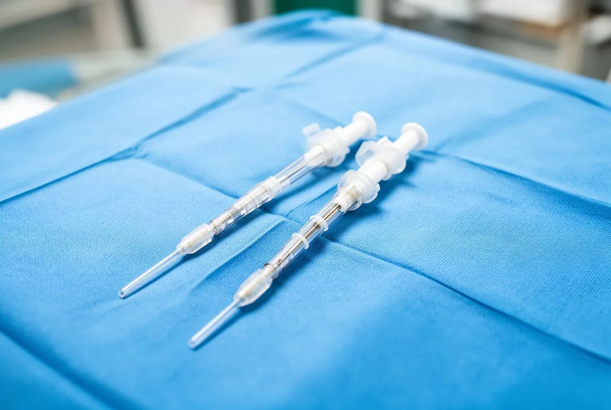
How size is calculated: The perfusionist calculates the required flow rate based on the patient's body surface area — a measurement combining height and weight — and targets approximately 2.2 to 2.5 liters per minute per square meter. Once the target flow is known, the team selects a cannula whose rated flow capacity exceeds that target with a safety margin. Most adult venous cannulas range from 24 French to 36 French in outer diameter, with larger sizes used for bigger patients or when higher flow rates are anticipated.
Pressure across the cannula is another critical factor. Venous drainage relies on gravity siphon and, in some setups, vacuum-assisted venous drainage to pull blood from the body into the reservoir. The internal diameter, length, and number of side holes all influence how much pressure is needed to move blood at the required rate. A well-sized cannula keeps the pressure gradient low, which protects blood cells from damage and reduces hemolysis — the breakdown of red blood cells that can occur when blood is forced through a restrictive space. Patients who may need a Blood Transfusion after surgery benefit from minimizing this cellular damage, as preserved red cells recover faster and reduce dependency on donor blood products.
Venous Cannula in ECMO: Life Support Beyond the Operating Room
Extracorporeal Membrane Oxygenation, or ECMO, is an advanced form of life support that uses a modified heart-lung machine to sustain patients whose heart or lungs are temporarily unable to function. The venous cannula plays a central role in ECMO just as it does in the operating room, but the context is different — ECMO patients may be in intensive care rather than surgery, and the cannula may remain in place for days or even weeks. This extended duration introduces unique challenges in monitoring, infection prevention, and patient comfort.
Two main ECMO configurations exist. In veno-venous ECMO, the venous cannula drains deoxygenated blood from the venous system, passes it through an artificial lung for gas exchange, and returns it to the venous side — this supports only lung failure. In veno-arterial ECMO, blood is drained venously but returned arterially, supporting both heart and lung failure. The latter configuration may serve as a Bridge to Recovery when the heart is expected to heal, a Bridge to Transplant when a donor heart is needed, or a Bridge to Decision when the medical team needs time to determine whether the patient is a candidate for definitive treatment.
ECMO cannulas are generally larger and more rigidly reinforced than standard surgical cannulas because they must maintain patency over long periods and withstand patient movement. Insertion is often performed percutaneously — through the skin with a needle and guidewire rather than an open incision — using the Seldinger technique familiar from routine IV placements but scaled up for large-bore cannulas. The femoral vein in the groin is the most common access point, though the internal jugular vein in the neck is used for upper-body drainage, particularly in pediatric patients. Because ECMO patients are critically ill, the venous cannula must be monitored constantly for migration, kinking, or clot formation, any of which could abruptly reduce flow and endanger the patient.
Venous Cannula vs Arterial Cannula: Key Differences
Every cardiopulmonary bypass circuit uses two cannulas: one to drain blood out and one to return it. The venous cannula removes deoxygenated blood from the body, while the arterial cannula returns oxygenated blood from the heart-lung machine back into the arterial system, usually at the ascending aorta. Understanding the difference between these two tubes helps families grasp the complete picture of how bypass works and why the surgical team pays close attention to both during an operation.
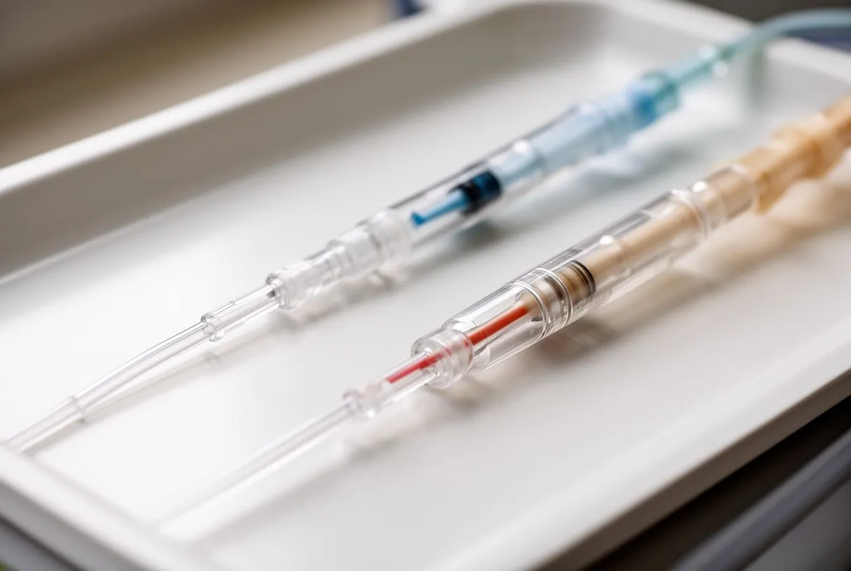
Structure and pressure handling: Venous cannulas are designed for low-pressure drainage and typically have thin walls with multiple side holes along the shaft to encourage gentle blood intake from a wide area. Arterial cannulas must withstand high pressure because they deliver blood back into the arterial system, so they have thick, reinforced walls and a single open tip. If the arterial cannula dislodges or fails, the consequence is immediate and severe — blood is pumped under pressure into the chest rather than the aorta. Venous cannula problems tend to manifest as gradual drainage reduction, giving the team more time to respond.
The insertion sites also differ. Venous cannulas enter the right atrium or vena cava through the right side of the heart, while arterial cannulas are sewn directly into the aorta with a reinforced purse-string technique. When a patient is on a Beta Blocker to control heart rate before surgery, the surgical team accounts for this medication in their cannulation plan, as beta blockers influence how the blood vessels respond to the sudden changes in flow that occur when bypass begins. Both cannulas are removed at the end of the procedure, and the purse-string sutures are tightened to close the insertion sites completely before the chest is closed.
Venous Cannula Complications and How Surgeons Prevent Them
While venous cannula placement is a routine and well-practiced step in cardiac surgery, complications can and do occur. The most common issues include inadequate drainage due to malposition, air entry into the bypass circuit, vessel injury during insertion, and postoperative bleeding at the cannulation site. Each of these problems has established prevention strategies that experienced surgical teams follow meticulously.
Air entry into the venous circuit is particularly dangerous because air can travel through the heart-lung machine and enter the arterial circulation, potentially causing a stroke. The team prevents this by ensuring the venous reservoir never runs dry, by carefully de-airing the circuit before connecting to the patient, and by positioning the cannula so that the drainage holes remain submerged in blood throughout the operation. Vessel injury is prevented through careful preoperative imaging — usually a CT scan — to identify anatomical variations and through gentle, controlled insertion techniques rather than forceful advancement.
Infection is another concern, particularly for ECMO patients whose cannulas remain in place for extended periods. The surgical team uses strict sterile technique during insertion and applies chlorhexidine-impregnated dressings at the exit site. If infection is suspected, Blood Cultures are drawn and the cannula may need to be removed and re-sited at a different location. Surgeons track C-reactive Protein levels — a blood marker of inflammation — to detect early signs of systemic infection. According to Healthline, infections related to invasive lines are among the most preventable hospital-acquired complications when evidence-based protocols are followed consistently. A major systematic analysis published in Global, regional, and national incidence, prevalence, and years lived with disability for 354 diseases and injuries for 195 countries and territories, 1990-2017 documents the significant global burden of cardiovascular conditions that ultimately require bypass support, underscoring why cannulation safety matters at a population level.
Pediatric Heart Surgery and Venous Cannula Considerations
Children undergoing cardiac surgery present unique challenges for venous cannula placement. Their veins and cardiac chambers are far smaller than adults', and many pediatric patients have congenital heart defects that alter normal anatomy in ways that require specialized cannulation strategies. A single-stage venous cannula may be appropriate for an adult, but a child with a complex congenital defect may need two carefully sized cannulas placed in the superior and inferior vena cava to achieve adequate drainage in a very small surgical field.
Cannula sizing in children is based on weight rather than body surface area, and the available sizes are correspondingly smaller — sometimes as fine as 12 or 14 French for infants. The perfusionist must calculate flow rates that account for the higher metabolic rate of children, who need proportionally more oxygen delivery per kilogram than adults. This means the cannula must drain efficiently despite its small diameter, and even minor malposition can have a significant impact on drainage quality. Surgeons at centers like Cirugía de Corazón, where Dr. Alejandro Molina leads the team, rely on the same principles of careful cannula selection; in children, these techniques are adapted to the specific anatomy each child presents.
Beyond the technical aspects, families should understand that pediatric cannulation is performed with the same principles of safety and precision as adult surgery but with additional attention to growth and development. The purse-string sutures used to seal the cannulation site are placed with fine, delicate technique to minimize scar formation on the heart. Parents are encouraged to discuss their child's specific anatomy and the surgical plan with the cardiac team so they understand which vessels will be cannulated and why. A validated national inpatient register study, External review and validation of the Swedish national inpatient register, demonstrates how systematic tracking of cardiac procedures across populations helps improve outcomes — a principle that applies equally to pediatric and adult care.
Venous Cannula Maintenance and Monitoring During Bypass
Once the venous cannula is placed and bypass is underway, the perfusionist becomes the primary guardian of drainage quality. The heart-lung machine console displays continuous readings of venous drainage pressure, arterial line pressure, blood flow rate, and oxygen saturation. If venous drainage drops, the perfusionist troubleshoots through a systematic checklist: checking for kinks in the tubing, verifying cannula position, adjusting the height of the drainage reservoir, and sometimes applying gentle vacuum assistance to improve siphon flow.
What the perfusionist watches in real time: The venous reservoir level is perhaps the single most important indicator of drainage health. A falling reservoir level signals that blood is not returning from the patient as quickly as it is being pumped out — a mismatch that must be corrected immediately. The perfusionist also monitors the color of venous blood, which should appear dark red. If it becomes brighter than expected, it may indicate that arterial blood is recirculating through the venous cannula, a problem that can occur if the cannula has migrated or if there is an unexpected communication between the arterial and venous sides of the circulation.
Temperature management is another aspect of bypass that involves the venous cannula. The heart-lung machine includes a heat exchanger that cools the blood as it leaves the patient and warms it as it returns. Cooling reduces the patient's metabolic rate, which protects the brain and other organs during the period when the heart is stopped. The venous cannula delivers the cooled, deoxygenated blood to the machine, where it is oxygenated, warmed, and returned through the arterial cannula. Patients who have been on Aspirin therapy before surgery may have slightly longer bleeding times, and the team accounts for this during cannulation site preparation and closure.
Venous Cannula Removal Procedure and Recovery After Heart Surgery
Removal of the venous cannula is one of the final steps before the chest is closed. Once the heart is beating normally and the team confirms that it can maintain circulation independently, the perfusionist gradually reduces bypass flow while the surgeon monitors the heart's filling and output. The venous cannula is withdrawn gently, and the purse-string sutures placed at insertion are tightened and secured to seal the opening. Any bleeding is controlled with additional sutures or topical sealants before the team proceeds to close the incision.
For most patients, the cannulation site heals without complications. The right atrium is muscular and well-suited to holding sutures, and the purse-string technique provides a reliable seal. Some patients, particularly those on anticoagulation medications or with thin atrial tissue, may require additional time and care to achieve complete hemostasis. The surgical team checks the site one final time before closing the sternum, and drainage tubes are placed in the chest to capture any residual fluid in the early postoperative period.
Recovery after heart surgery involves more than just the cannulation site — it encompasses the entire healing trajectory from the incision, the bypass run, and the underlying cardiac condition. Patients who had a Bioprosthesis valve implanted or a Bovine Pericardial Valve used in their repair will have specific follow-up schedules to monitor valve function. Those recovering from ECMO as a Bridge to Decision may face a longer and more complex recovery, depending on the underlying condition that required life support. Regardless of the specific procedure, the principles remain the same: attend follow-up appointments, take prescribed medications, report any new symptoms promptly, and maintain open communication with your cardiac team. If you or a loved one are facing heart surgery, we encourage you to schedule a consultation with a qualified cardiac surgeon to discuss your individual situation, the planned approach, and what to expect at every stage of your recovery.






