Valve or Device Explant: When an Implant Must Come Out
Heart device explant reasons explained by Guadalajara specialists — infection, failure, and recovery after pacemaker or defibrillator removal.
What Is a Heart Device Explant?
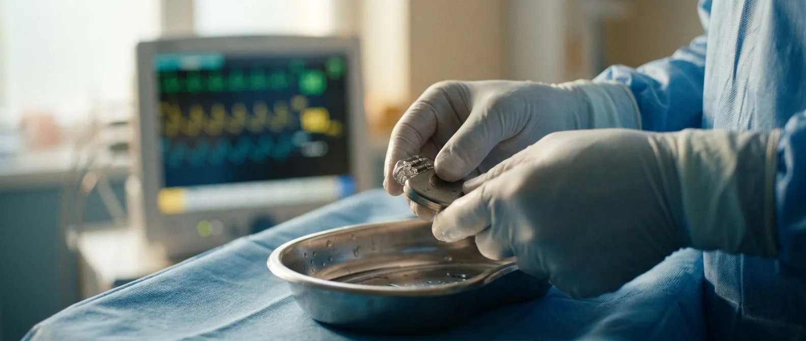
A heart device explant is the surgical removal of a previously implanted cardiac device — such as a pacemaker, defibrillator, or prosthetic heart valve — from the body. Patients usually learn about this possibility only when a problem surfaces, sometimes years or even decades after the original implantation. The procedure differs from a simple battery swap or lead revision: it means taking the hardware out entirely, often because the device has become infected, malfunctioned, or is no longer clinically necessary. For many patients, hearing that an implant must come out feels alarming, especially when the device has been keeping the heart functioning properly. Understanding what the procedure involves, why it becomes necessary, and what recovery looks like can help reduce that anxiety and give you a sense of control over the next steps.
The process of removing a cardiac device is more complex than placing one. When a pacemaker or implantable cardioverter defibrillator (ICD) sits in the chest for years, the body's tissues grow around the leads — the wires that connect the device to the heart muscle. Extracting those leads requires specialized tools and, in some cases, open surgical access to the heart itself. Similarly, a prosthetic heart valve that has deteriorated may need to be cut out and replaced, which involves reopening the chest and working on the ascending aorta or the heart's chambers directly. The complexity depends on how long the device has been in place, where the leads or structures are anchored, and whether infection has made the surrounding tissue fragile.
It is worth noting that explant surgery is not a sign that something went wrong with your care. Devices have lifespans, and the body changes over time. A valve placed twenty years ago may calcify; a pacemaker lead may fracture after millions of heartbeats; bacteria from a distant infection can settle on the device surface. Cardiac teams plan for these scenarios. According to MedlinePlus, device-related infections and mechanical failures are recognized complications of long-term implants, and removal — when indicated — is the standard of care, not a last resort.
Why Would a Pacemaker Need to Be Removed?
Device infection that has spread to the bloodstream: When bacteria colonize the pacemaker generator pocket or travel along the leads into the heart, antibiotics alone rarely resolve the problem. The device surface becomes a shield for bacteria, and the infection can seed deeper structures such as the atrium or the valve tissue. In these cases, the entire system — generator and leads — must be removed to clear the infection, followed by a course of intravenous antibiotics before any new device is considered. This is the most common reason a pacemaker comes out.
Lead fracture or insulation breakdown after years of wear: Leads flex with every heartbeat, and over time the metal conductor wires inside can fatigue and break. Insulation can also degrade, causing the lead to send inappropriate signals or fail to pace the heart at all. When a lead stops functioning and cannot be abandoned safely — because it poses a clot or arrhythmia risk — extraction becomes necessary. The decision to remove a fractured lead rather than cap it depends on the lead's location, how long it has been implanted, and whether a new lead can be placed alongside it.
Upgrade to a different device or system: Sometimes a patient's cardiac condition evolves — for example, heart failure progresses and a standard pacemaker no longer provides adequate support. The team may recommend upgrading to a cardiac resynchronization therapy (CRT) device, which requires new leads in specific positions. Removing old, non-functioning leads at the same time keeps the venous system from becoming crowded. In other situations, a patient's heart rhythm has fully recovered and the device is no longer needed, though true "no-longer-needed" explant is rare and carefully evaluated on a case-by-case basis.
Cardiac Device Infection: The Most Common Reason for Explant
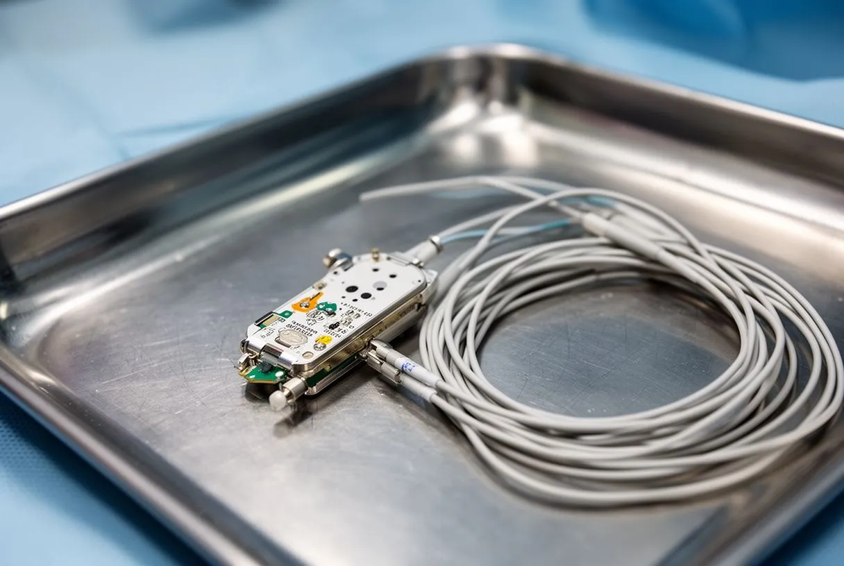
Device infection drives the majority of explant decisions, and understanding how these infections develop helps patients recognize warning signs early. Bacteria can enter the pacemaker pocket through the skin during the original surgery, through a later erosion where the device breaks through the skin surface, or by traveling through the bloodstream from another site in the body — a dental procedure, a urinary infection, or even a small skin abscess. Once bacteria settle on the device, they form a biofilm: a protective layer that shields them from antibiotics. This is why oral or intravenous antibiotics alone often fail to cure a device infection, and why physical removal of the hardware becomes essential.
The symptoms of a cardiac device infection vary, and some are subtle enough that patients dismiss them. Local signs include redness, swelling, warmth, or drainage at the generator site. A patient may notice arm pain if the infection travels along the lead into the venous system toward the heart. Systemic signs — fever, chills, night sweats, and fatigue — suggest the infection has reached the bloodstream, a condition called bacteremia. Blood cultures and an echocardiogram are the standard diagnostic tools. If the echo shows vegetation — a small cluster of bacteria and clot — on a lead or on a valve, the diagnosis of device-related endocarditis is confirmed, and explant surgery is scheduled urgently.
Treatment follows a staged approach. First, the infected device is removed completely: generator and all leads. The leads are extracted using laser sheaths or mechanical rotation tools that free them from the scar tissue that has formed around them inside the veins and heart. After removal, the patient receives several weeks of targeted intravenous antibiotics based on the specific organism identified in blood cultures. Only after the infection is cleared does the team discuss placing a new device — sometimes on the opposite side of the chest, to avoid re-contaminating the same pocket. This staged strategy has become the international standard, supported by cardiology guidelines worldwide.
How Defibrillator Removal Procedure Works
Removing an implantable cardioverter defibrillator (ICD) follows the same principles as pacemaker extraction but often involves additional complexity because ICD leads are thicker and sit deeper in the heart — typically anchored in the right ventricle with a shock coil along the lead body. The procedure is performed in a specialized cardiac catheterization or hybrid operating room, where the team has immediate access to emergency surgical backup if a lead tears a blood vessel or the heart wall. Laser-assisted extraction is the most common technique: a laser sheath slides over the lead and vaporizes the scar tissue binding it to the vein and heart wall, allowing the lead to be pulled free in a controlled manner.
Laser extraction for leads bound by scar tissue: After months or years in the body, leads become encased in fibrous sheaths along their entire length — from the subclavian vein entry point all the way to the heart. The laser sheath delivers controlled energy that cuts through this scar, freeing the lead millimeter by millimeter. For leads that have been in place over five years, the scar can be dense enough that the procedure takes significant time and carries a higher risk of vascular tearing.
Open surgical backup for high-risk extractions: In cases where a lead has been in place for a decade or more, or where infection has made the tissue friable, the extraction team works with a cardiac surgeon standing by. If the laser or mechanical extraction causes a tear in the superior vena cava or the right atrium — a rare but life-threatening complication — the patient is immediately transferred to an open surgical approach. An arterial cannula and venous drainage lines are placed for cardiopulmonary bypass, and the chest is opened to repair the damage and complete the lead removal under direct vision. This is why explant procedures are always performed in facilities equipped for emergency cardiac surgery.
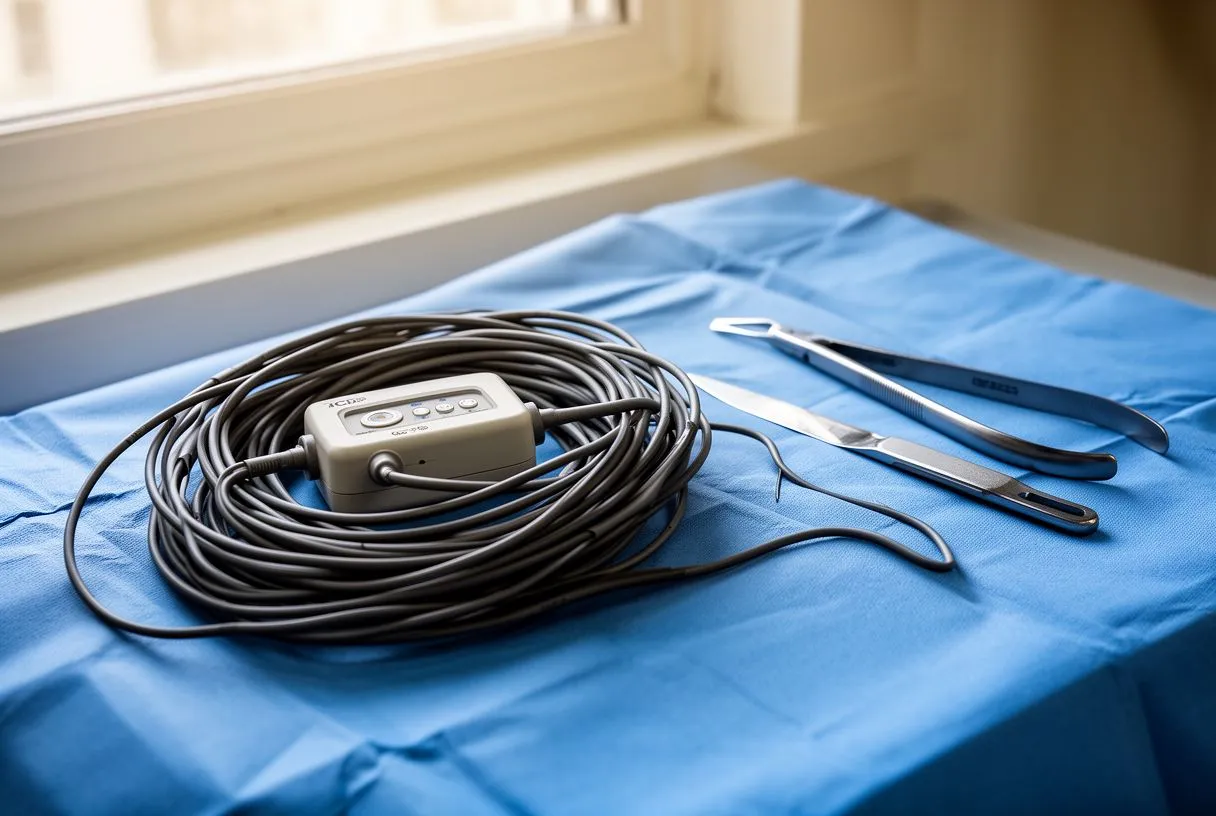
Heart Valve Explant: When Surgical Valves Fail
Heart valves — whether mechanical, biological, or transcatheter — are designed to last for many years, but none lasts forever. Biological tissue valves, made from cow or pig pericardium, typically begin to degenerate after ten to fifteen years. The leaflets calcify, become stiff, and eventually fail to open fully (stenosis) or close properly (regurgitation). Mechanical valves, made of carbon and metal, are far more durable but require lifelong blood thinners and carry their own set of complications, including clot formation on the valve surface. When a prosthetic valve fails, a heart valve explant is required: the old valve is cut out of the annulus — the ring of tissue where it sits — and a new valve is sewn in its place.
Surgical vs transcatheter valve-in-valve replacement: Reoperating on the heart to explant a failed surgical valve is a major procedure, involving reopening the sternum (resternotomy), clearing adhesions from the previous surgery, and working on a heart that may already have damage from the failed valve. For older or higher-risk patients, a transcatheter approach — placing a new valve inside the old one without open surgery — may be an option. This is sometimes called a valve-in-valve procedure, and it uses a balloon-expandable valve compressed onto a catheter, threaded through a blood vessel to the heart, and deployed inside the old prosthetic ring.
The choice between open surgical explant and a transcatheter approach depends on several factors: the type and size of the failing valve, the patient's overall surgical risk (calculated using tools like EuroSCORE II), the reason the valve failed, and whether other cardiac problems need to be addressed at the same time. For example, if a patient with a failing aortic valve also has an atheromatous plaque in the ascending aorta that needs repair, an open approach allows the surgeon to address both problems in one operation. The decision is always individualized, and a detailed discussion with your cardiac team — including review of imaging and risk scores — is essential before committing to either path.
Explant vs Revision: Which Does Your Situation Call For?
Patients facing a device problem often hear two terms used interchangeably — explant and revision — but they describe very different procedures with different implications. A revision is a targeted fix: replacing a battery, adding or repositioning a lead, or upgrading a generator while leaving the existing hardware in place. An explant is the complete removal of the device or valve, usually because the hardware itself has become the problem. Understanding which approach your situation calls for matters because explant carries significantly higher risk and a longer recovery.
- Revision: Generator change for battery depletion, lead addition for pacing optimization, or pocket relocation for discomfort. Typically outpatient or overnight stay. Lower risk profile.
- Explant: Complete removal for infection, lead fracture with extraction, valve failure requiring replacement, or device rejection. Requires specialized facility, surgical backup, and longer recovery.
- Combined approach: Sometimes a revision turns into an explant intraoperatively — for example, if the team discovers an unexpected infection during a generator change. Your consent should always cover both possibilities.
The distinction also affects how the procedure is scheduled and where it is performed. A revision can often be done in a standard cardiac procedure room with local anesthesia and sedation. An explant — especially one involving lead extraction or valve removal — requires a hybrid operating room, general anesthesia, and a surgeon present. If your team recommends explant, ask specifically why revision is not sufficient, what findings drove the decision, and what the alternative options are. These are reasonable questions, and any experienced cardiac team will welcome them.
What Are the Risks of Explant Surgery?
Every cardiac procedure carries risk, and explant surgery is no exception. The specific risks depend on the type of device being removed, how long it has been implanted, and the patient's overall health. For lead extraction procedures, the most serious complications involve tearing of the great veins or the heart wall. The superior vena cava, the large vein that returns blood from the upper body to the heart, runs alongside the pacemaker and ICD leads and is the most common site of vascular injury during extraction. If a tear occurs, blood fills the chest cavity rapidly, and emergency open surgery is needed within minutes to repair the tear and restore circulation.
Vascular and cardiac perforation during lead extraction: The risk of major complication during transvenous lead extraction is generally reported in the range of 1–2% in experienced centers, with mortality under 1%. Risk factors include leads implanted for more than ten years, ICD leads (which are thicker), female sex, and the presence of infection. These figures are not presented to frighten — they are the kind of numbers a good surgeon shares during the consent conversation. If your team cannot quote you their own institutional complication rates, that is a question worth asking before scheduling.
Incomplete extraction and retained lead fragments: Sometimes a lead cannot be fully removed safely — the distal tip may be deeply embedded in the heart muscle, or a portion may break off during extraction. A retained fragment is not always dangerous, but it can serve as a future site for infection or clot formation. The team documents any retained fragments in the operative report, and imaging follow-up is scheduled to monitor the area. Patients who have had an atypical infarction or who carry other cardiac risk factors may need closer surveillance. For valve explant, the risks parallel those of any reoperative cardiac surgery: bleeding, stroke, infection, and prolonged ventilator support, all of which are higher in second operations than in first-time valve surgery.
Device Explant Recovery: What to Expect Week by Week
Recovery from a heart device explant follows a different timeline depending on whether the procedure was a transvenous lead extraction (which is performed through small incisions at the pocket site) or an open surgical valve explant (which involves reopening the chest). For lead extraction patients, the hospital stay is typically two to five days — the first night in a monitored unit to watch for bleeding or arrhythmia, followed by continued intravenous antibiotics if infection was the reason for removal. Most patients return to normal activities within two to three weeks, with restrictions on heavy lifting and overhead arm movement for about a month to allow the venous entry site to heal.
For open valve explant, recovery mirrors that of the original valve surgery: five to seven days in the hospital, including one or two nights in the intensive care unit. The sternum needs six to eight weeks to knit back together, and during that time patients are advised to avoid driving, lifting anything over five kilograms, and any activity that places stress on the chest wall. Cardiac rehabilitation is typically recommended starting around four to six weeks postoperatively, gradually rebuilding strength and endurance. Pain management transitions from intravenous to oral medications within the first few days, and most patients are off prescription pain medication by the second or third week at home.
Several factors influence how smoothly recovery goes. Patients who had an autograft or other complex reconstruction will need closer monitoring. If the explant was performed for infection, the antibiotic course continues after discharge — often through a peripherally inserted central catheter (PICC line) for four to six weeks. Follow-up appointments include blood cultures to confirm the infection has cleared, echocardiograms to assess valve or heart function, and wound checks at the incision sites. Patients should be aware of signs that warrant an urgent call to their team: new fever, increasing chest pain, drainage from the incision, or swelling and redness at the pocket site.
Finding an Explant Surgeon in Guadalajara and Mexico
Choosing the right team for a heart device explant is arguably the single most important decision in this entire process. Not every cardiac surgeon performs lead extractions, and not every hospital has the hybrid operating room setup required for safe extraction with surgical backup. When evaluating a program — whether in Guadalajara, Querétaro, Hermosillo, or elsewhere — ask directly about the team's extraction volume, the availability of laser extraction equipment, and whether a cardiac surgeon is present in the room during the procedure. Programs that perform a high volume of extractions tend to have lower complication rates, a pattern seen across procedural medicine globally.
For international patients traveling to Mexico for cardiac care, the logistics can feel daunting, but many programs are experienced in coordinating care across borders. Ask about language support, pre-operative telemedicine consultations, estimated length of stay, and what happens if complications require a longer hospitalization than planned. Some patients worry about whether their insurance will cover a procedure performed abroad — this is a question to explore with your insurer before committing, as policies vary widely.
Technology is also changing how cardiac teams plan these procedures. Advanced imaging — CT scans that map the exact position of leads within the venous system and heart — allows surgeons to anticipate challenges before the first incision. Some centers are exploring machine learning approaches to predict extraction difficulty based on lead age, type, and patient anatomy, drawing on analytical methods described in fields as diverse as big data and genomics — as noted in a survey of transfer learning. While these tools are still emerging in cardiac device management, they represent the direction in which personalized procedural planning is heading.
Explant Long-Term Outcomes and Quality of Life
The long-term outlook after a heart device explant depends heavily on why the device was removed and whether a replacement was needed. For patients who had an infected pacemaker or ICD removed and a new device placed after antibiotic treatment, survival and quality of life generally return to baseline — assuming the underlying rhythm condition remains managed. For patients who had a valve explant and replacement, the outcomes parallel those of the original valve surgery, with durability of the new valve and freedom from reoperation being the key long-term metrics. Patients who receive a biological valve replacement after explant should understand that the new valve will also eventually wear out, typically in ten to fifteen years, and may require another intervention.
- Infection cleared: With complete device removal and a full antibiotic course, cure rates for device infection exceed 95% in most series. Recurrence is rare when the protocol is followed properly.
- Valve function restored: After valve explant and replacement, most patients see improvement in symptoms like shortness of breath and fatigue within weeks. Echocardiogram follow-up at six months and annually confirms valve function.
- Quality of life: Patients often report improved peace of mind after a problematic device is removed and a functioning replacement is in place. Anxiety about device malfunction or infection resolves once the source is eliminated.
It is also worth noting that the field continues to evolve. Genetic testing is increasingly used to identify patients at risk for certain cardiac conditions that may have led to device placement in the first place, reflecting advances described in a review on ten years of next-generation sequencing technologies. Understanding the genetic underpinnings of conditions like atrial flutter