

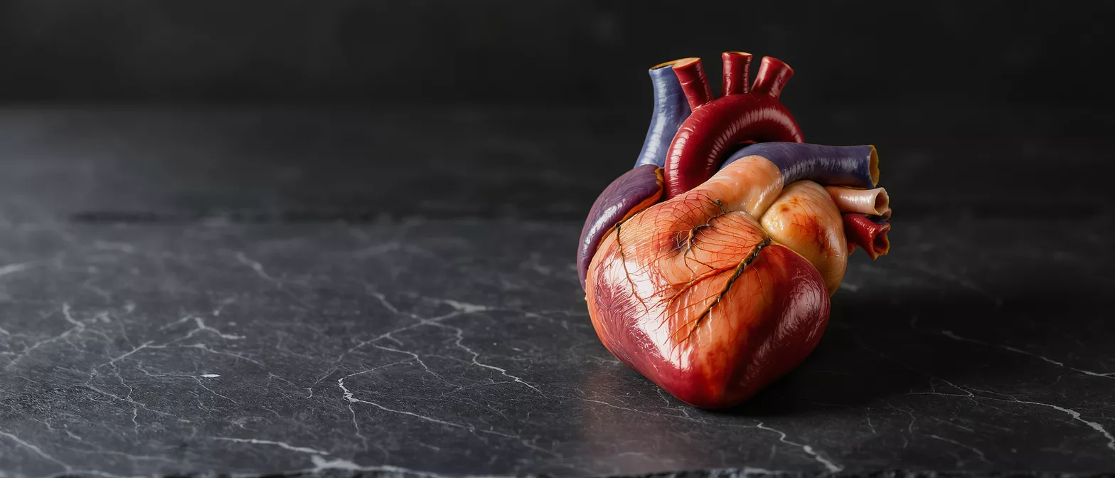
Superior Vena Cava
Learn what the superior vena cava is, how blood flow works, and related conditions.
What is the superior vena cava
The superior vena cava is one of the two largest veins in the human body, responsible for carrying oxygen-poor blood from the upper half of the body back to the heart. When medical professionals discuss the superior vena cava definition, they are referring to a major cardiovascular vessel that collects blood from the head, neck, arms, and upper chest. It acts as a crucial biological highway, ensuring that deoxygenated blood is efficiently returned to the heart so it can be pumped to the lungs for fresh oxygen.
Anatomy relative to the heart: This large vein is located in the upper mediastinum, the central compartment of the chest cavity. It sits just to the right of the midline and drains directly into the right upper chamber of the heart, known as the right atrium. Because of its central placement and considerable size, it is a primary focus during many forms of cardiac surgery.
Understanding this vessel is particularly important for patients preparing for heart surgery. Surgeons must carefully navigate around the superior vena cava heart connection when repairing valves or correcting structural defects. Knowing how this vessel functions helps patients better visualize their own anatomy and the surgical approach their medical team will take.
How superior vena cava blood flow works
The mechanics of superior vena cava blood flow rely on normal breathing and pressure changes within the chest rather than a dedicated pump. As you inhale, the chest cavity expands and creates a slight vacuum effect. This negative pressure naturally draws deoxygenated blood downward from the head, neck, and upper limbs toward the heart, ensuring a continuous and steady stream of circulation.
Role in the circulatory loop: Blood entering from the upper body is low in oxygen because the tissues have already extracted the oxygen they need. The superior vena cava body drainage system delivers this oxygen-poor blood into the right atrium. From there, the blood moves into the right ventricle and is promptly pumped out to the lungs to release carbon dioxide and pick up fresh oxygen.
Any disruption to this natural flow can lead to noticeable symptoms. Because the vein operates under relatively low pressure compared to the arteries, it is vulnerable to compression from surrounding structures in the chest. A patient facing cardiac surgery often requires imaging to ensure that this specific blood flow pathway is clear and functioning optimally before the operation begins.
Superior vena cava anatomy and structure
In terms of physical structure, the superior vena cava anatomy is characterized by its short but remarkably wide tube. It is formed by the joining of the left and right brachiocephalic veins, which are also known as the innominate veins. These two large vessels converge just behind the right first rib, creating a single, robust channel that runs vertically down into the right atrium.
Wall composition and structural integrity: Unlike the thick, muscular walls of the arteries that carry high-pressure blood away from the heart, the superior vena cava veins have relatively thin walls. They contain smooth muscle and elastic tissue but lack the heavy muscular layers found in arteries. This thinner structure allows the vessel to expand easily and accommodate variations in blood volume.
Because the walls are thin, the vessel is somewhat delicate and can be easily compressed or displaced by abnormal growths in the chest. Surgeons must handle this vessel with extreme care during operations. A thorough understanding of its precise structural layout and anatomical relationships allows cardiovascular specialists to perform complex operations without inadvertently tearing or collapsing this vital vein.
Superior vena cava function in the heart
The primary superior vena cava function is to act as the main collecting vessel for deoxygenated blood returning from the upper portion of the body. It gathers blood from the brain, face, neck, arms, and upper chest wall. This function is vital for maintaining balanced circulation, as it ensures that blood continuously cycles through the lungs to be reoxygenated.
Impact on overall cardiac workload: The heart relies on a steady and unimpeded return of blood to maintain an efficient pumping rhythm. If the superior vena cava cardiovascular connection is partially blocked, blood can back up in the upper body. This backup forces the heart to work harder to pull blood through the narrowed pathway, increasing stress on the cardiac muscle over time.
Patients undergoing heart surgery should understand that maintaining the integrity of this vessel is just as important as repairing the heart valves. If the return flow is compromised, the entire circulatory system suffers. Preserving or restoring normal function in this area is a key priority for any successful cardiac operation.
Superior vena cava drainage and connected vessels
The superior vena cava drainage network is an intricate web of smaller vessels that eventually converge into this main vein. Understanding the superior vena cava vessels that feed into it helps clarify how the body manages blood return. The most prominent tributaries include the internal jugular veins, which drain the brain and face, and the subclavian veins, which drain the arms.
- Jugular veins: Drain blood from the brain, face, and neck, emptying directly into the brachiocephalic veins.
- Subclavian veins: Receive blood from the arms and shoulders, merging with the internal jugular veins to form the brachiocephalic veins.
- Azygos vein: An alternative drainage pathway that runs up the spine and connects directly into the superior vena cava, offering a crucial backup route if the main pathway is blocked.
The presence of the azygos system is particularly important in medicine. If a blockage occurs in the superior vena cava chest region, blood can often reroute through the azygos vein to find its way back to the heart. This natural bypass system prevents immediate total failure, allowing medical professionals time to diagnose and treat the underlying cause of the obstruction.
Types of superior vena cava obstruction
When examining the types of superior vena cava obstruction, medical professionals generally categorize them as either intrinsic or extrinsic. Intrinsic obstructions occur when a problem originates inside the vein itself, such as a blood clot or a tumor growing directly within the vessel. Extrinsic obstructions happen when an outside structure, like an enlarged lymph node or a tumor, presses against the vein and collapses it.
Thrombosis and catheter-induced narrowing: Deep vein thrombosis can develop in the upper body, especially in patients with pacemaker wires or long-term central venous catheters. These internal blood clots can suddenly block the vessel. In some cases, scar tissue forms around the catheter site, gradually narrowing the vessel diameter and restricting the return of blood to the heart.
For a cardiac surgery patient, distinguishing between these types of obstruction is critical. Extrinsic compression often requires addressing the outside mass, while intrinsic blockages may require blood thinners or surgical removal of the clot. A clear diagnosis allows the surgical team to plan the safest and most effective intervention for the patient.
Superior vena cava syndrome signs and causes
Superior vena cava syndrome is a clinical condition that occurs when this major vein becomes severely compressed or blocked. The signs of this syndrome are usually highly visible because the blood cannot drain properly from the head and arms. Patients often experience facial swelling, a feeling of fullness in the head, and distended veins across the chest and neck.
Common symptoms patients experience: People with this condition frequently notice shortness of breath, particularly when lying flat, and a bluish tint to the skin. Vision changes, dizziness, and a persistent cough are also common due to the increased pressure in the head. In severe cases, the swelling can become so pronounced that it obstructs the airway, making it a medical emergency.
The causes of this syndrome range from malignant tumors, such as lung cancer pressing on the vein, to benign conditions like blood clots from a central line. Because the symptoms can be frightening and mimic other heart conditions, prompt evaluation by a cardiovascular specialist is essential.
How superior vena cava obstruction is diagnosed
Diagnosing a blockage in this vessel requires advanced imaging techniques that allow doctors to see the structures inside the chest. A standard chest X-ray might show a mass, but a contrast-enhanced CT scan is the gold standard for visualizing the superior vena cava obstruction. This scan uses a special dye to highlight the blood vessels, clearly revealing exactly where the blood flow is restricted.
- CT Angiography: Provides a 3D view of the chest, showing both the blood vessel and any surrounding masses compressing it.
- Venography: A targeted test where dye is injected directly into the arm veins, offering a precise map of the blockage.
- Ultrasound: A non-invasive tool often used to check for blood clots in the neck and arm veins feeding into the chest.
Magnetic resonance imaging may also be used if a patient cannot tolerate the contrast dye used in CT scans. Once the diagnosis is confirmed, the medical team can determine whether the blockage is a clot requiring blood thinners or a physical compression requiring surgical intervention. Understanding what to expect during a vascular diagnostic appointment helps reduce patient anxiety before the tests begin.
Treatment for superior vena cava cardiovascular issues
Treating conditions affecting this vital vessel depends entirely on the underlying cause of the problem. If a blood clot is the issue, patients are typically prescribed anticoagulant medications to prevent further clotting and allow the body to naturally dissolve the existing blockage. In more severe cases, doctors may use a specialized catheter to physically break up or remove the clot.
Stents and surgical bypass: When the vein is compressed by a tumor or scar tissue, doctors often insert a stent, which is a small mesh tube, to prop the vessel open and restore immediate blood flow. For complex blockages that cannot be stented, surgeons may perform a bypass procedure, creating a new pathway around the blocked section using a healthy synthetic graft.
Post-operative care is highly important to ensure the long-term success of these interventions. Following a strict cardiac rehabilitation routine, taking all prescribed medications, and maintaining a post-surgery compression garment if advised, all contribute to a smoother and safer recovery process.
Superior vena cava definition and patient takeaways
To summarize the superior vena cava definition, it is the essential large vein that returns oxygen-depleted blood from the upper body directly to the right side of the heart. Its anatomy is uniquely designed to handle low-pressure blood return, and it features crucial tributaries like the azygos system that can act as natural bypass routes if a blockage occurs. Understanding its function helps patients grasp how their circulatory system remains balanced.
Key points to remember: If you or a family member is facing cardiac surgery, knowing about this vessel helps you understand the complexity of the surgical map the doctors will use. A blockage here, leading to superior vena cava syndrome, presents with visible symptoms like facial swelling and requires immediate imaging and intervention. The thin walls of this vessel make it vulnerable to external pressures but also highly adaptable when healthy.
Ultimately, being informed about your heart anatomy transforms a frightening medical procedure into a comprehensible process. The superior vena cava is a major player in keeping your body oxygenated and healthy. By recognizing its role and the treatments available for its obstruction, you can approach your cardiovascular care with greater confidence and clarity.






