

Incidental Finding on a Heart Scan: What Happens Next
An incidental finding heart scan result can feel overwhelming.
What Is an Incidental Finding on a Heart Scan?
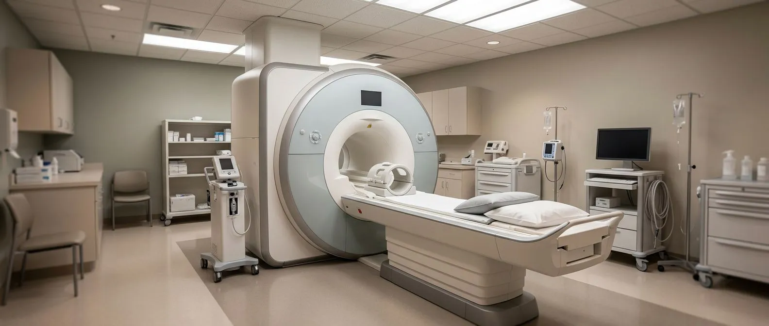
An incidental finding heart scan result occurs when an imaging test performed for one specific reason reveals an unexpected anomaly in a nearby area. When doctors image the lungs to check for a respiratory infection, or perform a routine chest CT scan in the emergency room, they frequently capture clear views of the heart and its surrounding structures. These incidental discoveries can be a double-edged sword: they provide valuable information about asymptomatic issues, but they can also introduce sudden medical anxiety to a patient who otherwise felt completely fine. It is crucial to recognize that most of these findings are not immediately life-threatening and that there are well-established pathways for evaluating them.
Sometimes, a scan ordered for an unrelated complaint, such as a persistent cough, might show an enlargement of the left or right Atrium, the upper chambers of the heart that receive blood returning from the body and lungs. Before the advent of detailed imaging, physicians relied heavily on Auscultation, the practice of listening to heart sounds and blood flow murmurs through a stethoscope. While auscultation remains the cornerstone of initial physical exams, modern imaging catches subtle structural variations that produce no audible murmurs, leading to these incidental discoveries.
Understanding what these unexpected results truly mean requires a structured, methodical approach rather than immediate panic. Radiologists and cardiologists are specifically trained to distinguish between harmless anatomical variants and clinical pathologies that require monitoring or intervention. The goal is to determine if the finding is a benign anomaly or a clue to an underlying condition that needs proactive management, ensuring that decisions are made based on solid clinical evidence.
How Common Are Incidental Findings on Heart Imaging?
The frequency of these discoveries has increased dramatically because high-resolution computed tomography (CT) and magnetic resonance imaging (MRI) are now a standard part of medical care. Decades ago, obtaining a detailed anatomical view of the heart required invasive procedures or a specialized cardiac catheterization. Today, a rapid outpatient scan performed for a completely different reason can visualize the aorta, heart valves, and coronary arteries with incredible clarity.
Researchers publishing in The Lancet note that advanced imaging modalities routinely detect subclinical pathologies that would have gone unnoticed decades ago. This shift in diagnostic capability means we are now catching early signs of aortic aneurysms or coronary plaque long before a catastrophic event like severe Arm Pain or an Atypical Infarction occurs. However, the discovery of these early-stage anomalies requires a careful balance between beneficial early intervention and the avoidance of over-treating benign findings.
Recognizing that a large percentage of adults have minor structural quirks helps put the initial shock of an abnormal heart scan result into perspective. Finding an anomaly is not a harbinger of an immediate cardiac event; it is often just a marker of normal aging or a previously undiagnosed but stable minor condition. The challenge for the cardiologist is to stratify the few clinically significant findings that require attention from the majority that simply warrant observation.
Types of Cardiac Incidentalomas and Their Meaning
Structural and vessel anomalies: Unexpected results often involve the aorta or the coronary arteries rather than the heart valves themselves. For example, an Atheromatous Plaque, a fatty deposit within the artery wall, might be observed early before it causes any significant restriction to blood flow. Identifying whether this plaque is soft and vulnerable or hardened and calcified plays a major role in assessing a patient's future risk of a heart attack.

Another common discovery is a slight dilation of the Ascending Aorta, the very first segment of the body's main artery as it exits the heart. While small to moderate dilations are simply monitored carefully through regular echocardiograms, significantly enlarged dimensions eventually trigger surgical thresholds. When the aortic diameter reaches a dangerous level, preventive surgical repair is necessary to avoid the risk of a life-threatening dissection or rupture.
What to Expect After an Abnormal Heart Scan Result
After receiving an abnormal heart scan result, the immediate next step is a detailed consultation with a qualified cardiologist. The physician will carefully review the imaging, correlate it with your specific symptoms and medical history, and may order more targeted tests. For instance, if the incidental finding suggests possible coronary artery disease, they might schedule a cardiac stress test or a specialized coronary CT angiogram to see exactly how much blood flow is restricted.
Guiding these follow-up decisions are rigorous clinical trials and published medical protocols. For example, the Multifactorial intervention to prevent cardiovascular disease in patients with early rheumatoid arthritis: protocol for a multicentre randomised controlled trial illustrates how comprehensive cardiovascular risk assessments are structured. While that trial focuses on patients with rheumatoid arthritis, the methodology reflects how cardiologists use multifactorial data—including age, blood pressure, and cholesterol—to contextualize an incidental finding.
Your doctor will look for clinical signs that provide context to the scan, determining if the anomaly is merely an incidentaloma or a clue to an underlying condition that requires prompt medical management. This phase requires patience, as additional testing might be necessary to establish a baseline. Establishing an accurate baseline is a crucial step to ensure that any future changes in the heart's structure or function can be measured reliably over time.
Does an Incidental Finding on a Heart Scan Mean I Need Surgery?
Discovering an anatomical anomaly does not automatically mean you need immediate surgery. In fact, the most common response to an incidental cardiac finding is a strategy of watchful waiting. Many heart abnormalities are minor and stable, managed effectively with medication, lifestyle adjustments, and routine surveillance to ensure they do not progress or cause symptoms over time.
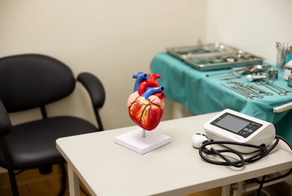
Treatment decisions are strictly guided by established clinical thresholds. For example, an aortic diameter greater than 5.5 cm generally warrants surgical repair because the statistical risk of rupture at that size outweighs the risks of the operation. If an intervention is indeed required, options range from minimally invasive procedures like Balloon Angioplasty, which uses a tiny inflated balloon to open a narrowed artery, to traditional open-heart surgeries that may require an Arterial Graft to bypass a severe blockage.
Heart Scan Result Anxiety and Coping With Uncertainty
It is entirely normal to feel significant heart scan result anxiety after receiving unexpected news about your cardiovascular health. The sudden uncertainty can be highly stressful, especially when the medical terminology in the imaging report feels overwhelming or alarming. This anxiety often leads patients to excessive internet searches late at night, which typically highlight the worst-case scenarios rather than the most likely, benign outcomes.
- Acknowledge the uncertainty: Recognize that waiting for a follow-up appointment is a standard part of the diagnostic process, not a sign of imminent danger. Give the imaging results a few days to be properly reviewed by a specialist.
- Focus on what you control: Maintain your normal daily routine, avoid excessive internet symptom-checking, and instead write down specific questions to ask your cardiologist during your consultation.
A clear management plan developed with your medical team is the most effective way to reduce this anxiety. Knowing exactly what will happen next, when to return for follow-up imaging, and under what specific circumstances to seek urgent medical help restores a sense of control over your health. The cardiologist can also translate the actual meaning of the scan findings, stripping away frightening medical jargon to reveal the true, often much less severe, clinical significance.
Heart Scan Second Opinion: When to Seek Another Look
Seeking a heart scan second opinion is highly recommended if you are unsure about the recommended treatment path or if the initial interpretation of the imaging lacks clear clinical context. Because incidental findings can be subtle and subject to interpretation, a second review by an experienced interventional cardiologist or cardiothoracic surgeon can confirm the size, location, and true significance of the anomaly.
Complex decisions, particularly those involving potential interventions, benefit greatly from a multidisciplinary review. Clinical evidence, such as the Cost-effectiveness of stepwise provisional versus systematic dual stenting strategies in patients with distal bifurcation left main stem lesions: economic analysis of the EBC MAIN trial, shows how nuanced treatment choices can be in cardiology. Deciding between an aggressive intervention and conservative management when major vessels are involved requires a careful consideration of long-term clinical outcomes and risks.
When seeking a second opinion, ensure you bring your original scan images and the initial radiologist's report. This allows the consulting physician to independently evaluate the raw data rather than relying solely on another doctor's written summary. Sometimes, a second opinion reveals that what was initially flagged as a problem is actually a benign anatomical variant, entirely preventing unnecessary further testing or treatment.
Follow Up Care for Asymptomatic Heart Abnormalities
Follow up care for asymptomatic heart abnormalities typically involves serial imaging. This means repeating the heart scan, CT, or echocardiogram at regular intervals set by your physician, such as six months or a year. The goal is to closely track any changes in structural size or valve function, ensuring the anomaly remains stable and is not growing at a dangerous rate.

During this surveillance period, your doctor might prescribe medications to manage underlying cardiovascular risk factors. For instance, if there is evidence of early coronary disease, a daily regimen of Aspirin might be recommended to prevent blood clots, alongside medications that target lowering Apolipoprotein B levels, a protein that plays a core role in plaque buildup in the arteries, to stabilize the arterial walls and prevent further damage.
Incidental Heart Valve Finding: What Patients Should Know
An incidental heart valve finding, such as mild regurgitation (a leak) or mild stenosis (narrowing), is a very frequent discovery on echocardiograms. The heart's four valves control the one-way flow of blood through the heart's chambers, and minor leaks are often a normal part of the aging process as the delicate leaflets thicken slightly over decades of use.
If a valve disease progresses to the point where intervention is required, modern cardiology offers highly effective transcatheter solutions. For example, a Balloon-expandable Valve can be implanted via a catheter threaded through a blood vessel, a procedure commonly known as TAVR (transcatheter aortic valve replacement). This advanced procedure avoids the need for open-chest surgery in suitable patients, offering a lower risk profile and a much faster recovery time for those who qualify.
- Monitor for progression: While the incidental finding may be asymptomatic at first, symptoms like new shortness of breath, unusual fatigue, or chest tightness indicate the valve issue may need active treatment rather than just observation.
- Understand the interventions: Before a valve replacement is considered, less severe narrowing might temporarily be treated with Balloon Aortic Valvuloplasty, a procedure that uses a small inflatable balloon to gently stretch the valve open.
Coronary Calcium Incidental Finding: Assessing Your Risk
A coronary calcium incidental finding is frequently discovered during a CT scan of the chest performed for lung health screening. Calcium deposits within the coronary arteries indicate the presence of atherosclerosis, or plaque buildup, even if you currently have no symptoms of chest pain or exercise intolerance. This is a structural change that happens slowly over time; having an Automated External Defibrillator at home does not prevent this calcification from forming, only proper medical management can slow its progression.

In conclusion, an incidental finding on a heart scan is a starting point for careful evaluation, not an immediate crisis. By working closely with a qualified cardiac team, you can determine the clinical significance of the anomaly and create a safe, evidence-based management plan tailored to your specific situation. If you have received an unexpected scan result, we encourage you to discuss your case with a specialist and book a consultation to ensure your heart health is properly managed rather than self-diagnosing.






