Ventricular Fibrillation: Emergency Treatment and Prevention
Ventricular fibrillation treatment starts with fast CPR and defibrillation.
Ventricular fibrillation treatment is a race against the clock. In this rhythm, the heart's lower chambers — the ventricles — quiver chaotically instead of pumping blood. Within seconds, the person collapses and stops breathing normally. It is frightening to read about, and more frightening to witness. But here is the hopeful part: fast action with CPR and a defibrillator saves lives, and modern cardiology offers real options for preventing the next episode. This guide walks through what ventricular fibrillation is, what happens in those first critical minutes, and how specialists evaluate and treat the heart afterward — in plain language, for patients and families in Mexico and beyond.
What Is Ventricular Fibrillation?
The rhythm itself. Ventricular fibrillation is a heart rhythm disorder in which the ventricles receive chaotic electrical signals. Instead of contracting in one coordinated squeeze, thousands of muscle fibers twitch independently. The heart produces almost no forward blood flow. Brain, heart, and every other organ are starved within seconds.
Why it is a medical emergency. This is not a rhythm the heart can sustain or correct on its own. Without defibrillation — an electrical shock that resets the heart — ventricular fibrillation leads to sudden cardiac arrest and death within minutes. It is one of the most time-critical emergencies in all of medicine.
Where it fits among rhythm disorders. Atrial Fibrillation, a much more common condition, affects the heart's upper chambers and is serious but rarely immediately fatal. Ventricular fibrillation is a different category of threat. When our team explains this difference to patients, we use one simple image: atrial fibrillation is an engine idling badly; ventricular fibrillation is an engine that has stopped entirely.
What Causes Ventricular Fibrillation?
Underlying heart disease. Most episodes arise in hearts that are already diseased. A prior heart attack leaves scar tissue that disrupts electrical pathways. Other contributors include Ventricular Hypertrophy (thickening of the heart muscle), heart failure, and inherited electrical conditions such as long QT syndrome. Structural problems like a Ventricular Septal Defect — a hole in the wall between the ventricles — can also play a role over a lifetime.
Acute triggers. Sometimes the trigger is a fresh event: a new coronary artery blockage, a severe electrolyte imbalance (especially low potassium), or a drug toxicity. In postoperative settings, Postoperative Atrial Fibrillation can appear after surgery and, rarely, deteriorate into dangerous ventricular rhythms if other risk factors are present.
Triggers doctors look for. The evaluation after an episode usually focuses on finding the cause, because treatment depends on it:
- Scar from a previous heart attack: the most common substrate for ventricular arrhythmias.
- Acute ischemia: a blocked artery starving heart muscle right now.
- Electrolyte disturbance: potassium, magnesium, or calcium out of range.
- Inherited channelopathies: genetic electrical diseases such as Brugada syndrome.
What Is the Difference Between Coarse and Fine Ventricular Fibrillation?
What the words mean. On the heart monitor, ventricular fibrillation can look "coarse" — large, tall, disorganized waves — or "fine" — small, low-amplitude squiggles. Coarse VF usually means the heart muscle still has energy and is fibrilling vigorously. Fine VF suggests the muscle is weakening and losing electrical amplitude as oxygen deprivation continues.
Why it matters in resuscitation. Coarse fibrillation responds better to defibrillation. As minutes pass without treatment, coarse waves typically decay into fine waves, and shock success drops sharply. This visual decline is one reason rescuers are trained to minimize any delay before the first shock.
Fine VF and asystole. If treatment is very late, fine VF can deteriorate into asystole — a flat line. At that point, defibrillation no longer helps; the priority becomes high-quality CPR and medications to try to restore an organized rhythm. For families watching a monitor, this is why speed matters more than anything else.
Ventricular Fibrillation vs Ventricular Tachycardia on an ECG: What Are the Differences?
Ventricular Tachycardia on the tracing. Ventricular Tachycardia (VT) is a fast but organized rhythm. The ECG shows wide, regular, rapid beats — often 150 to 250 per minute — that look like repeated, similar shapes. The ventricles are beating too fast, but they are still beating together.
Ventricular fibrillation on the tracing. VF shows no organized beats at all. The tracing is a chaotic, irregular wobble with no distinguishable QRS complexes. If VT is a car engine revving uncontrollably, VF is the engine shaking itself apart.
Why the distinction changes treatment. Many VT episodes are treated with medications, synchronized cardioversion, or ablation. VF cannot be treated with synchronized shocks — the machine cannot find a wave to synchronize with — so it requires an unsynchronized shock. These two rhythms sit on a continuum: VT can degenerate into VF, which is why new, sustained VT is treated urgently rather than watched. This overlap is one of the questions we take time to explain in consultation, because patients often have both rhythms documented on the same monitor strip.
What Is Ventricular Fibrillation Emergency Treatment?
The immediate goal: restore circulation. Emergency treatment for ventricular fibrillation follows one unbreakable sequence: call emergency services, start CPR, and apply a defibrillator as soon as one is available. Every minute without defibrillation lowers the chance of survival meaningfully. Bystander CPR acts as a bridge — it keeps oxygen flowing to the brain until a shock can be delivered.
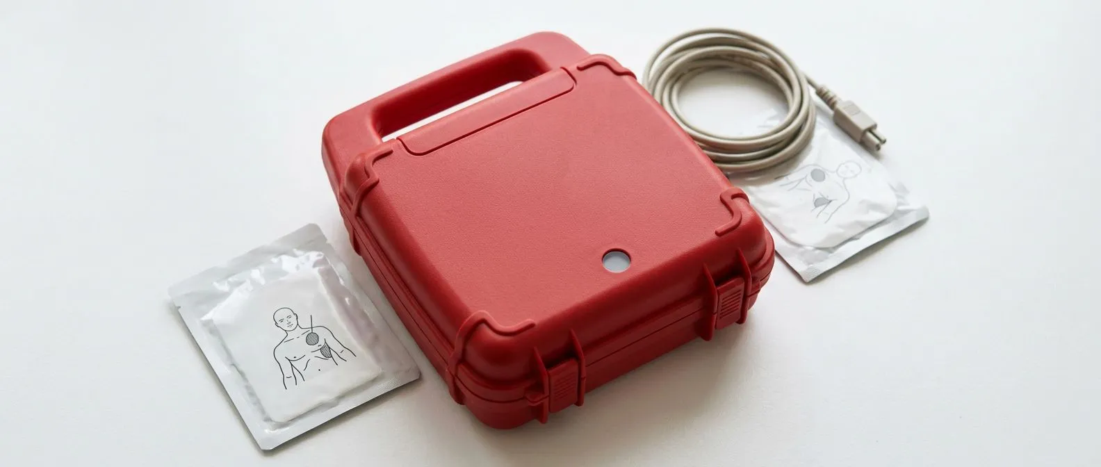
What bystanders should do. You do not need medical training to save a life here. Public AEDs (automated external defibrillators) are designed to analyze the rhythm and give voice instructions:
- Call emergency services and, if possible, send someone for the nearest AED.
- Push hard and fast in the center of the chest — about 100 to 120 compressions per minute.
- Attach the AED pads and let the device analyze; deliver the shock if it advises one.
- Resume compressions immediately and continue until professionals arrive.
What paramedics do on arrival. Ambulance teams confirm the rhythm, deliver shocks, insert an airway, give adrenaline and antiarrhythmic drugs such as amiodarone, and transport the patient to a hospital capable of cardiac care. In our experience, patients who arrive at the hospital with a spontaneous pulse have received the fastest possible field response — which is why community awareness of CPR matters as much as any hospital technology.
How Do Defibrillation Procedure Steps Work?
What the shock actually does. A defibrillation shock delivers a brief, powerful electrical current across the heart. It does not "restart" the heart like a battery. Instead, it momentarily depolarizes every heart cell at once — wiping the chaotic activity clean — so the heart's natural pacemaker can restart an organized rhythm.
How an AED guides you. Automated external defibrillators analyze the rhythm and only allow a shock when it is appropriate, which makes them safe for untrained bystanders. Pads are placed on the upper right chest and lower left side. The machine charges and instructs everyone to stand clear. One shock is followed immediately by two minutes of CPR before the next analysis.
Inside the hospital. Advanced teams use manual defibrillators with greater control over energy and timing, and they may combine shocks with medications and with catheter-based reperfusion if a heart attack is the trigger. Survival after out-of-hospital cardiac arrest depends heavily on this chain: recognition, CPR, early shock, and rapid advanced care. A study of anticoagulated patients with atrial fibrillation in the European PREFER in AF registry illustrates how underlying rhythm disease, clot risk, and bleeding risk must all be balanced when planning long-term therapy — the same principle of individualized balance applies to patients rescued from VF (BMJ Open).
What Happens in Hospital After Resuscitation?
Stabilization. Once a pulse is restored, the patient is admitted to an intensive care unit. The first priorities are supporting blood pressure, oxygen, and body temperature, and checking for injury from CPR or the arrest itself. Therapeutic cooling is sometimes used to protect the brain after prolonged arrest.
Finding the cause. The team then works to identify why the episode happened. Tests typically include an electrocardiogram, blood tests for cardiac enzymes and electrolytes, an echocardiogram (an ultrasound of the heart), and a coronary angiogram — a dye study of the heart arteries. A new blockage may be opened urgently with a stent.
The decision about an ICD. If the episode occurred without a reversible cause, most patients are candidates for an implantable cardioverter defibrillator before discharge. When patients reach our consulting rooms after an event like this, our team reviews every study against international practice guidelines and explains, in plain words, what each one shows before any recommendation is made.
Is an Implantable Cardioverter Defibrillator for VFib Right for You?
What the device does. An implantable cardioverter defibrillator (ICD) is a small device placed under the skin near the collarbone, with wires leading into the heart. It constantly watches the rhythm. If it detects ventricular fibrillation or fast VT, it delivers therapy automatically — usually a paced sequence or an internal shock — within seconds, wherever the patient happens to be.
Who needs one. International guidelines recommend an ICD for survivors of VF or sustained VT without a reversible cause, and for certain patients with weakened heart function even before any documented episode. The device does not prevent the arrhythmia from starting, but it prevents it from being fatal — an important distinction for patients and families to understand.
What implantation involves. The procedure itself is usually straightforward: local anesthesia with sedation, an incision below the collarbone, and leads guided through a vein into the heart. Most patients go home within a day or two.
Can Cardiac Ablation Treat Ventricular Fibrillation?
What ablation is. Cardiac ablation uses thin catheters threaded through veins into the heart to find and destroy small areas of tissue that trigger or maintain dangerous rhythms. Energy — usually radiofrequency heat or cryo (freezing) — creates tiny scars that block the abnormal electrical circuits.
When it is used for VF. Ablation is not typically the first treatment for ventricular fibrillation, but it plays a growing role for patients whose arrhythmias are driven by identifiable triggers, especially the premature beats that spark VF in hearts with scar. In those patients, catheter ablation can reduce the number of ICD shocks dramatically, protecting both quality of life and heart function.
Choosing the right combination. For many patients the answer is not one therapy but a combination: an ICD for protection, medication to suppress episodes, and ablation when triggers can be mapped. I often tell patients in our Guadalajara and Querétaro consulting rooms that the best plan is the one matched to their own heart's anatomy and scar pattern — not a one-size-fits-all recipe.
How Can Ventricular Fibrillation Be Prevented?
Protecting the heart muscle. The most powerful prevention is avoiding the diseases that create the substrate for VF. That means controlling blood pressure, cholesterol, and diabetes, not smoking, staying physically active, and treating coronary artery disease early. A heart attack treated promptly leaves less scar, and less scar means fewer abnormal circuits.
Correcting reversible factors. Regular monitoring of potassium and magnesium matters for patients on diuretics or kidney disease. Medications that prolong the QT interval — certain antibiotics, psychiatric drugs, and others — need review in susceptible patients. Family members of anyone who died suddenly at a young age may benefit from cardiac genetic evaluation.
Devices and follow-up. For patients with known risk — prior myocardial infarction with reduced pumping function, heart failure, or inherited conditions — guideline-directed therapy and regular follow-up substantially reduce risk. Our multidisciplinary team reviews every such case together, comparing the patient's situation against international guidelines before recommending any intervention, and we actively encourage a second opinion as a normal part of that decision.
When Should You Seek Help for Heart Rhythm Symptoms?
Warning signs to take seriously. Fainting during exercise, a racing heartbeat with dizziness, chest pain, or unexplained breathlessness deserve evaluation — promptly, not eventually. Patients with known heart disease should discuss any new palpitations with their cardiologist. These symptoms are often benign, but the rare dangerous causes are best found before they cause an emergency.
How specialists evaluate risk. A thorough evaluation may include an ECG, Holter monitoring (a wearable recorder), an echocardiogram, and in selected cases an electrophysiology study, where catheters map the heart's electrical system directly. Scar imaging such as cardiac MRI adds detail. The first contact at our practice is a free video consultation: we get to know the patient, talk through the condition together, and identify exactly which studies are still needed — so nothing unnecessary is ordered.
Building the treatment plan. Once the picture is complete, the team proposes a plan with options and trade-offs: medication, device therapy, catheter procedures, or surgery where indicated. Because Dr Molina trained in cardiothoracic surgery at the Universidad de Guadalajara, in cardiac, minimally invasive, robotic and structural surgery at the University of Montreal, and in structural cardiology and TAVR at CHU Bordeaux, our patients can access conventional, minimally invasive, robotic and transcatheter options — and we recommend the less invasive route only where the evidence truly supports it for that individual.
Conclusion: Ventricular Fibrillation Treatment Offers Real Hope
Ventricular fibrillation treatment has two halves. The first half belongs to everyone: recognize the emergency, start CPR, and use a defibrillator fast. The second half belongs to cardiology teams: find the cause, protect the patient with an ICD when indicated, and address the underlying disease with medication, ablation, or surgery. The survival rate after VF depends most on the speed of the first response — and the long-term outlook depends on the quality of the evaluation that follows. If you or a loved one has survived this event, or if heart rhythm symptoms worry you, the most useful next step is a calm, thorough conversation with a qualified cardiac team. Our team in Guadalajara, Querétaro and Hermosillo is ready to review your case, explain your options clearly, and help you decide — the decision always remains yours.