After a Transient Ischaemic Attack: Heart Checks to Ask For
Transient ischaemic attack heart screenings prevent strokes.
What Is a Transient Ischaemic Attack and Why Heart Checks Matter
A transient ischaemic attack, sometimes called a mini stroke, happens when blood flow to part of the brain is briefly interrupted. The symptoms — which can include sudden weakness on one side of the body, slurred speech, or vision changes — usually resolve within minutes to a few hours. Because recovery seems complete, many people assume the danger has passed and carry on as before. In reality, a TIA is a powerful warning sign that something in the cardiovascular system needs urgent attention, and the heart is very often where the investigation begins. A transient ischaemic attack heart evaluation looks for the underlying source of the problem before a more serious, permanent event occurs.
The reason heart checks are so important after a TIA is straightforward: the heart and brain share the same blood supply. A clot that forms inside the heart can travel through the bloodstream and lodge in a brain artery, producing exactly the kind of temporary symptoms a TIA causes. Irregular heart rhythms, damaged valves, or narrowed coronary arteries can all reduce blood flow in ways that affect the brain first. Identifying which of these factors played a role is the key to preventing a full stroke, and it is why doctors treat a TIA as a medical emergency rather than a minor episode to forget.
Many patients feel entirely normal by the time they reach a doctor, which makes it tempting to delay or skip testing. However, cardiac specialists at institutions such as the Cleveland Clinic emphasise that the period immediately after a TIA is when the risk of a recurrent event is highest. Prompt evaluation — ideally within days, not weeks — gives your medical team the best chance to find and treat the cause before it progresses to something far more dangerous.
How the Heart and Brain Share Blood Vessels After a TIA
Understanding why a heart check matters so much after a TIA requires a basic picture of how blood moves through the body. The heart pumps oxygen-rich blood upward through the carotid arteries in the neck and into the brain. If anything disrupts that flow — whether a clot that breaks loose from the heart, a narrowed artery, or an irregular heartbeat that allows blood to pool and clot — the brain is often the first organ to show symptoms. This shared circulation is why cardiologists and neurologists work together after a TIA, each examining a different part of the same connected system.
Cardiovascular and cerebrovascular diseases together represent one of the largest causes of disability worldwide, as documented in the Global Burden of Disease Study 2015. The connection between heart and brain disease is not coincidental. Conditions such as atherosclerosis — the gradual buildup of fatty deposits inside artery walls — affect blood vessels throughout the body simultaneously. A patient who has narrowing in the carotid artery supplying the brain is very likely to have similar narrowing in the coronary arteries supplying the heart. Finding one problem almost always means searching for the other.
The Aortic Arch as a Clot Source: The aortic arch, the curved top portion of the body's largest artery, sits just above the heart and gives rise to the vessels that feed the brain. Plaque or debris here can dislodge and travel upward, causing a TIA. In some cases, significant disease in this area may eventually require Aortic Arch Replacement, a surgical procedure that removes the diseased segment and restores healthy blood flow. More commonly, though, the arch is assessed with imaging and monitored over time, and the findings help your team decide whether medication alone is sufficient or whether a surgical consultation is warranted.
Transient Ischaemic Attack Heart Screening: What Every Patient Should Know
When you arrive for a post-TIA cardiac evaluation, your medical team will follow a structured pathway designed to identify the most likely sources of interrupted brain blood flow. The specific tests ordered depend on your symptoms, medical history, age, and overall risk profile, but most patients can expect a combination of electrical monitoring, imaging, and blood tests. The goal is to build a complete picture of how well your heart is functioning and whether any structural or rhythm abnormalities could have contributed to the episode. Understanding what each test looks for can help you feel more prepared and less anxious about the process.
Why Comorbidity Matters: Patients with multiple cardiovascular risk factors face a compounded threat, and research has consistently shown that underlying heart disease frequently accompanies other serious illnesses. A study published in The Lancet documenting clinical characteristics of severely ill patients in Wuhan, China, found that cardiovascular comorbidities were among the most common pre-existing conditions. This pattern reinforces why a TIA should trigger a comprehensive heart assessment rather than a narrow one — the aim is to uncover all contributing factors, not simply the most obvious.
A typical screening pathway begins with non-invasive tests and progresses to more detailed imaging if initial results suggest a problem. Here is a summary of the tests most commonly requested after a TIA:
- Electrocardiogram (ECG): Records the heart's electrical activity to detect irregular rhythms or signs of previous heart attacks.
- Echocardiogram: Uses sound waves to create moving images of the heart's chambers, valves, and pumping strength.
- Blood tests: Check cholesterol, blood sugar, clotting factors, and markers of inflammation that may reveal underlying conditions.
- Carotid ultrasound: Examines the arteries in the neck for narrowing or plaque that could reduce blood flow to the brain.
- Extended rhythm monitoring: Uses a wearable device to capture intermittent irregular heartbeats that a brief ECG might miss.
Electrocardiogram and Heart Rhythm Monitoring After a TIA
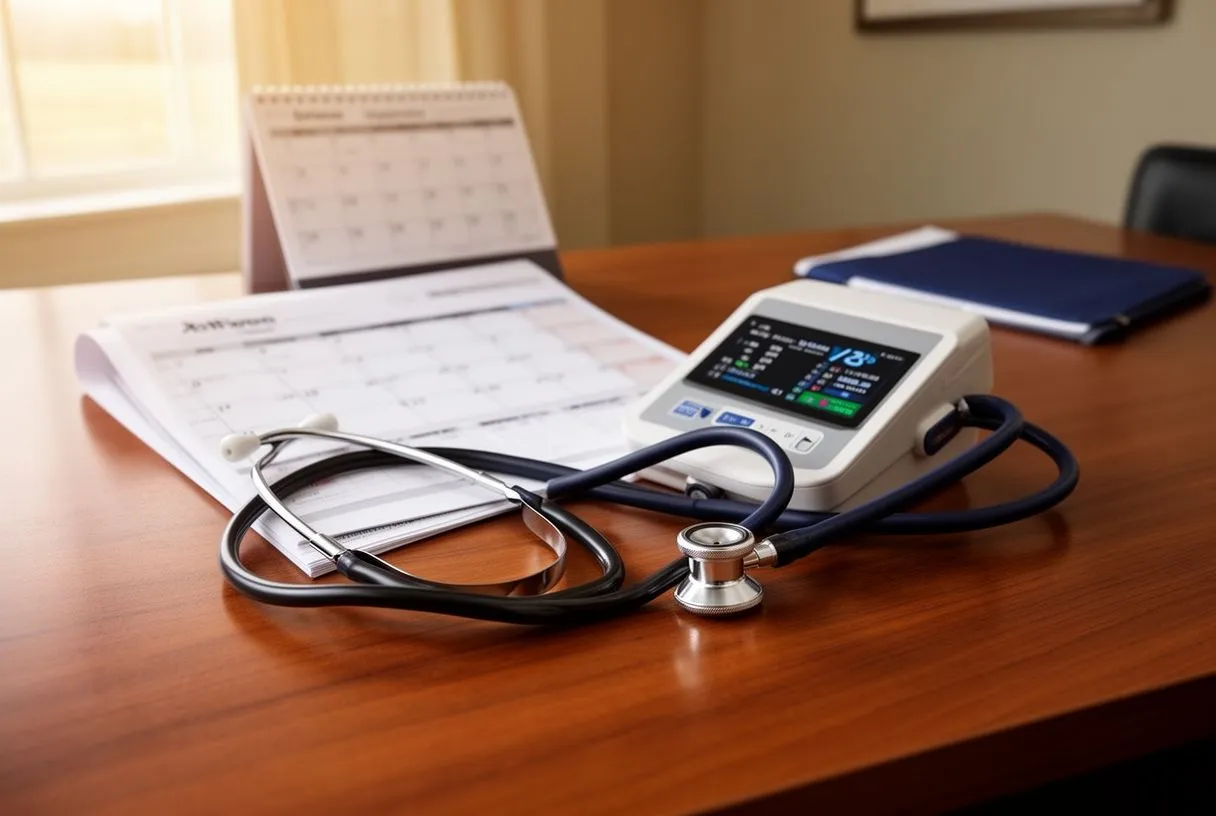
An electrocardiogram, usually called an ECG or EKG, is almost always the first heart test performed after a TIA. This painless recording measures the electrical activity of your heart through small sensors placed on the chest, arms, and legs. It takes only a few minutes but can reveal irregular rhythms, previous silent heart attacks, or signs that the heart muscle is under strain. Because some rhythm problems come and go unpredictably, a single ten-second ECG may not capture them, which is why longer monitoring is frequently recommended as a follow-up step.
Detecting Atrial Fibrillation: The most common rhythm disorder linked to TIA is atrial fibrillation, a condition where the upper chambers of the heart beat irregularly and often too fast. When the heart beats erratically, blood can pool in a small pouch called the left atrial appendage, forming clots that may travel to the brain. Detecting atrial fibrillation after a TIA can take days or even weeks of monitoring, sometimes using a small wearable device or an implantable recorder placed just under the skin. If identified, treatment may include medications such as Amiodarone to help restore a steady rhythm, or blood thinners like warfarin to reduce the likelihood of future clot formation.
It is worth noting that rhythm monitoring is not a one-size-fits-all process. Some patients need only a 24-hour Holter monitor, while others benefit from a device that records for several weeks at a time. The choice depends on how frequently the suspected rhythm problem occurs and how strongly the medical team suspects it as the cause of the TIA. Your cardiologist will explain the reasoning behind the chosen monitoring duration and will review the results with you in detail, so you understand exactly what was found and what it means for your ongoing treatment plan.
Heart Ultrasound After TIA: What an Echocardiogram Reveals
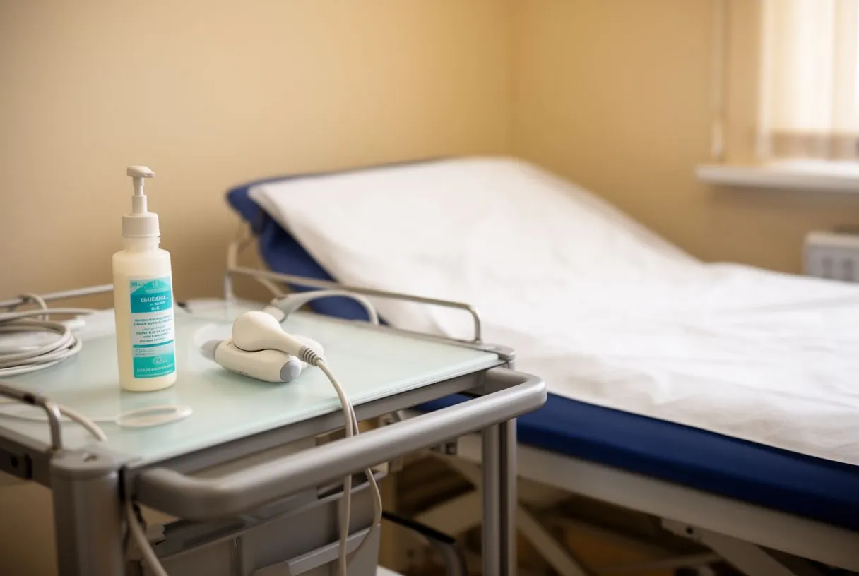
An echocardiogram is a heart ultrasound that uses sound waves to create detailed moving images of the heart's structure and function. After a TIA, this test is essential because it allows your medical team to see whether the heart's chambers, valves, and walls are working correctly. The test is completely painless — a technician places a small probe on the chest, and the images appear on a screen in real time. No radiation is involved, and the procedure typically takes about thirty to forty-five minutes from start to finish.
What the Ultrasound Looks For: The echocardiogram can reveal several issues that might cause a TIA. Blood clots inside the heart, especially in the left atrial appendage, are a prime target. Valve problems such as Aortic Regurgitation — where the aortic valve does not close properly, allowing blood to leak backward — or Aortic Insufficiency can create conditions where clots form or blood flow becomes turbulent. The ultrasound can also assess how forcefully the heart pumps, whether the chambers are enlarged, and whether there are congenital defects such as a patent foramen ovale, a small hole between the upper chambers that can allow clots to bypass the lungs and travel directly to the brain.
In some cases, a standard echocardiogram through the chest wall may not provide enough detail, and a transoesophageal echocardiogram may be recommended. This involves a thin probe passed down the throat while the patient is lightly sedated, giving the sonographer a much closer view of the heart's structures from behind. While the idea may sound uncomfortable, patients are given medication to relax and usually remember very little of the procedure. The additional detail can be invaluable when standard imaging leaves questions unanswered, and your team will only recommend it if they believe the extra information could genuinely change your treatment.
Blood Clot and Heart Valve Checks After a Mini Stroke
When a TIA occurs, one of the most urgent questions is whether a blood clot formed inside the heart and travelled to the brain. This is why blood clot detection is a central part of the cardiac workup. Alongside echocardiography, your team may order blood tests to check for conditions that make clotting more likely — such as elevated cholesterol, high blood sugar, or markers of inflammation. Anaemia, a condition where the blood carries less oxygen than normal, may also be identified during these tests and addressed, as it can compound the effects of reduced blood flow. If a clot source is found, Antiplatelet Therapy — medications that make platelets less sticky and less likely to clump together — is typically started immediately to reduce the risk of recurrence.
Heart Valve Assessment: The heart's valves play a critical role in keeping blood moving in one direction, and damaged valves can contribute to TIA risk in several ways. Valve disease can cause turbulent blood flow that encourages clot formation, or it can allow bacteria to settle on the valve surface if an infection develops elsewhere in the body. For patients who have artificial valves or certain congenital defects, Antibiotic Prophylaxis before dental or medical procedures may be recommended to prevent bacteria from entering the bloodstream. Your cardiologist will assess whether your valve condition warrants this precaution based on the specific findings from your echocardiogram and other imaging.
Valve evaluation also includes checking the aortic valve and the surrounding structures. The Aortic Annulus — the ring of tissue where the valve attaches — is measured to assess whether the valve is structurally sound. In some cases, Annulus Sizing helps determine whether a valve repair or replacement might be needed in the future. For patients with significant valve disease who are not yet surgical candidates, an Annual Echocardiogram allows the team to track changes over time and intervene before a problem becomes urgent. The key message is that valve health is monitored proactively, not reactively, after a TIA.
Coronary Artery and Atherosclerosis Screening After TIA
Atherosclerosis — the gradual narrowing of arteries by fatty deposits — is the single most common underlying cause of both TIA and heart attacks. After a TIA, screening the coronary arteries for blockages is essential because the same disease process affecting the brain's blood supply is very likely affecting the heart's as well. This screening may begin with non-invasive tests such as a stress test or coronary CT angiogram, which can reveal narrowed segments without the need for catheters or a hospital stay. If these initial tests are reassuring, your team may focus on medical management and lifestyle changes; if they raise concerns, further investigation is arranged promptly.
When More Detailed Imaging Is Needed: If non-invasive tests suggest significant coronary artery disease, a cardiac catheterization may be recommended. This procedure involves threading a thin tube through a blood vessel in the wrist or groin and advancing it to the heart, where dye is injected to make the coronary arteries visible on X-ray. While more invasive than other tests, it provides the most precise images of blockages and allows the cardiologist to measure exactly how narrowed each artery is. The information gathered can determine whether medication, stenting, or bypass surgery is the most appropriate next step, and your team will discuss each option with you in plain language before any decision is made.
Atherosclerosis screening also extends beyond the coronary arteries themselves. The Aortic Diameter may be measured to check for aneurysms — areas where the artery wall has weakened and bulged outward. If an aneurysm is found, monitoring its size over time is critical, and Aortic Aneurysm Repair may eventually be recommended if it reaches a dangerous diameter. Your team will also be alert for signs of Aortic dissection: a true cardiovascular emergency that occurs when the inner layer of the aorta tears. The same imaging that evaluates the heart can often assess the aorta and other major vessels, making the post-TIA workup a comprehensive vascular evaluation rather than a narrowly focused heart test.
How Long Should Cardiac Monitoring Continue After a TIA
One of the most common questions patients ask after a TIA is how long the heart monitoring will last. The honest answer is that it varies considerably from person to person, depending on what the initial tests reveal and whether a clear cause has been identified. For some patients, a few days of rhythm monitoring and a single echocardiogram provide all the answers needed. For others — particularly those whose TIA cause remains uncertain after standard testing — monitoring may continue for weeks or even months, using implantable devices that record the heart's rhythm continuously and transmit data back to the cardiology team.
The reasoning behind extended monitoring is sound. Atrial fibrillation, the irregular rhythm most commonly linked to TIA, can be highly intermittent. Some patients experience episodes only once every few weeks, meaning a short recording period will almost certainly miss them. Studies have shown that the longer monitoring continues, the more likely intermittent atrial fibrillation is to be detected — and once found, it can be treated effectively with blood thinners that dramatically reduce stroke risk. Your cardiologist will weigh the inconvenience of longer monitoring against the potential benefit of finding a treatable rhythm problem, and will involve you in that decision.
Even after the intensive monitoring phase ends, follow-up cardiac care does not stop. Patients who have had a TIA are typically enrolled in ongoing cardiovascular surveillance, which may include periodic echocardiograms, blood tests, and clinical reviews. Ankle Swelling, breathlessness, or new palpitations should be reported promptly, as these can signal worsening heart function that needs assessment. The goal of long-term follow-up is not simply to watch and wait, but to actively manage risk factors — blood pressure, cholesterol, rhythm stability, and valve function — so that the conditions that produced the first TIA are controlled before they can produce a second.
When a TIA Leads to Preventive Heart Surgery
For most TIA patients, the cardiac evaluation identifies a medical problem that can be managed with medications, lifestyle changes, and monitoring. However, in a minority of cases, the workup reveals a structural heart or vessel problem significant enough to warrant surgery. This is not as alarming as it may sound — the entire purpose of the post-TIA evaluation is to find such problems while there is still time to intervene preventively, before a major stroke or heart attack occurs. When surgery is recommended, it is because the treating team believes that fixing the underlying structural issue offers better protection than medication alone.
Surgical Scenarios After TIA: Several cardiac surgical procedures may arise from a TIA workup. If severe carotid artery narrowing is found alongside coronary disease, combined surgery may be considered. Significant aortic arch disease may lead to Aortic Arch Replacement, a complex operation that uses an Aortic Cross-clamp to temporarily stop blood flow while the diseased segment is replaced, often employing Antegrade Cerebral Perfusion to keep the brain supplied with oxygen during the procedure. Severe valve disease may require repair using an Annuloplasty Ring to support a leaking valve, or valve replacement through open surgery or transcatheter aortic valve replacement (TAVR) for patients who are not suitable for conventional surgery. In each case, the recommendation follows careful imaging, discussion, and multidisciplinary review.
Patients preparing for cardiac surgery after a TIA will go through a thorough pre-operative process. This includes detailed imaging, anaesthetic assessment, and sometimes an Antimicrobial Shower protocol to reduce infection risk before surgery. The surgical team will explain the procedure, the expected recovery time, and the long-term benefits in terms of stroke prevention. It is natural to feel anxious about the prospect of heart surgery, but remember that the TIA has done something valuable: it has provided an early warning that allows the problem to be addressed proactively, often with excellent outcomes, rather than discovered in an emergency setting where options may be far more limited.
Working With Your Cardiology Team: Next Steps and Questions to Ask
After a TIA, the most important step you can take is to engage actively with your cardiology team and ensure you understand every aspect of your evaluation and treatment plan. A good cardiac specialist will welcome your questions and will explain findings in plain, accessible language. If you do not understand a test result, a medication, or a recommendation, ask for clarification — there is no such thing as a silly question when it