Aneurysm Surveillance: How Often to Scan
Aneurysm surveillance scans keep you safe with the right timing.
If you or someone close to you has been told there is an aneurysm — a balloon-like widening of a blood vessel — the weeks after that diagnosis can feel uncertain. The good news is that most aneurysms grow slowly and can be watched safely with a planned schedule of aneurysm surveillance scans. This guide explains how often those scans happen, what each test shows, and how you and your care team decide together when watching is enough and when treatment becomes the safer path.
What Is Aneurysm Surveillance?
Surveillance means watching, not waiting blindly. Aneurysm surveillance is a structured program of imaging tests performed at set intervals to measure the size of an aneurysm and track whether it is changing. The most common location is the aorta, the body's main artery that runs from the heart down through the chest and abdomen. An Aortic Aneurysm that measures below a certain diameter carries a low risk of rupture, and careful monitoring allows surgery to be offered only to the people who genuinely need it.
The idea is simple: blood vessels change slowly, so repeated measurements over months or years reveal the true growth rate far better than any single scan. A picture taken today tells you where the aneurysm is; a series of pictures tells you where it is going. That direction and speed are what drive every decision about Aortic Aneurysm Repair versus continued observation.
In our practice, every case is reviewed by the multidisciplinary team against international practice guidelines before any recommendation is made, and the patient receives a clear proposal with the alternatives explained in plain language. The final decision is always theirs — our role is to make sure it is an informed one.
What Size Aortic Aneurysm Requires Surveillance?
The answer depends on where the aneurysm sits. For an Abdominal Aortic Aneurysm — one in the section of the aorta running through the belly — most guidelines recommend surveillance for aneurysms measuring roughly 3.0 to 5.4 centimeters in diameter, with repair generally offered at 5.5 centimeters or larger, or faster growth. A Thoracic Aortic Aneurysm, located in the chest, follows slightly different thresholds that also account for the patient's body size, underlying connective-tissue conditions, and family history.
Diameter is not the only number that matters. Shape, growth speed, the presence of a family history of rupture, and conditions such as bicuspid aortic valve or Marfan syndrome all shift the thresholds. This is why a size that is safe to watch in one person may deserve earlier repair in another.
- 3.0–3.9 cm: typically monitored with less frequent imaging, often annually or longer
- 4.0–5.4 cm: closer surveillance, usually every six to twelve months depending on location and growth
- 5.5 cm or faster growth: most teams discuss repair, because rupture risk begins to outweigh the risk of an operation
How Are Aneurysms Monitored Over Time?
The main tool is imaging at fixed intervals. Aneurysms are monitored through repeat scans — ultrasound, CT, or MRI — performed on a schedule set by the aneurysm's size and growth rate. Each scan is compared with the previous one, and measurements are taken at the same points on the vessel so that change is real, not an artifact of angle or technique. This is what an aneurysm monitoring schedule actually looks like in practice: the same test, the same landmarks, every time.
Between scans, your team also watches for symptoms and manages the conditions that drive aneurysm growth — high blood pressure above all. Blood pressure control is one of the few things patients can actively do to slow the process, alongside not smoking, since tobacco use is one of the strongest known accelerators of aneurysm expansion.
When we review a patient's scans in consultation, we take the time to explain what the images actually show in everyday words, and to answer every question before any decision. We also encourage second opinions and treat seeking one as a normal part of deciding about heart surgery.
What Is the Most Accurate Test for Aneurysm?
Accuracy depends on the location you are measuring. For abdominal aneurysms, ultrasound is the workhorse of aneurysm surveillance scans: it is painless, uses no radiation, and is reliable for tracking diameter over time. For the chest, and for planning any operation, CT angiography offers the most detailed picture of the vessel wall, its branches, and the exact anatomy a surgeon would face. MRI is a strong alternative when radiation exposure needs to be limited over years of monitoring.
There is no single "best" test for everyone. A young patient facing decades of follow-up may favor MRI to avoid cumulative radiation. An older patient whose aorta has branches a surgeon must map before an Endovascular Aneurysm Repair will almost certainly need CT. The most accurate test is the one that answers the specific question being asked at that moment.
Our team performs conventional, minimally invasive, robotic and transcatheter procedures, and we recommend the less invasive route only where the evidence supports it for that patient. In our practice, we are a multidisciplinary team of cardiac surgeons led by Dr Alejandro Molina, with consulting rooms in Guadalajara, Querétaro and Hermosillo, and every imaging recommendation is made with that full picture in view.
Abdominal Aneurysm Ultrasound Timing and Screening
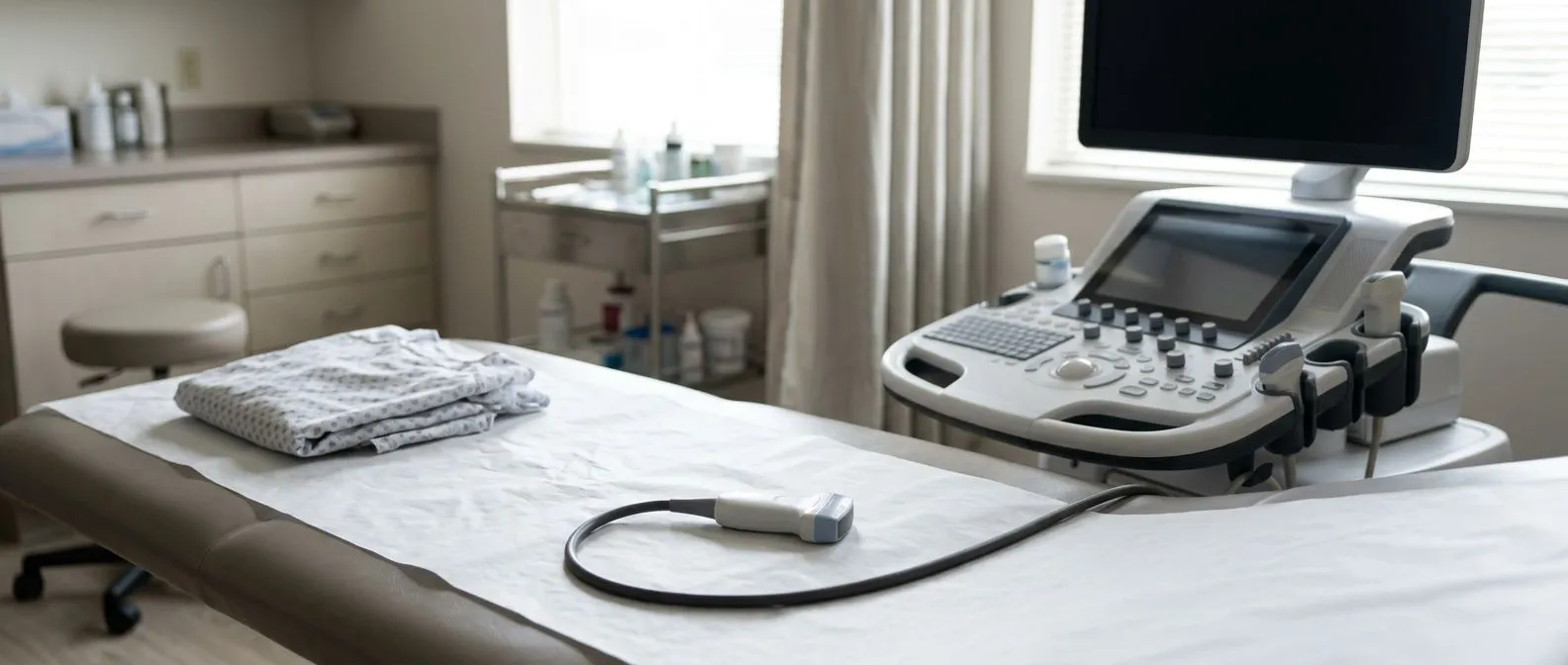
Ultrasound is the standard first test for the abdomen. An Abdominal Aortic Aneurysm is usually found or confirmed with an abdominal ultrasound, a scan in which a technician moves a small probe over the belly and measures the aorta from outside the body. It takes minutes, involves no needles or radiation, and is accurate enough to anchor years of follow-up. Screening programs in several countries offer this scan to older men precisely because it is simple and effective at finding silent aneurysms.
Timing follows size. A small aneurysm found on screening may be rescanned after a year or longer; a larger one moves to a six-month cycle. The interval is not arbitrary — it is set so that any meaningful growth is caught well before it reaches a dangerous size.
A "watch and wait" plan can create real anxiety, especially when the next scan is months away. We explain the reasoning behind the interval so the waiting feels like a plan, not a pause. Patients who understand the schedule report far less distress between appointments.

Aneurysm CT Scan Interval and MRI Surveillance
CT and MRI take over when detail matters. Once an aneurysm approaches a size where repair becomes a live possibility, or when it sits in the chest near the heart's valve and artery origins, the aneurysm CT scan interval typically shortens and CT angiography replaces simple ultrasound. The scan uses contrast dye and X-rays to build a detailed three-dimensional map of the vessel. MRI surveillance offers a similar level of detail without radiation, which matters for patients facing many years of follow-up.
Each test has trade-offs worth knowing. CT is fast and widely available but delivers a radiation dose, so teams try to avoid unnecessary repeats. MRI takes longer, costs more in many centers, and is not suitable for some implanted devices. Ultrasound remains the anchor for abdominal disease because it avoids both. A sensible long-term plan often mixes them: ultrasound for routine tracking, CT before decisions.
Every case we review is discussed by the full team before a recommendation is given, and where a less invasive approach — including Endovascular Aneurysm Repair, performed through small groin incisions rather than open surgery — might suit the anatomy, we say so alongside the conventional option. The patient receives both choices, with the evidence for each.
Early Warning Signs of an Aneurysm
Most unruptured aneurysms cause no symptoms at all. This is the hardest truth for many patients to sit with: an aneurysm can grow for years without a single warning sign, which is exactly why surveillance scans matter more than symptoms. When signs do appear, they depend on location — a deep, steady abdominal or back pain for an Abdominal Aortic Aneurysm, chest or upper back discomfort for a Thoracic Aortic Aneurysm, or a pulsating feeling in the abdomen.
Certain symptoms demand immediate attention. A sudden, severe, tearing or ripping pain in the chest, back, or abdomen; fainting; signs of shock such as pale, clammy skin and rapid weak pulse — these suggest rupture or dissection, and the response is an emergency call, not a scheduled appointment. Emergency Treatment of Ruptured Abdominal Aneurysm describes how survival in these events depends heavily on how quickly the patient reaches a team capable of repair, which is why the warning signs deserve to be memorized by the whole family.
Never wait for symptoms to confirm what a scan already knows. If you have a known aneurysm, the surveillance schedule is your early warning system — it works precisely because it acts before the body ever signals trouble. We tell every patient: the scan you keep when you feel fine is the one doing its job.
Aneurysm Growth Tracking and When to Repeat a Scan
Growth rate changes everything. Aneurysm growth tracking is the practice of comparing each scan to the last and calculating how quickly the diameter is expanding. A stable aneurysm measured at 4.2 centimeters for three years is a different clinical situation than one that grew from 4.0 to 4.5 in a single year, even though both are "under 5.5." Rapid growth is itself a signal, and it can move a patient toward repair earlier than diameter alone would.
So when to repeat an aneurysm scan? The general rhythm is: small aneurysms roughly every year, larger ones every six months, and any time new symptoms appear — do not wait for the scheduled date if pain or new sensations develop. After repair, a different schedule begins with Surveillance Imaging to confirm the repair is holding, since even a successfully treated aneurysm needs long-term checking of the repaired segment.
Research continues to refine how we monitor. Relationship Between Intraluminal Thrombus Volume and Circulating ADAMTS-13 Activity in Abdominal Aortic Aneurysms explores blood-based markers that may one day complement size measurements in predicting which aneurysms are most active. We follow this literature closely, because tomorrow's monitoring may be smarter than today's — but the schedule you follow now is built on solid, established evidence.

When Watch and Wait Turns Into Treatment
Surveillance has a finish line. The purpose of all this scanning is to identify the moment when repair becomes the lower-risk option — for most abdominal aneurysms, that threshold is around 5.5 centimeters, and for thoracic aneurysms it varies with location and underlying conditions. Crossing that line does not mean emergency. It means a planned, elective operation, performed in controlled conditions, with far better outcomes than an emergency repair after rupture.
Two main repair routes exist. Open Aortic Aneurysm Repair replaces the weakened segment with a synthetic graft through a chest or abdominal incision. Endovascular Aneurysm Repair threads a stent-graft through small incisions in the groin to reinforce the vessel from inside, usually with a shorter hospital stay. Some patients with a Thoracic Aortic Aneurysm near the heart's valve may also need valve attention at the same time — a situation where a multidisciplinary team earns its keep.
When we reach this decision point with a patient, we lay out both routes with their real trade-offs, and we say plainly which one we believe carries the lowest risk for their situation. In our practice Dr Molina performs conventional, minimally invasive, robotic and transcatheter procedures, so the recommendation is shaped by what the patient needs, not by what we happen to do.
Planning Your Aneurysm Imaging Schedule
A few practical points make the journey smoother. Keep every scan report and, where possible, have scans performed at the same facility or at least with the same technique, so measurements are comparable. Ask for your actual diameter numbers in centimeters, not vague words. Confirm your next appointment before you leave each visit — the most common failure of surveillance is not a bad scan but a missed one. And know that imaging costs and availability vary, so if you are traveling for care or weighing options for aneurysm imaging near Guadalajara, Querétaro or Hermosillo, ask in advance which studies are genuinely needed.
This is where a first conversation helps. Our first contact is a free video consultation: we get to know the patient, talk through the condition, and say which studies are still needed so no unnecessary ones are ordered. A face-to-face visit at the consulting rooms follows only when the patient decides to go ahead. If your existing scans already answer the questions, we will not ask you to repeat them.
Aneurysm surveillance scans are, in the end, a quiet partnership between you and your medical team: a measured vessel, a known growth rate, and a plan with clear thresholds. If you have been diagnosed with an aneurysm, or you are overdue for a follow-up scan, bring your reports to a qualified cardiac team and talk it through — with us or with anyone. The schedule you keep today is the emergency you may never have.