Atrial Flutter Treatment: Cardioversion and Ablation
Atrial flutter treatment options explained: cardioversion and catheter ablation compared.
What Is Atrial Flutter and How Does It Affect Your Heart?
Atrial flutter treatment begins with understanding what is happening inside the heart. Atrial Flutter is an abnormal heart rhythm that originates in the upper chambers of the heart, known as the atrium. Instead of beating in a steady, coordinated way, the atrial muscle fires electrical signals in a rapid, organized circular pattern — often around 300 times per minute. The lower chambers (the ventricles) cannot keep up with this speed, so they usually respond at a fraction of that rate, producing a heart rate that may feel very fast and uncomfortable but is more controlled than the raw atrial signal.
What does atrial flutter feel like for the patient? Many people describe a sensation of the heart racing, thudding, or skipping beats. Some feel shortness of breath, chest tightness, fatigue, or lightheadedness. Others may notice reduced exercise tolerance — activities that once felt easy now leave them winded. In some cases, atrial flutter is discovered incidentally during a routine electrocardiogram (ECG) when the patient had no symptoms at all. Because the rhythm can come and go, it is not unusual for symptoms to appear unpredictably, which makes documenting the rhythm with a monitor an important early step.
The core concern with atrial flutter is not only the symptoms it produces but also the risk it carries for stroke. When the atrium beats too fast, it does not empty fully with each cycle. Blood that lingers in the left atrial appendage can form clots. If a clot dislodges and travels to the brain, it can cause a stroke. This is why every atrial flutter treatment plan addresses both rhythm control and stroke prevention — the two are inseparable. According to Harvard Health, managing the arrhythmia and reducing clot risk together is the foundation of care.
Atrial Flutter vs Atrial Fibrillation: Key Differences in Treatment
Patients often confuse atrial flutter with atrial fibrillation because both involve rapid rhythms in the upper chambers and both increase stroke risk. However, they are electrically distinct. Atrial fibrillation produces chaotic, disorganized electrical activity — the atrial walls quiver rather than contract. Atrial flutter, by contrast, produces a single, organized electrical loop, typically traveling around a specific anatomical structure in the right atrium called the cavotricuspid isthmus. This organized nature is precisely what makes atrial flutter more amenable to catheter ablation as a definitive treatment.
Why does this distinction matter for your treatment plan? Because the flutter circuit follows a predictable path, an electrophysiologist can often map and eliminate it with a high degree of precision. In atrial fibrillation, the electrical chaos is spread across multiple areas of the atrium, making ablation more complex and less predictably curative. For atrial flutter, the ablation target is usually a narrow corridor of tissue that, once interrupted with radiofrequency energy, permanently breaks the circuit. This is why atrial flutter ablation success rate is generally higher than that for atrial fibrillation, and why many patients are offered ablation earlier in their treatment journey.
Medication strategies also differ. Rate-control medications such as beta-blockers or calcium channel blockers may be used in both conditions to slow the ventricular response. Rhythm-control medications (antiarrhythmics) can help maintain a normal sinus rhythm, but they tend to be less reliable for long-term flutter control than ablation. In both conditions, stroke prevention with blood thinners — including Aspirin in very low-risk patients or anticoagulants in higher-risk patients — remains a central pillar. Your cardiac team will assess your individual stroke risk using validated scoring systems before recommending the appropriate strategy.
How Atrial Flutter Treatment Begins: Diagnosis and Medication
Before any procedure is planned, the diagnosis must be confirmed and the patient's overall cardiac health assessed. Diagnosis typically begins with a 12-lead ECG, which captures the characteristic sawtooth flutter waves that distinguish this rhythm from other arrhythmias. If the flutter is intermittent, a Holter monitor or event recorder may be worn for days or weeks to capture episodes. An echocardiogram uses ultrasound to evaluate the structure of the heart — checking valve function, chamber sizes, and pumping strength. This imaging is essential because structural heart disease can influence which treatment path is safest and most effective.
What medications are used first in atrial flutter management? The initial approach often focuses on controlling the heart rate to relieve symptoms and reduce strain on the heart muscle. Beta-blockers (such as metoprolol) and calcium channel blockers (such as diltiazem) slow the electrical conduction through the atrioventricular node, reducing how many flutter signals reach the ventricles. If rate control alone does not resolve symptoms or if the rhythm is poorly tolerated, antiarrhythmic medications may be introduced to attempt to restore and maintain normal sinus rhythm. These drugs carry their own risks and require monitoring, so they are usually prescribed with careful follow-up.
Anticoagulation is a critical consideration from the outset. Because atrial flutter carries a real stroke risk, blood thinners may be started even before a cardioversion or ablation is scheduled. The decision depends on how long the flutter has been present (if longer than 48 hours, the clot risk is higher) and the patient's overall stroke risk profile. In some cases, a transesophageal echocardiogram is performed to check for existing clots in the left atrial appendage before any rhythm-restoring procedure. The comprehensive analysis of case series methodology in methodological quality and synthesis of case series and case reports underscores how individualized these treatment decisions must be, based on the best available evidence rather than one-size-fits-all protocols.

Electrical Cardioversion for Atrial Flutter: What to Expect
Electrical cardioversion is a procedure that uses a synchronized electric shock to reset the heart's rhythm back to normal sinus rhythm. It is one of the most direct forms of atrial flutter treatment and is often used when medication alone has not restored a normal rhythm or when symptoms are severe and persistent. The procedure is performed in a hospital setting, typically in a cardiac procedure room or intensive care unit, with the patient under brief sedation or general anesthesia. Pads are placed on the chest, and the defibrillator delivers a precisely timed shock synchronized to the heart's own R wave to avoid inducing a more dangerous rhythm.
Is cardioversion painful, and how long does it take? The patient is fully sedated for the brief shock — typically lasting a fraction of a second — so there is no sensation of pain during the delivery. The entire appointment usually takes a few hours, including preparation, sedation, the procedure itself, and recovery monitoring. Most patients go home the same day. The success rate for electrical cardioversion in atrial flutter is relatively high because the flutter circuit is organized and responsive to a reset impulse. However, cardioversion does not prevent the flutter from returning — it restores the rhythm in the moment, but the underlying electrical substrate remains.
For this reason, cardioversion is often paired with longer-term strategies. If flutter has been present for more than 48 hours, or if the onset time is unknown, anticoagulation is typically continued for at least three weeks before and four weeks after the procedure to minimize stroke risk. Some patients undergo cardioversion as a bridge to ablation — restoring normal rhythm temporarily while awaiting a more definitive procedure. Others may need only one cardioversion and remain in normal rhythm with the help of medications. The approach depends on how frequently flutter recurs, the patient's symptoms, and underlying heart structure.
Atrial Flutter Ablation: How Catheter Ablation Works
Catheter ablation for atrial flutter is a minimally invasive procedure designed to permanently interrupt the electrical circuit causing the arrhythmia. Unlike cardioversion, which resets the rhythm temporarily, ablation targets the anatomical pathway the signal travels along and creates a line of scar tissue that blocks it. For the most common type of atrial flutter — known as typical or cavotricuspid isthmus-dependent flutter — the target is a narrow band of tissue between the inferior vena cava, the tricuspid valve annulus, and the coronary sinus ostium. By ablating this corridor, the circular electrical loop is permanently broken.
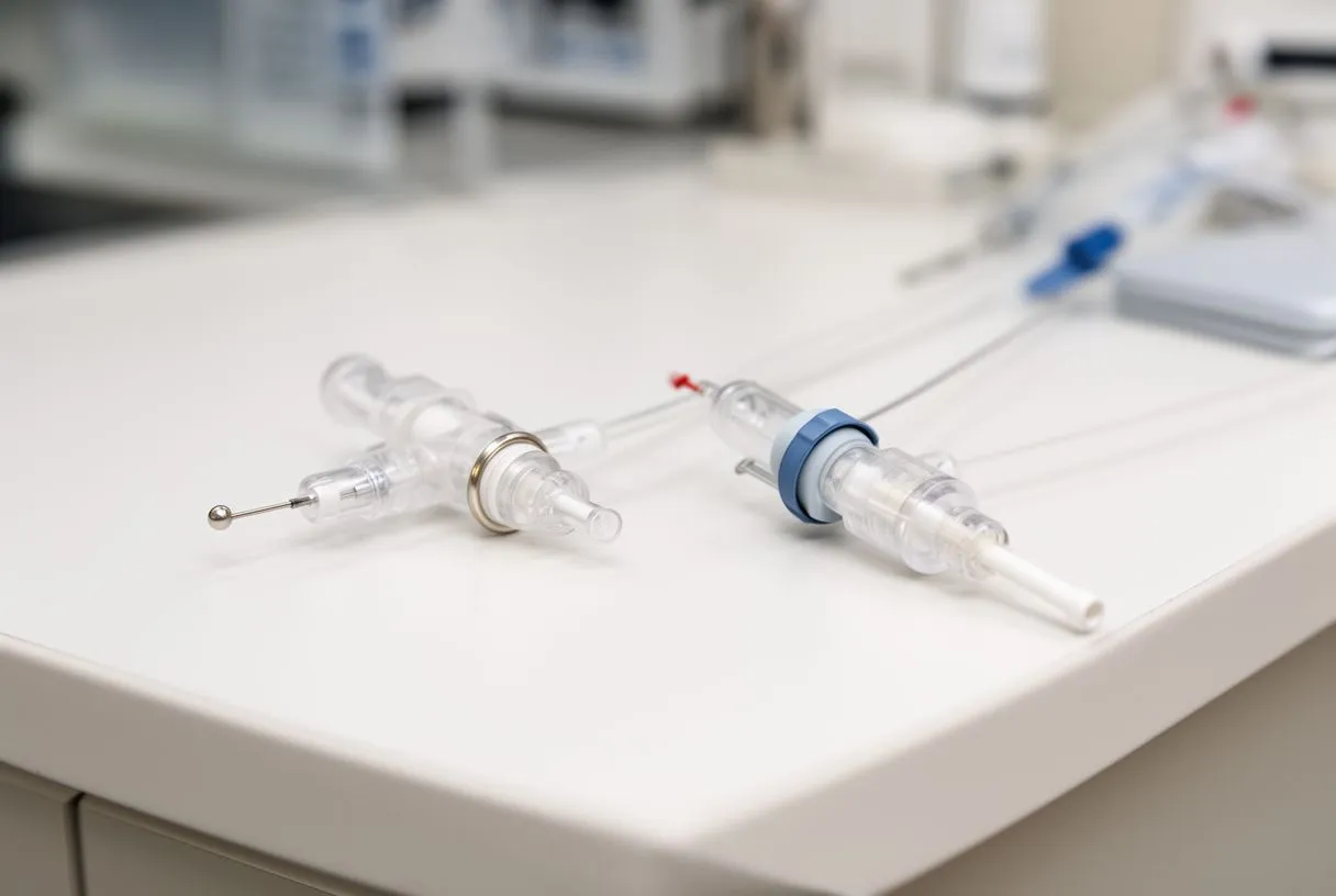
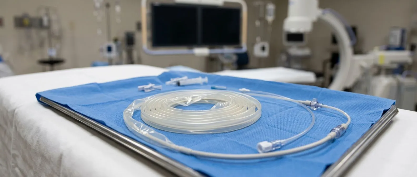
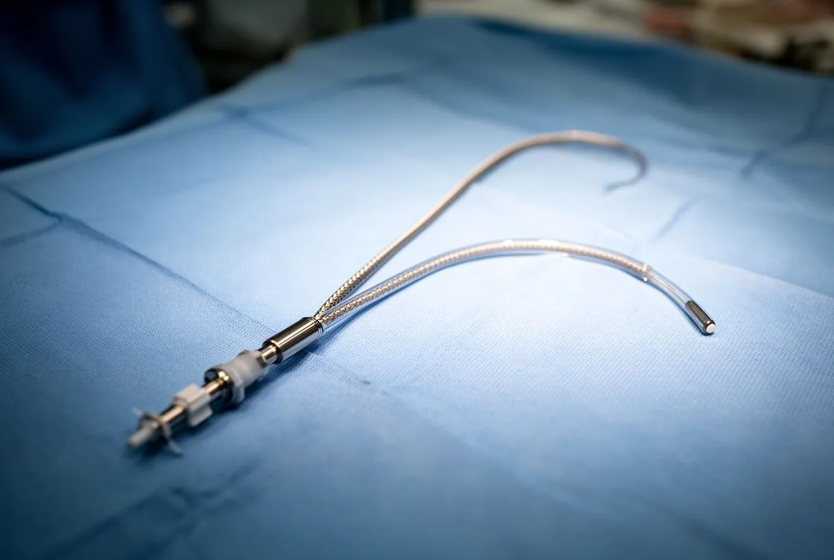
How does the catheter reach the heart? The electrophysiologist inserts thin, flexible catheters through small punctures in the groin — typically the femoral vein — and threads them up through the venous system into the right atrium. No large incisions are needed, and the procedure does not require opening the chest. Once the catheters are positioned, the electrophysiologist uses electrical mapping to identify exactly where the flutter signal is traveling. This mapping is done in real time, often while the patient is in the flutter rhythm, so the circuit can be visualized and precisely targeted.
The procedure is typically performed under conscious sedation or light anesthesia, meaning the patient is relaxed and comfortable but not deeply unconscious. The duration varies depending on anatomy and the complexity of the flutter circuit, but most atrial flutter ablations are completed in one to two hours. Because the ablation target in typical flutter is well-defined and anatomically predictable, the procedure tends to be shorter and more straightforward than ablations for atrial fibrillation. Patients are usually monitored for a few hours afterward and discharged the same day or after an overnight stay, depending on the center's protocol.
Radiofrequency Ablation for Atrial Flutter: The Procedure Explained
Atrial flutter radiofrequency ablation uses controlled heat energy delivered through the tip of the catheter to create precise lesions in the heart tissue. The radiofrequency energy heats the cells at the catheter tip to approximately 50–60 degrees Celsius, causing a small, controlled area of scar formation. This scar does not conduct electricity, so once the isthmus line is completed, the flutter circuit can no longer propagate. The energy is applied in a series of point-by-point lesions or as a continuous drag along the target corridor, and the electrophysiologist monitors the electrical signals throughout to confirm that conduction across the isthmus has been eliminated.
What confirms that the ablation worked during the procedure? After the initial ablation line is created, the electrophysiologist tests whether the flutter is still inducible and whether electrical signals can still cross the ablated corridor. This is done by pacing from one side of the line and checking for conduction to the other. If signals still cross, additional lesions are applied until the line is electrically complete — a state called bidirectional block. Achieving bidirectional block across the cavotricuspid isthmus is the procedural endpoint that predicts long-term success. Without this confirmation, the flutter may recur, which is why intraprocedural testing is a critical quality measure.
Patients sometimes ask whether ablation hurts. Most report feeling little to no pain from the ablation energy itself, though some may feel warmth or mild discomfort in the chest during lesion delivery. The catheter manipulation in the groin area can cause pressure or mild soreness afterward. Sedation levels are adjusted to keep the patient comfortable throughout. Because no chest incision is made, recovery from the access site is typically quick. The detailed clinical understanding of pulmonary vascular and cardiac conditions, as explored in studies like Chronic thromboembolic pulmonary hypertension, reflects the broader importance of precise catheter-based interventions in modern cardiac care.
Atrial Flutter Ablation Success Rate and Long-Term Outcomes
The atrial flutter ablation success rate is one of the most encouraging aspects of this treatment. For typical cavotricuspid isthmus-dependent flutter, acute procedural success — defined as achieving bidirectional block — exceeds 90% in experienced centers. Long-term freedom from flutter recurrence is also high, with many studies reporting rates above 85% at one year and sustained benefit over multiple years. This is substantially better than the long-term results of cardioversion alone, which does not prevent recurrence, or medication-only strategies, which may fail over time or produce side effects that limit their use.
What factors influence whether ablation stays successful? Several variables affect long-term outcomes. The experience and volume of the electrophysiology center matters — higher-volume centers tend to report better results. Patient factors also play a role: the presence of structural heart disease, left atrial enlargement, or concurrent atrial fibrillation can reduce the durability of the result. In some patients, atrial fibrillation may emerge after a successful flutter ablation because the underlying atrial substrate that predisposed to flutter can also support fibrillation. This is not a failure of the flutter ablation but rather a separate rhythm problem that may need its own management plan.
It is important for patients to have realistic expectations. Ablation for typical atrial flutter is considered one of the most effective arrhythmia procedures available, and for many patients it functions as a cure — they no longer experience flutter and may be able to stop rhythm-control medications. However, ongoing follow-up is essential. Some patients require a second ablation if the initial line heals incompletely or if a different flutter circuit develops. Stroke risk assessment should also continue, as the need for anticoagulation depends on the patient's overall risk profile, not solely on whether flutter has been eliminated. Your cardiac team will tailor follow-up based on your specific anatomy and rhythm history.
Cardiac Ablation Recovery: Timeline and What Patients Experience
Recovery from atrial flutter ablation is generally straightforward, but understanding the timeline helps patients know what to expect and when to seek help. Immediately after the procedure, the patient remains flat for several hours while the groin access site heals and the sedation wears off. Vital signs and heart rhythm are monitored continuously. Most patients are discharged the same day or after one night in the hospital. Upon returning home, patients are advised to avoid heavy lifting, strenuous exercise, and immersion of the access site in water for several days to a week.
- First 24–48 hours: Mild groin soreness, fatigue, and occasional palpitations are common as the heart adjusts and irritated tissue heals.
- First 1–2 weeks: Energy levels gradually return; light walking is encouraged. Some patients experience skipped beats or brief palpitations, which are usually benign but should be reported if persistent.
- 4–8 weeks: Full recovery of normal activity. A follow-up ECG and sometimes a Holter monitor confirm rhythm stability. Medications may be adjusted at this stage.
When should you contact your doctor during recovery? Patients should seek immediate medical attention if they experience significant chest pain, shortness of breath, fever, increasing redness or swelling at the access site, or fainting. These could indicate a complication such as pericardial effusion (fluid around the heart), infection, or a vascular issue at the puncture site. While serious complications are uncommon, early evaluation is always the safest course. Between scheduled follow-ups, patients should keep a simple log of any palpitations or symptoms to share with their cardiac team, as this information helps guide decisions about medication adjustments or additional testing.

Are Atrial Flutter Treatments Safe? Risks and Complications
Atrial flutter treatment — whether cardioversion, ablation, or medication — is generally safe when performed by experienced teams, but no cardiac procedure is without risk. Understanding these risks allows patients to make informed decisions rather than proceeding with unrealistic expectations. For electrical cardioversion, the most significant immediate risk is stroke if a clot is present in the left atrial appendage and dislodges when normal rhythm is restored. This is why anticoagulation and transesophageal echocardiography are used to screen for clots before the procedure. Skin irritation at the pad sites and transient rhythm irregularities after the shock are also possible but typically resolve quickly.
What are the specific risks of catheter ablation? The most serious complications are rare but include pericardial effusion or tamponade (blood or fluid accumulating around the heart, which can compress it), damage to blood vessels at the access site, blood clots, and injury to the phrenic nerve or the artery that runs near the ablation target. The need for an Arterial Line or more invasive monitoring during ablation is uncommon but may be used in complex cases. Most centers report major complication rates below 2% for typical flutter ablation, and many of these complications can be managed without surgery if recognized promptly.
- Vascular complications: Bleeding, hematoma, or pseudoaneurysm at the groin puncture site — usually managed with compression or ultrasound-guided repair.
- Arrhythmia recurrence: The flutter circuit may resume if the ablation line heals incompletely — addressed with a repeat procedure if needed.
- Pericardial effusion: Fluid around the heart — rare but potentially serious; recognized and drained if it occurs.
Medications also carry risks that deserve honest discussion. Antiarrhythmic drugs can have proarrhythmic effects — meaning they can paradoxically worsen arrhythmias in some patients — and may affect other organs, requiring regular blood tests. Rate-control medications can cause low blood pressure, fatigue, or slow heart rates that limit activity. Anticoagulants carry bleeding risk, which must be balanced against stroke prevention benefit. Your cardiac team will help you weigh these risks against the benefits in the context of your overall health, your symptoms, and your goals. The aim is not to eliminate all risk but to choose the path with the lowest risk for your situation and the greatest likelihood of improving your quality of life.
Choosing the Right Atrial Flutter Treatment for Your Situation
There is no single correct atrial flutter treatment for every patient. The right choice depends on how long the flutter has been present, how severe the symptoms are, whether structural heart disease exists, the patient's stroke risk, and personal preferences about procedures versus medications. For a patient with new-onset, symptomatic flutter and no structural disease, electrical cardioversion may quickly restore normal rhythm, followed by a discussion about whether ablation is warranted to prevent recurrence. For a patient with recurrent flutter despite cardioversion or medication, catheter ablation is often the most effective long-term solution and may be offered as first-line therapy.
How does the decision differ for patients with other cardiac conditions? When atrial flutter coexists with other heart problems — valve disease, coronary artery disease, or heart failure — the treatment plan must account for the full clinical picture. For example, a patient with an enlarged left Atrium due to long-standing valve disease may have a higher risk of atrial fibrillation developing after flutter ablation. In these cases, the electrophysiologist and the cardiac surgeon may coordinate care, considering whether surgical intervention for the underlying structural problem should take priority or proceed in parallel with rhythm management. Every decision should be grounded in guideline-based thresholds and individualized risk assessment.
For patients traveling from outside Mexico or within Mexico to seek care with Dr Alejandro Molina and his team, the consultation process includes a thorough review of any existing ECGs, echocardiograms, and medication history. This allows the team to provide a clear recommendation — whether that means medication optimization, cardioversion planning, or scheduling an ablation — along with a candid discussion of expected outcomes, recovery timelines, and any logistical considerations. The goal is always to ensure you understand your options, the reasoning behind each recommendation, and the evidence supporting it. If you or a loved one is experiencing symptoms of atrial flutter, we encourage you to book a consultation with our cardiac team rather than self-diagnosing — every heart rhythm story is unique, and the right treatment begins with understanding yours.