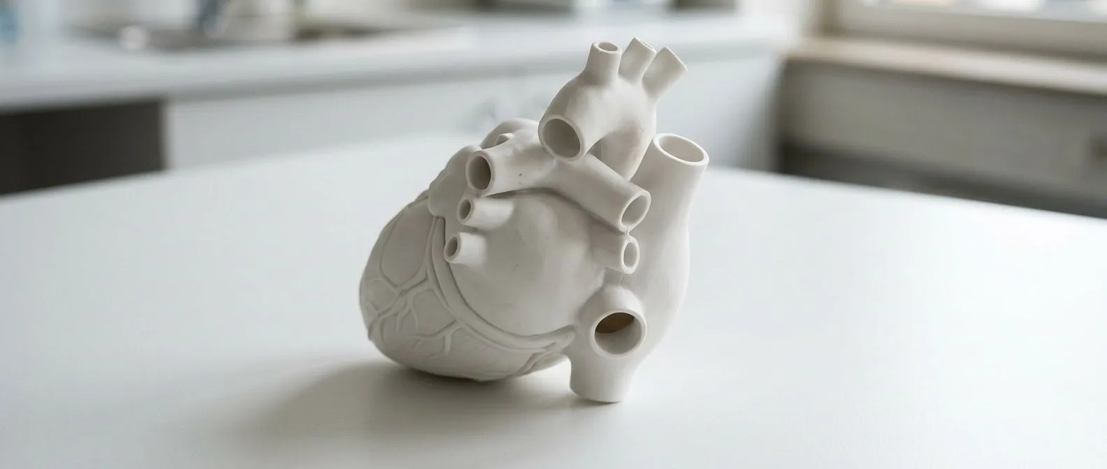
Base of the Heart: Where the Great Vessels Attach
Base of the heart anatomy explained by expert cardiac surgeons in Guadalajara.
What Is the Base of the Heart Anatomy?
Understanding the base of the heart anatomy is essential for patients navigating complex cardiac diagnoses. Contrary to what the name suggests, the base of the heart is not the bottom or the downward-pointing tip. Anatomically, it is the superior (upper) and posterior (back) portion of the heart. It sits just behind the sternum and is tilted slightly toward the right shoulder. This region is where the major blood vessels—the aorta, pulmonary trunk, superior vena cava, inferior vena cava, and pulmonary veins—enter and exit the cardiac chambers. Because the great vessels converge here, the base acts as the central plumbing junction for the entire circulatory system.
Anatomical orientation defined: When cardiac surgeons discuss the heart's orientation, they describe it as a cone resting on its side. The pointed end of this cone, which rests on the diaphragm and points toward the left hip, is the apex. The flat, broader end pointing toward the right shoulder is the base. The base is firmly anchored by the great vessels and the pericardium, a double-walled sac that surrounds the heart. This anchoring prevents the powerful, beating muscle from shifting violently within the chest cavity during exertion. Auscultation, the medical act of listening to the internal sounds of the body, is often performed away from the base, as the apex offers the clearest projection of valve closures.
Why the base matters clinically: Many structural heart diseases and vascular complications originate at this upper junction. Because the aortic and pulmonary valves are housed near the base, conditions like valve stenosis or regurgitation heavily impact this area. If you are facing heart surgery, understanding this layout helps you visualize why certain incisions are made or why specific imaging angles are required. Knowing that the base is the structural anchor for all incoming and outgoing blood flow clarifies why surgeons must approach this area with meticulous precision, ensuring the lifelong stability of your circulatory system.
Where Do the Great Vessels Attach to the Heart?
The great vessels are the largest arteries and veins in the body, and they all attach directly to the base of the heart. There are four primary great vessels that manage the continuous cycle of blood flow: the aorta, the pulmonary trunk, the superior vena cava, and the inferior vena cava. In addition to these, the four pulmonary veins also attach to the base, bringing oxygen-rich blood back from the lungs to the left atrium. This complex network of vessels is arranged in a highly specific, layered architecture at the heart's base, ensuring that oxygen-depleted blood and oxygen-rich blood remain completely separate. Disruptions to this precise arrangement can lead to severe congenital or acquired cardiac conditions.
The functional layout of attachments: At the base of the heart anatomy, the attachments are grouped by function. The superior vena cava empties oxygen-poor blood from the upper body into the right atrium, while the inferior vena cava does the same from the lower body. From the right ventricle, the pulmonary trunk carries this oxygen-poor blood out to the lungs. Conversely, the pulmonary veins deliver freshly oxygenated blood to the left atrium, which is then pumped out to the entire body through the aorta. These structures overlap and weave around one another, creating a dense, fibrous foundation known as the cardiac skeleton, which stabilizes the valves and electrical pathways. According to the American Heart Association, understanding this vascular network is critical for diagnosing and treating structural heart defects. heart.org
Clinical implications of vessel attachment: When these attachments become diseased, the entire cardiovascular system suffers. Aneurysms, or abnormal bulges, frequently occur in the aorta right where it attaches to the heart base. If the attachments stretch or tear, blood can leak backward into the heart chambers, causing volume overload and eventual heart failure. For patients preparing for intervention, imaging these attachments determines whether a surgeon can repair a valve natively or if a full replacement is necessary. The geometry of the base dictates what mechanical or biological replacements can physically fit without obstructing the other great vessels, making preoperative planning essential for long-term recovery.
How the Ascending Aorta Connects to the Heart Base
The ascending aorta is the first segment of the largest artery in the human body, originating directly from the left ventricle at the heart base. It rises upward from the left ventricle, curving slightly forward and to the right, before arching over the heart to distribute blood to the head and body. The connection point, known as the aortic root, houses the aortic valve. This root is a complex structure that must withstand the highest pressure in the circulatory system, as it receives blood forcefully ejected during every heartbeat. When surgeons evaluate the base of the heart anatomy, the health of the ascending aorta is a primary focus, as structural failures here are immediately life-threatening.
Aortic root dynamics and disease: The aortic root expands and contracts with every pulse, acting as a shock absorber for the blood ejected from the heart. However, this constant stress can lead to the buildup of Atheromatous Plaque, a fatty deposit that narrows the artery and restricts blood flow. Plaque in the ascending aorta can be dangerous if fragments break off and travel to the brain, causing a stroke. Furthermore, genetic conditions or chronic hypertension can cause the aortic root to dilate. When the diameter of the ascending aorta exceeds clinical thresholds (often measured at greater than 5.5 centimeters), the risk of a fatal dissection or rupture increases dramatically. In these cases, prophylactic surgical intervention is necessary to replace the weakened segment of the vessel before it fails.
Surgical stabilization of the aortic root: Repairing the connection between the aorta and the heart base requires stopping the heart and using cardiopulmonary bypass. Surgeons must carefully excise the damaged tissue while preserving the coronary arteries, which branch off the ascending aorta just above the valve. The aortic root is then replaced with a synthetic graft, often incorporating a mechanical or biological valve. This procedure, known as a Bentall operation, permanently restores the structural integrity of the heart base. Recovery from this type of open-heart surgery is substantial, requiring weeks of restricted activity, but it provides a definitive cure for aortic root aneurysms, allowing patients to return to their normal lives without the fear of sudden aortic rupture.
The Pulmonary Trunk Origin and Right Ventricle
Adjacent to the aorta at the base of the heart is the pulmonary trunk, the great vessel responsible for carrying oxygen-depleted blood to the lungs. It originates from the right ventricle and extends upward and slightly to the left before splitting into the left and right pulmonary arteries. The pulmonary valve sits at this origin point, ensuring that blood flows only in one direction—from the right ventricle into the pulmonary trunk—and preventing backflow during the heart's relaxation phase. Understanding the relationship between the right ventricle and the pulmonary trunk is vital for patients diagnosed with right-sided heart failure or pulmonary hypertension, as these conditions heavily tax this specific anatomical region.
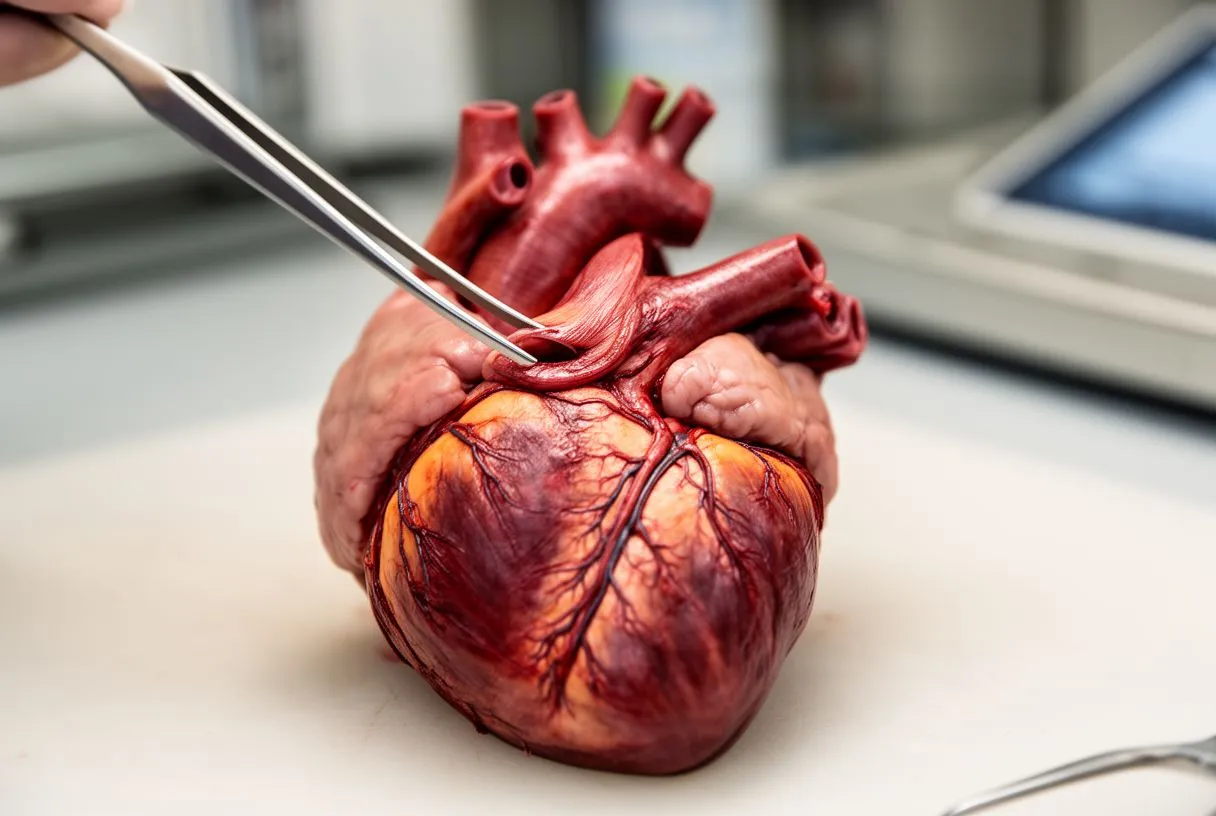
Pulmonary valve function and challenges: The pulmonary valve operates under lower pressures than the aortic valve, but it is equally important for efficient circulation. If the pulmonary valve becomes stenotic (narrowed), the right ventricle must work harder to push blood through the obstructed opening, leading to muscle thickening and eventual fatigue. Alternatively, if the valve is incompetent and allows backward leakage, the right ventricle becomes volume overloaded. Both scenarios strain the right side of the heart, leading to symptoms like fluid retention in the legs and abdomen, severe fatigue, and shortness of breath. Imaging the base of the heart anatomy allows cardiologists to measure the velocity of blood across this valve, determining the severity of the dysfunction.
Treatment of pulmonary trunk disorders: When the pulmonary trunk or its valve fails, interventions range from balloon-based therapies to open surgical replacement. In some cases, transcatheter approaches can be used to implant a new valve without opening the chest, though this is highly dependent on the patient's specific anatomy. For severe structural anomalies or congenital defects present at the base of the heart, surgeons may need to completely reconstruct the right ventricular outflow tract. Patients considering these procedures must discuss the durability of biological versus mechanical conduits with their surgical team, as replacements in this lower-pressure system often have different longevity profiles than those placed in the high-pressure aortic system.
Superior and Inferior Vena Cava Entry Points
The right side of the heart base is defined by the entry points of the superior and inferior vena cava, the two largest veins in the body. These vessels are responsible for returning oxygen-depleted blood from the systemic circulation back to the heart. The superior vena cava collects blood from the upper half of the body—head, neck, and arms—while the inferior vena cava collects from the lower half. Both empty directly into the right Atrium, the upper right chamber of the heart. The seamless entry of these vessels into the base of the heart anatomy ensures a continuous, low-pressure return of blood that is ready to be sent to the lungs for oxygenation.
Flow dynamics at the venous return: Unlike the high-pressure ejection seen in the aorta, venous return is a passive, steady process driven by skeletal muscle pumps and breathing mechanics. When a person inhales, negative pressure in the chest draws blood upward through the inferior vena cava. Disruptions at the entry points of these vessels, such as a tumor compressing the superior vena cava, can cause dramatic symptoms like facial swelling and visible vein distension in the neck. In the context of the base of the heart anatomy, surgeons must carefully navigate these entry points during right-sided heart surgeries. Any distortion of the vena cava attachments can impede venous return, leading to a dangerous drop in cardiac output. In severe cases of tricuspid valve disease affecting this venous inflow region, structural repair is essential. Transcatheter Edge-to-Edge Repair for Treatment of Tricuspid Regurgitation
Clinical assessment of vena cava function: Echocardiograms are frequently used to assess the inferior vena cava's size and collapsibility, which indicates a patient's fluid volume status and right heart function. A dilated, non-collapsible vena cava suggests elevated right atrial pressure, often a sign of right-sided heart failure or significant tricuspid valve regurgitation. For patients with pacemakers or implantable defibrillators, the leads are threaded down the superior vena cava to attach at the base of the heart; over time, these leads can cause scarring or stenosis at the entry point. Recognizing these potential complications highlights why regular follow-up with a cardiac specialist is crucial for long-term device management.
Where Do the Pulmonary Veins Enter the Heart?
On the posterior aspect of the heart base, four pulmonary veins deliver oxygen-rich blood from the lungs to the left atrium. There are typically two veins from the right lung (superior and inferior) and two from the left lung. Unlike most veins in the body, pulmonary veins do not have valves; they rely on pressure gradients between the lungs and the left atrium to keep blood flowing in the correct direction. The entry points of these veins are clinically significant because they are intimately connected to the heart's electrical system. Understanding where these veins enter the base of the heart anatomy is critical for treating arrhythmias, specifically atrial fibrillation.
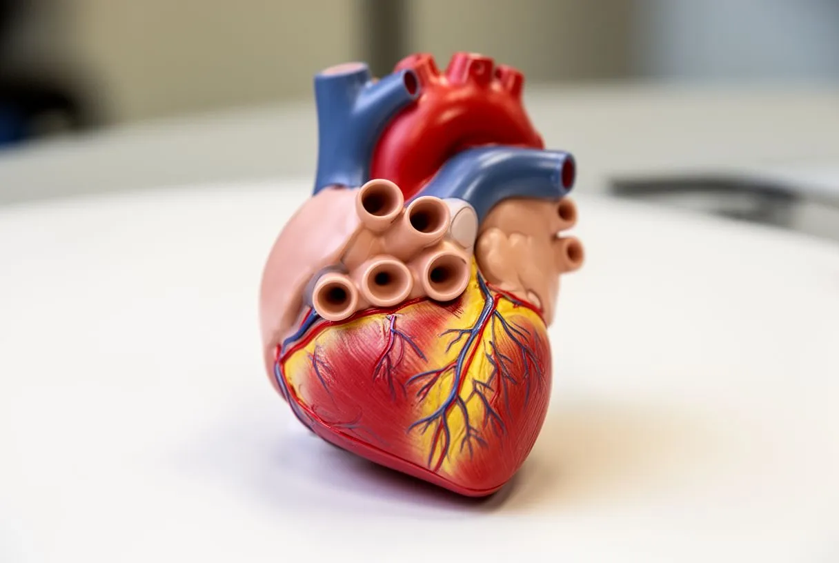
The pulmonary veins and cardiac arrhythmias: The tissue where the pulmonary veins connect to the left atrium is a frequent source of chaotic electrical signals. These rogue signals can cause the atria to quiver rapidly, a condition known as Atrial Flutter or atrial fibrillation. Cardiologists often perform ablation procedures at the base of the heart, threading catheters through the venous system into the left atrium to cauterize the tissues around the pulmonary vein entry points. By isolating these electrical signals, normal rhythm can often be restored. If untreated, persistent atrial fibrillation originating near the pulmonary veins increases the risk of stroke and can lead to progressive weakening of the heart muscle over time.
Anatomical variations in venous entry: While most people have four pulmonary veins entering the left atrium, anatomical variations are common. Some individuals may have a common trunk where the veins merge before entering the heart, or an extra right middle pulmonary vein. These variations are usually benign but become highly relevant during structural heart interventions, such as left atrial appendage occlusion or pulmonary vein isolation for arrhythmia management. If a patient requires surgery on the mitral valve, which sits just below the left atrium, surgeons must avoid manipulating or narrowing the pulmonary vein entry points. Preoperative CT imaging of the base of the heart anatomy is routinely used to map these variations and prevent inadvertent vascular compromise.
Heart Base vs Apex: Understanding the Difference
Distinguishing between the base and the apex of the heart is a fundamental concept in cardiology and crucial for patients trying to understand diagnostic reports. The base is the superior, broad end where the great vessels attach, while the apex is the inferior, pointed tip of the heart, located at the fifth intercostal space, resting just above the diaphragm. The base is structurally passive, heavily anchored by dense connective tissue and vascular attachments; the apex is highly mobile, thrusting forward against the chest wall with every heartbeat. When evaluating the base of the heart anatomy versus the apex, it is the difference between the stable "plumbing junction" and the actively pumping "motor" of the heart.
How location affects symptom presentation: Because the apex makes direct contact with the chest wall, the forceful thrust of the left ventricle is felt most strongly here. Patients experiencing a classic heart attack (myocardial infarction) often feel pain radiating from the chest down the left Arm Pain pathway, a symptom heavily associated with ischemia at the apex and anterior heart wall. Conversely, an Atypical Infarction affecting the posterior or basal regions of the heart may present as vague indigestion, jaw pain, or shortness of breath without classic chest pain. The location of the heart disease—whether at the mobile apex or the anchored base—directly influences how the patient perceives symptoms, making clinical history-taking incredibly important for an accurate diagnosis.
Diagnostic focus: base versus apex: During a physical examination, a physician places a stethoscope directly over the apex to hear the mitral valve (the loudest point of the heart) and over the base to assess the aortic and pulmonary valves. If a murmur is loudest at the base, it suggests a problem with the outflow of blood; if loudest at the apex, it suggests an inflow or mitral valve issue. When reading an echocardiogram report, understanding this orientation helps patients visualize their condition. If a report notes "basal hypokinesis," it means the upper portion of the heart muscle is not contracting well, often due to disease in the vessels supplying that specific region. Knowing the difference empowers patients to have more informed discussions with their cardiologists regarding their specific structural issues.
What the Heart Base Looks Like on an Echocardiogram
An echocardiogram is the primary tool used to visualize the base of the heart anatomy in a clinical setting. Using high-frequency sound waves, this non-invasive imaging test provides real-time, moving pictures of the heart's structure and function. When the ultrasound probe is placed on the chest, specifically in the "parasternal" view, the sonographer captures a cross-section of the heart base. This view clearly shows the aortic valve, the pulmonary valve, the left atrium, and the right atrium. For patients, understanding what the echocardiogram reveals about the heart base can demystify the diagnostic process and clarify why specific interventions are recommended.
Visualizing valves and chambers at the base: The heart base is the anchor for the aortic and pulmonary valves, which appear as thin, highly mobile flaps on the monitor. If these valves are thickened or calcified, it indicates stenosis, meaning they cannot open fully to allow blood to pass. Blood flow is represented by color Doppler imaging—red and blue hues moving across the dark chambers. If blood leaks backward through a valve at the base of the heart, the color map will show a regurgitant jet flowing backward into the atrium. This visualization helps surgeons determine the exact repair strategy needed. Furthermore, the chambers at the base, particularly the left atrium, are measured; if the left atrium is severely enlarged, it often indicates chronic pressure overload or untreated valve disease.
Systemic factors affecting the heart base: The health of the base of the heart anatomy is often a reflection of systemic conditions. For example, elevated levels of Apolipoprotein B, a protein involved in lipid metabolism, correlate strongly with the buildup of plaque in the aortic root and coronary arteries branching off the base. Patients with atrial fibrillation originating at the base are frequently managed with anticoagulants to prevent stroke. Clinical trials like ARISTOTLE have guided how medications such as Apixaban are used to safely manage these patients. Additionally, for those with vascular disease at the base, daily low-dose Aspirin is often prescribed to inhibit platelet aggregation and reduce the risk of acute blockages.
- Transesophageal Echo (TEE): A specialized probe passed down the esophagus provides a posterior, highly detailed view of the heart base, crucial for surgical planning.
- Color Doppler: Visualizes blood flow velocities at the base, instantly revealing valve leaks or stenotic jets that require surgical correction.
- Chamber Dimensions: Measuring the left atrium at the base helps quantify the severity of chronic valve regurgitation and atrial fibrillation.
Surgical Approaches to the Base of the Heart
Accessing the base of the heart anatomy requires highly specialized surgical techniques, as it is densely packed with critical vessels and anchored to the sternum. The most common approach is a median sternotomy, where the surgeon makes a vertical incision down the center of the chest and separates the breastbone to directly visualize the heart base. This provides the maximum exposure required to safely manipulate the aorta, pulmonary trunk, and atrial chambers. Once the chest is open, the patient is placed on cardiopulmonary bypass, where a machine takes over the work of the heart and lungs, allowing the surgeon to operate on a still, bloodless field. The complexity of these interventions requires the highest level of surgical expertise.
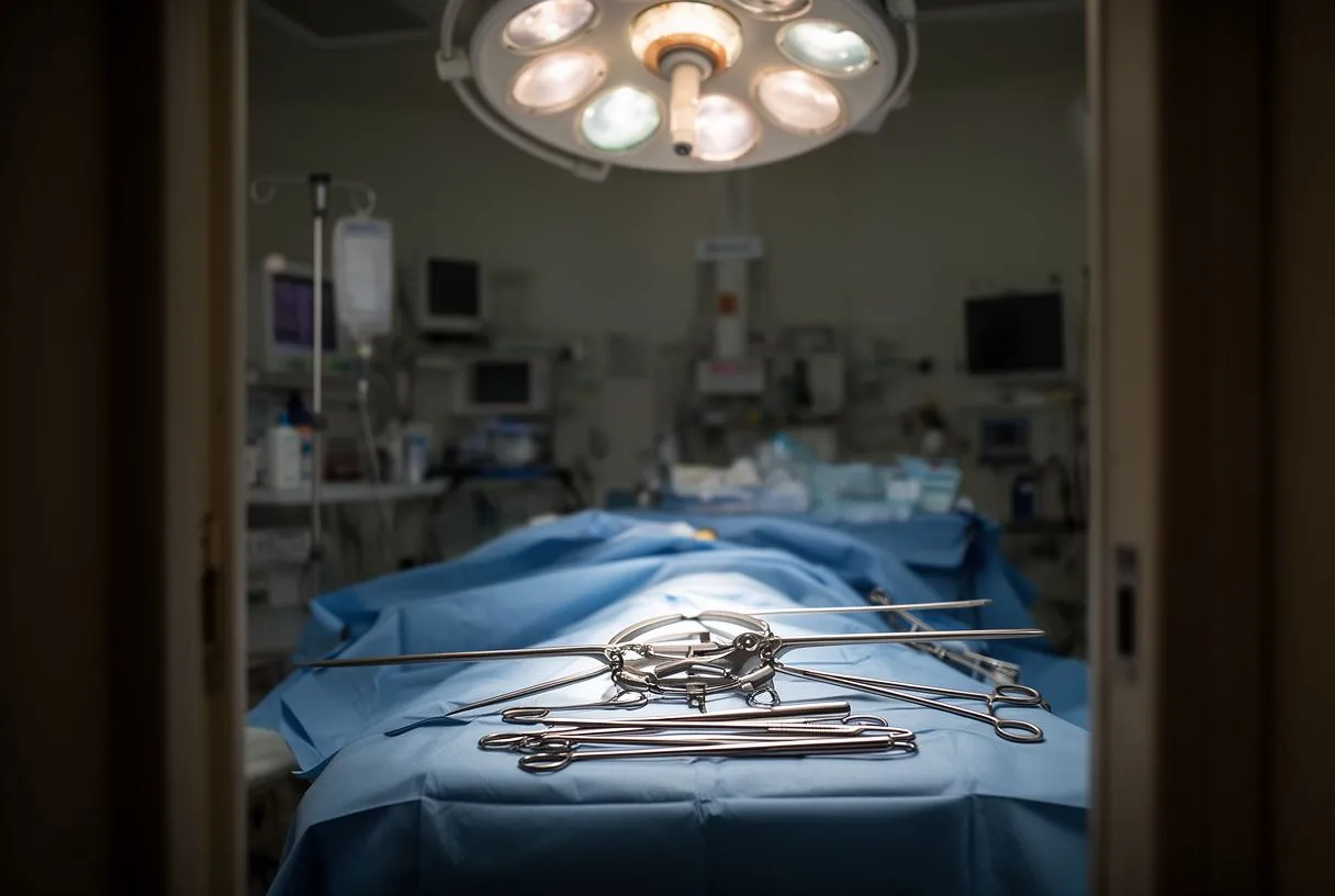
Bypass and cannulation at the base: Establishing cardiopulmonary bypass involves placing an Arterial Cannula into the ascending aorta and a venous cannula into the right atrium or superior vena cava at the base of the heart. An Arterial Line is also placed to continuously monitor systemic blood pressure during the procedure. The surgeon then administers a specialized solution to stop the heart (cardioplegia), protecting the muscle while the structural repairs are made. In modern cardiac surgery, efforts are made to use Autologous Blood, the patient's own blood, whenever possible to minimize transfusion risks and promote faster recovery. Advanced centers may even use Awake ECMO (Extracorporeal Membrane Oxygenation) for temporary support in critically ill patients before definitive surgery, allowing the heart and lungs to rest while stabilizing the patient.
Repairing and replacing structures at the base: Once the heart base is exposed, surgeons address the specific pathology. If the aortic valve is diseased, it can be replaced with a Balloon-expandable Valve (used in TAVR procedures) or a traditional mechanical or biological valve. Prior to valve replacement, some patients undergo Balloon Aortic Valvuloplasty to temporarily stretch a narrowed valve. Similarly, narrowed coronary arteries branching from the base may be treated with Balloon Angioplasty to open the vessel, followed by the placement of a stent. When multiple vessels are blocked, a surgeon may perform a bypass using an Arterial Graft, such as the internal mammary artery, or even an Autograft, using the patient's own pulmonary valve to replace a diseased aortic valve (the Ross procedure). The specific technique chosen depends entirely on the patient's anatomy and the extent of the disease at the heart base. Recent evaluations of tricuspid interventions show promising outcomes for high-risk surgical patients. Outcomes After Current Transcatheter Tricuspid Valve Intervention
When to Seek Help for Heart Base Conditions
Conditions affecting the base of the heart anatomy—such as aortic aneurysms, severe valve regurgitation, or atrial fibrillation—often develop slowly and silently. Because the heart is incredibly adaptive, patients may not experience noticeable symptoms until the disease has progressed to an advanced stage. However, early detection is critical. Symptoms that should prompt an immediate medical evaluation include unexplained chest pressure, severe shortness of breath, fainting during exertion, or palpitations that feel like a fluttering or pounding in the chest. If you have a family history of aortic disease or sudden cardiac death, or if you have been diagnosed with a connective tissue disorder, proactive monitoring of your heart base is essential.

Emergency symptoms of base pathology: The sudden onset of severe, tearing chest pain radiating to the back is a hallmark sign of an acute aortic dissection, a catastrophic failure at the base of the aorta. This is a surgical emergency requiring immediate transport to a hospital with cardiac surgical capabilities. In cases of sudden cardiac arrest caused by a massive arrhythmia originating at the pulmonary vein entry points, an Automated External Defibrillator (AED) can be life-saving by shocking the heart back into a normal rhythm. Recognizing these emergency signs and understanding the anatomical reality of the heart base empowers patients and families to act decisively. Do not wait for symptoms to subside if they are sudden and severe; immediate intervention at the base of the heart is often the difference between life and death.
Long-term management and follow-up: For patients with known, stable conditions affecting the base of the heart, consistent monitoring is the key to preventing sudden deterioration. This means keeping regular appointments with your cardiologist, undergoing scheduled echocardiograms to measure the size of the aortic root or the function of the basal valves, and adhering strictly to prescribed medication regimens. The base of the heart anatomy is resilient, but when it fails, the entire circulatory system is compromised. We encourage you to discuss your specific risks and symptoms with a qualified cardiac team. If you are experiencing structural heart concerns, do not self-diagnose; book a consultation with Dr Alejandro Molina and the team at Cirugía de Corazón to receive a personalized, evidence-based assessment of your cardiac health.
- Red Flag Symptoms: Sudden tearing chest pain, rapid irregular heartbeat, and unexplained syncope require immediate emergency room evaluation.
- Monitoring Needs: Patients with known aortic dilation should undergo annual imaging to track the diameter of the heart base and prevent rupture.
- Expert Consultation: Discussing surgical risks, recovery timelines, and lifestyle adjustments with a specialist ensures the lowest risk strategy for your specific situation.