Bioprosthetic Valve: How Long a Tissue Valve Lasts
Bioprosthesis valve lifespan: learn how long tissue heart valves last, what affects durability, and when reoperation may be needed.
When a cardiac surgeon tells you that your heart valve needs replacing, one of the first questions that comes to mind is how long the replacement will last. Understanding bioprosthesis valve lifespan is essential because it shapes every decision that follows — whether you choose a tissue valve or a mechanical one, when you might need reoperation, and how you plan your life around follow-up care. This guide walks patients and families through what current clinical understanding tells us about tissue heart valve longevity, what drives valve wear and tear, and how to recognise the signs that a valve may need attention. Our goal is to give you clear, evidence-based information so that when you speak with a qualified cardiac team, you can ask the right questions and make decisions that fit your specific situation.
What Is a Bioprosthesis Valve Lifespan and Why It Matters
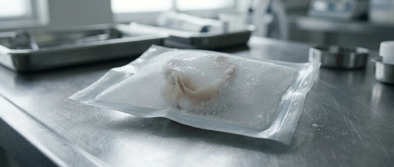
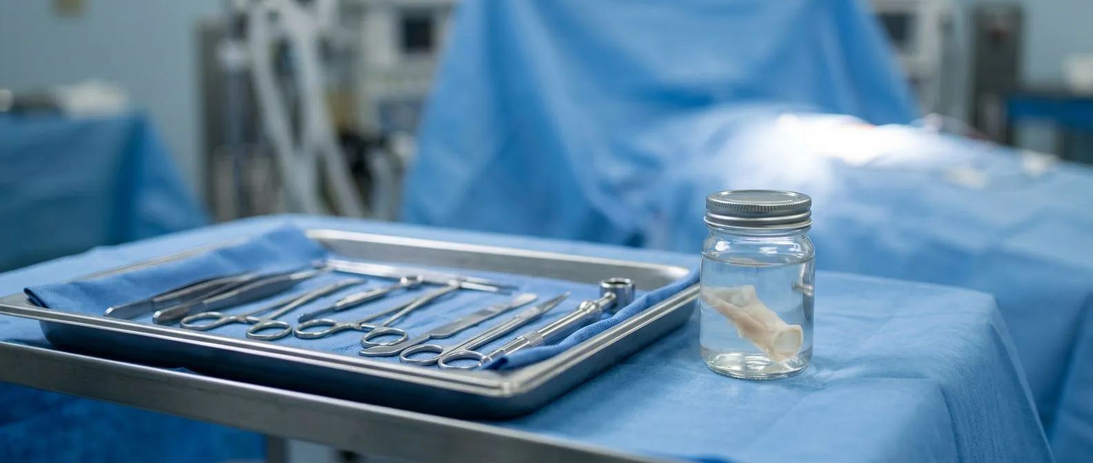
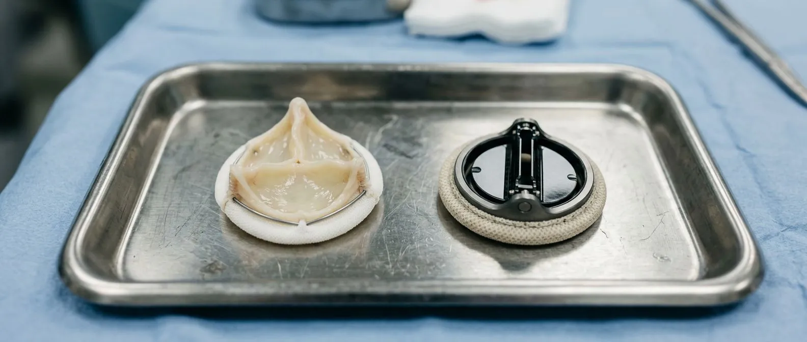
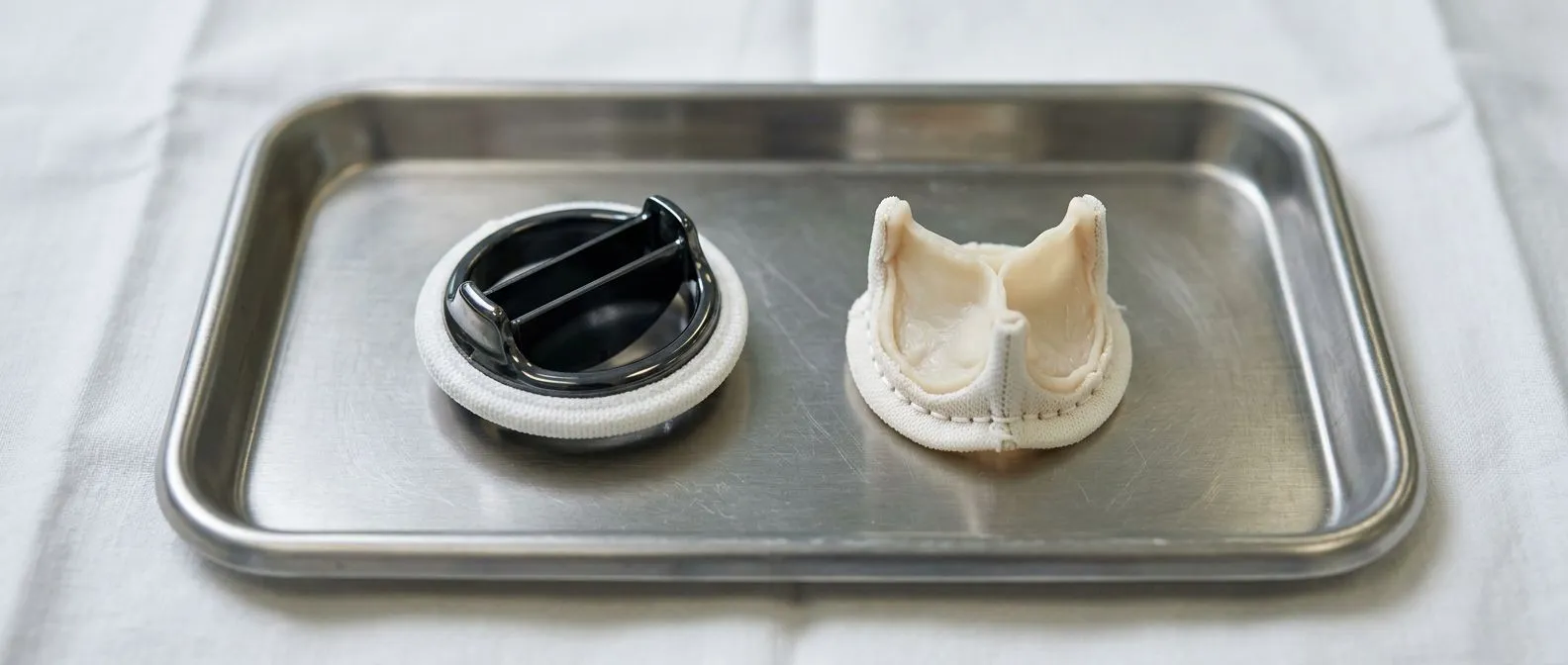
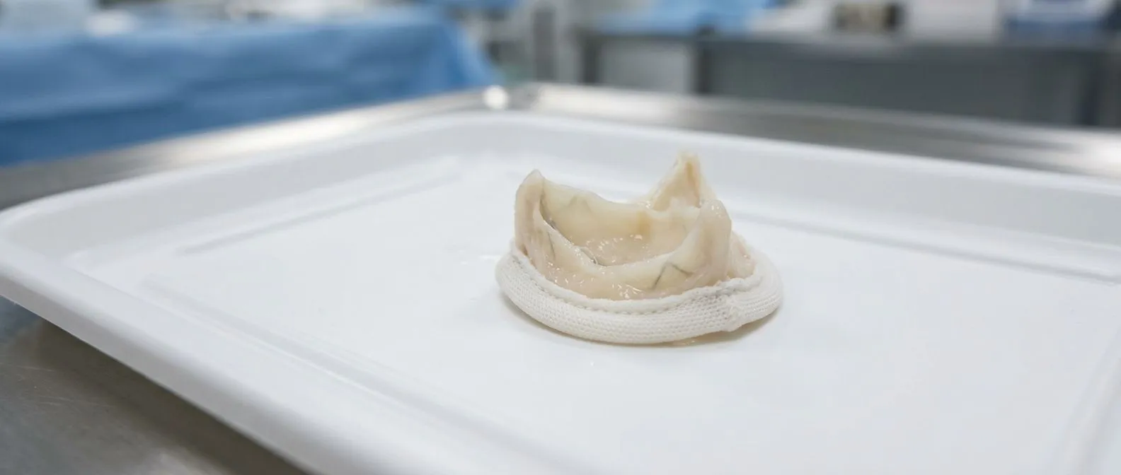
A bioprosthetic valve — sometimes called a tissue valve — is a replacement heart valve made from animal tissue, most commonly cow pericardium (the sac around the heart) or pig valve tissue. Unlike mechanical valves made of carbon and titanium, tissue valves are biological, which means they behave much like your native valve but, like all living or once-living tissue, they age. Bioprosthesis valve lifespan refers to the period during which the valve continues to function effectively before structural deterioration begins to compromise its performance. This is not a single fixed number — it varies based on valve position, patient age, and the specific design of the prosthesis.
Why does lifespan matter so much for patient decisions? The durability of your replacement valve directly influences whether you may need a second surgery later in life. For a younger patient, a valve that lasts fifteen years might mean facing reoperation in their fifties or sixties. For an older patient, the same valve may last the rest of their natural life without any further intervention. This is why surgeons consider your age, overall health, and life expectancy when recommending valve type. A tissue valve avoids the need for lifelong blood-thinning medication (such as Aspirin or warfarin), which is a major quality-of-life advantage, but the trade-off is that biological tissue eventually degenerates.
It is also important to understand that "lifespan" in clinical terms does not mean the valve suddenly stops working one day. Deterioration is typically gradual. The valve may develop small tears, calcium deposits, or thickening of the leaflets — the flaps that open and close to regulate blood flow. These changes slowly reduce how efficiently the valve opens (a problem called stenosis) or how well it closes, allowing blood to leak backward (called regurgitation). The clinical question is not just how long the valve lasts but how long it continues to function well enough that you remain free of symptoms and your heart remains healthy. Research published by The Lancet has contributed significantly to our understanding of long-term valve performance, helping clinicians set realistic expectations for patients.
How Long Do Bioprosthetic Valves Last in Real Patients
Patients often ask for a simple number, and while no single figure applies to everyone, clinical experience gives us useful ranges. For surgical bioprosthetic aortic valves — those placed through open-heart surgery at the aortic position — the expected functional lifespan is generally in the range of 10 to 15 years for patients in their sixties or older. For patients who are younger, say in their forties or fifties, the same valve may show signs of deterioration sooner, sometimes within 7 to 10 years. This is one of the most important findings in valve research: tissue valves tend to degrade faster in younger patients, likely because of more active calcium metabolism and greater haemodynamic demand over time.
What do long-term studies actually show about tissue valve longevity? Published clinical data consistently demonstrate that the majority of bioprosthetic aortic valves remain functional for at least a decade, with many continuing to perform well beyond 15 years in older recipients. The variability is significant, though. Some patients experience early structural valve deterioration within five to eight years, while others have valves that function well for over 20 years. Factors such as patient age at implantation, valve position (aortic versus mitral), valve size, and underlying conditions like kidney disease all influence the outcome. It is worth noting that the ascending aorta — the first portion of the body's largest artery as it leaves the heart — sometimes needs simultaneous repair or replacement, and the health of that surrounding tissue can affect long-term results.
For mitral valve bioprostheses, the longevity picture is somewhat different. The mitral valve is located between the left atrium and the left ventricle and operates under different pressure conditions than the aortic valve. Published series suggest that bioprosthetic mitral valves may have a somewhat shorter functional lifespan than their aortic counterparts, though the difference is not dramatic for most patients. What matters most is that your surgeon discusses the expected durability specific to your valve type and position, grounded in the data for that particular prosthesis design rather than broad generalisations. No surgeon can guarantee a specific number of years, but they can give you an evidence-based estimate that accounts for your individual circumstances.
What Affects Bioprosthetic Valve Durability Over Time
Several well-established factors influence how long a bioprosthetic valve continues to function well. The single most significant is patient age at the time of implantation. Younger patients — generally under 60 — tend to experience faster structural deterioration, a phenomenon thought to be related to more active calcium metabolism and a stronger immune response to the animal tissue. Older patients tend to have slower rates of calcification and tissue wear, which is why tissue valves are more commonly recommended for patients over 65 or 70. This does not mean younger patients cannot receive bioprosthetic valves, but they should understand the higher likelihood of earlier reoperation.
How do other health conditions influence valve wear? Chronic kidney disease accelerates calcium deposition on valve leaflets, which can stiffen the tissue and reduce valve mobility sooner than in patients with normal kidney function. Similarly, patients with conditions that affect calcium metabolism, such as hyperparathyroidism, may see faster degeneration. Lifestyle factors like smoking and poorly controlled hypertension can also contribute to faster wear by increasing the mechanical stress on the valve and promoting vascular disease more broadly. The presence of an atheromatous plaque in nearby vessels does not directly damage the valve, but it signals broader cardiovascular risk that may influence overall outcomes.
Valve design and manufacturing also play a role. First-generation bioprosthetic valves used different tissue preservation techniques than modern valves, and durability has improved over time. Current-generation valves benefit from advanced anti-calcification treatments that slow the deposition of calcium on the tissue. The way the valve is mounted — whether it is stented (supported by a frame) or stentless — can also affect longevity and haemodynamic performance. Your cardiac surgeon will select a valve prosthesis based on your anatomy, the size of your aortic annulus or mitral annulus, and the expected durability profile. Advances in tissue engineering, including the use of collagen scaffolds, are being explored to improve valve longevity further. Research on collagen scaffold applications in tissue engineering highlights the potential for future valve designs that may resist deterioration more effectively.
Types of Tissue Heart Valves and Their Longevity Differences
Not all tissue valves are the same, and understanding the differences helps explain why durability varies. The two main categories are porcine (pig) valves and bovine (cow) pericardial valves. Porcine valves are typically taken from the pig's natural aortic valve, treated and mounted on a stent or frame. Bovine pericardial valves are constructed from the pericardial tissue of cows, shaped and attached to a frame to mimic the function of a natural heart valve. Both types have been used for decades, and both have demonstrated good long-term performance, but they have somewhat different durability profiles in clinical studies.
Which tissue valve type tends to last longer? Bovine pericardial valves have generally shown slightly better durability in some published series, though the differences are not large enough to make a universal recommendation. Modern bovine pericardial valves benefit from advanced tissue fixation processes that reduce calcification, and their design can be optimised to provide excellent haemodynamics. Porcine valves have a long and well-established track record and continue to be used widely. The choice between the two often comes down to the surgeon's experience, the specific valve sizes available, and the anatomical fit for the patient rather than a dramatic difference in expected lifespan.
- Porcine valves: Taken from pig aortic tissue, well-established track record with decades of clinical data supporting their use.
- Bovine pericardial valves: Made from cow pericardium, some studies suggest marginally better durability and excellent haemodynamic profiles.
- Stentless valves: No rigid frame, allowing more natural blood flow; sometimes used in younger patients for better haemodynamics, though implantation is more complex.
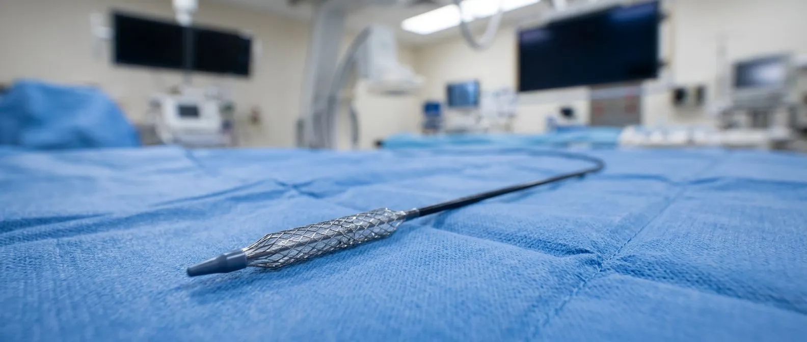
- TAVR valves: Transcatheter bioprosthetic valves delivered via catheter; long-term durability data still maturing but encouraging for older, higher-risk patients.
Beyond the tissue source, the construction method matters. Stented valves have a rigid or semi-rigid frame that supports the tissue leaflets, making implantation more straightforward and providing consistent valve geometry. Stentless valves lack this frame and rely on the patient's own tissue for support, which can provide better blood flow characteristics — particularly beneficial for patients with a small aortic annulus — but require more surgical expertise to implant correctly. Each design has trade-offs, and the right choice depends on your anatomy, your surgeon's experience, and the expected durability of the specific valve model being considered.
Biological Valve vs Mechanical Lifespan: Which Lasts Longer
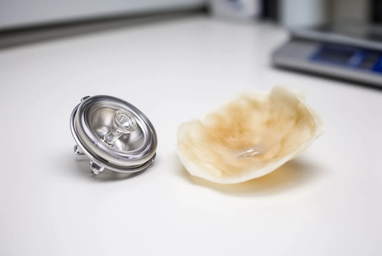
The comparison between biological and mechanical valves is one of the most common topics in pre-surgical consultations, and it is central to understanding bioprosthesis valve lifespan in context. Mechanical valves, made from durable materials like pyrolytic carbon, are designed to last a lifetime. There is no expected structural deterioration — the valve will not wear out in the way that tissue does. This makes mechanical valves the standard recommendation for younger patients who would otherwise face multiple reoperations. However, the durability advantage comes with a significant trade-off: mechanical valves require lifelong anticoagulation therapy to prevent blood clots from forming on the artificial surface.
What does lifelong blood thinning mean in daily life? Patients with mechanical valves must take warfarin (or a similar anticoagulant) every day for the rest of their lives. This requires regular blood tests to monitor the international normalised ratio (INR), ensuring the blood is thinned enough to prevent clots but not so thin that bleeding becomes dangerous. Dietary restrictions, medication interactions, and the risk of bleeding during accidents or future surgeries all become ongoing considerations. For some patients, this burden is manageable; for others — particularly those who live in areas with limited access to regular blood monitoring — it can be a major factor. This is especially relevant for our international patients who may travel long distances for follow-up care and need to consider whether reliable anticoagulation monitoring is available to them at home.
The bioprosthetic valve trade-off is essentially the opposite: you accept a shorter functional lifespan in exchange for freedom from lifelong anticoagulation. Most patients with tissue valves take Aspirin or a similar antiplatelet medication for a period after surgery, but they do not need the ongoing warfarin therapy that mechanical valves require. This means no regular INR blood tests, fewer dietary restrictions, and a lower long-term risk of bleeding complications. The cost of this freedom is the possibility of reoperation when the valve eventually degenerates. For many older patients — and increasingly for younger patients who prioritise quality of life — this trade-off is acceptable. The decision should always be made in conversation with your cardiac surgeon, weighing your age, lifestyle, access to monitoring, and willingness to undergo potential future surgery.
Bioprosthetic Valve Failure Rate and What Causes Deterioration
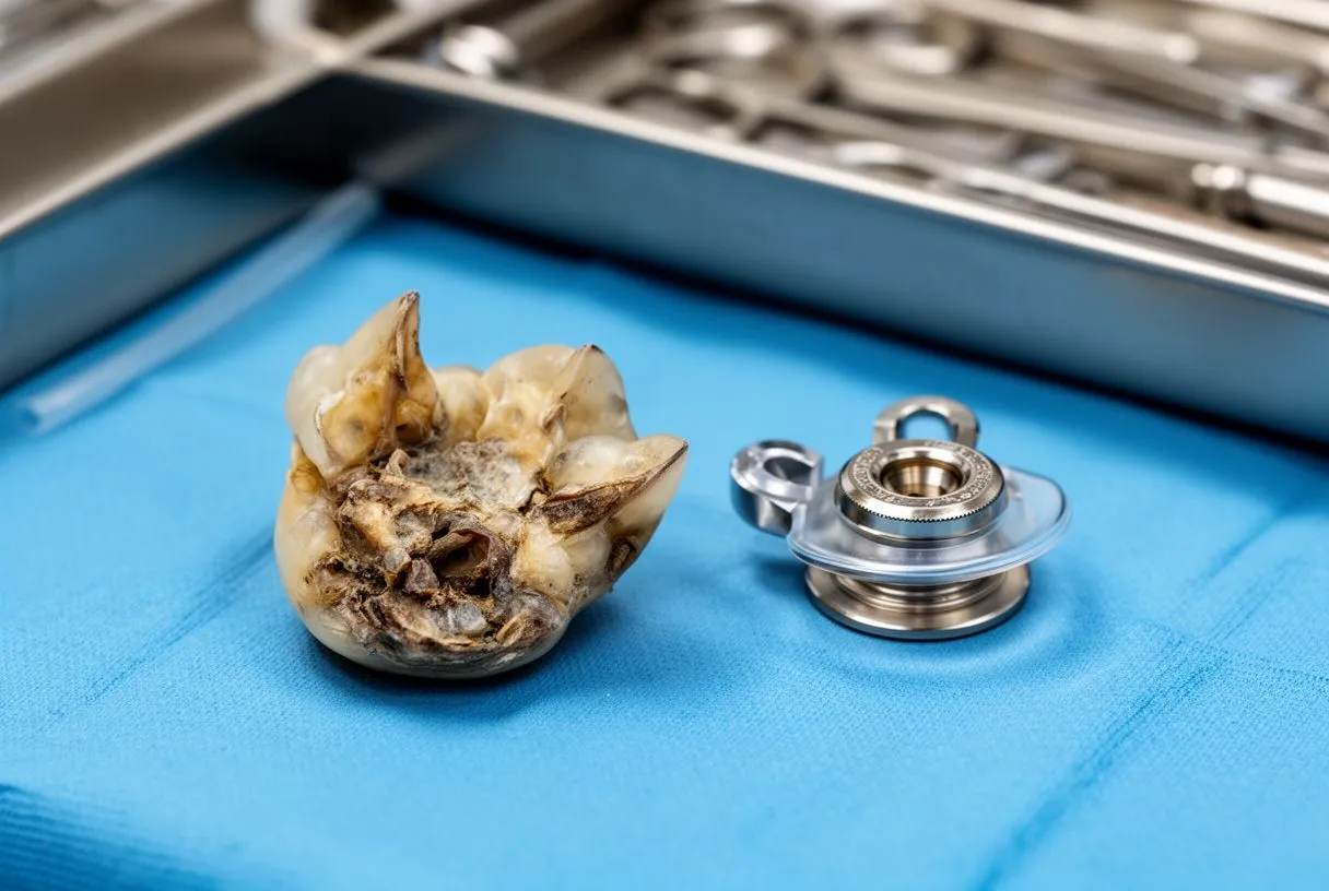
Understanding the bioprosthetic valve failure rate requires distinguishing between structural and non-structural deterioration. Structural deterioration is the gradual wear and tear of the valve tissue itself — the process that defines the end of the valve's functional lifespan. This includes calcification (calcium deposits on the leaflets), leaflet tears, and thickening or fibrosis of the tissue. Non-structural dysfunction includes problems not related to the tissue itself, such as valve thrombosis (a clot on the valve), endocarditis (infection of the valve), or paravalvular leak (leaking around the edge of the valve rather than through it). These non-structural issues can occur at any time and are not necessarily related to valve age.
What is the typical timeline for structural valve deterioration? For most patients, structural deterioration does not become clinically significant within the first 5 to 8 years after implantation. The risk increases progressively after that point, with a steeper rise after 10 years. By 15 years, a significant proportion of bioprosthetic valves show some degree of structural change, though not all of these changes require immediate intervention. The rate of deterioration follows a curve rather than a cliff edge — there is no single year at which valves suddenly fail. Instead, the probability of needing reoperation increases gradually, which is why regular echocardiographic follow-up is essential to monitor valve function over time.
Calcification is the primary mechanism of structural deterioration in tissue valves. Calcium deposits on the leaflets make them stiff, preventing them from opening fully (stenosis) or closing completely (regurgitation). The process is similar in some ways to the calcification that occurs in native aortic valve stenosis, though the timeline is different. Leaflet tears can also occur, particularly in areas where the tissue is subjected to repeated mechanical stress. Infection of the valve — endocarditis — can dramatically accelerate deterioration and may require urgent valve replacement regardless of the valve's age. Patients should be aware that good dental hygiene, prophylactic antibiotics before dental procedures when recommended, and prompt treatment of infections all help protect the valve from this non-structural cause of failure.
Tissue Valve Degeneration Symptoms to Watch For
One of the most important things a patient with a bioprosthetic valve can do is remain alert to symptoms that might indicate their valve is beginning to deteriorate. Because structural deterioration is gradual, symptoms often develop slowly and may be subtle at first. The most common symptom is increasing shortness of breath, particularly with exertion. You might notice that activities you previously managed without difficulty — climbing stairs, walking uphill, or carrying groceries — begin to cause breathlessness. This happens because the narrowing or leaking valve reduces the heart's ability to pump blood efficiently, causing fluid to back up into the lungs.
What other warning signs should prompt a cardiac review? Beyond breathlessness, watch for new or worsening fatigue, which can develop so gradually that you attribute it to ageing rather than valve function. Chest discomfort or pressure, especially with activity, may indicate that the heart is working harder to pump through a narrowing valve. Palpitations — a sense of fluttering or irregular heartbeats — can occur if the strain on the heart triggers rhythm disturbances such as atrial flutter. Some patients develop swelling in the ankles or legs, which can signal that the heart is struggling to maintain adequate circulation. Any of these symptoms in a patient with a known bioprosthetic valve warrant prompt evaluation with an echocardiogram to assess valve function.
It is worth emphasising that many of these symptoms can also be caused by conditions unrelated to the valve, which is why self-diagnosis is never appropriate. What you should do is report new symptoms to your cardiologist or cardiac surgeon promptly. Regular follow-up echocardiograms — typically annually or as recommended by your team — are designed to catch valve changes before they cause symptoms. In some cases, your team may detect early deterioration on a routine scan before you notice anything at all. This is the ideal scenario, as it allows planned intervention rather than emergency surgery. For patients who develop arrhythmias related to valve dysfunction, a wearable cardioverter-defibrillator may be considered in specific clinical situations while awaiting definitive treatment, as discussed in research on wearable cardioverter-defibrillators in suspected myocarditis.
Bioprosthetic Valve Reoperation: When and Why It Happens
The possibility of reoperation is the most significant trade-off of choosing a bioprosthetic valve, and understanding what that involves helps patients make informed decisions. Reoperation becomes necessary when the valve has deteriorated to the point that it causes symptoms or shows evidence of significant dysfunction on echocardiography. The timing varies enormously — some patients never need reoperation because their valve lasts the rest of their life, while others face it within a decade. The decision to reoperate is based on clinical findings, not on the calendar. Your team will assess the degree of stenosis or regurgitation, the effect on heart function, and your overall health before recommending surgery.
Is reoperation riskier than the first surgery? Reoperative cardiac surgery is generally more complex than the initial operation. The heart and surrounding tissues have scar tissue from the first surgery, which makes access more difficult and can increase the technical challenge. However, experienced cardiac centres perform reoperations regularly, and outcomes are generally good when the surgery is planned rather than emergent. The risk is higher than for a first-time valve replacement, but for many patients, it remains within an acceptable range. Surgical risk is commonly estimated using scoring systems such as EuroSCORE II, which accounts for age, cardiac function, and other health factors to provide a personalised risk estimate. Your surgeon should discuss your specific risk profile openly.
For some patients, reoperation can now be performed using minimally invasive or transcatheter techniques. If the failing valve is an aortic bioprosthesis, a transcatheter valve can sometimes be implanted inside the existing degenerated valve — a procedure known as valve-in-valve TAVR. This approach avoids open-heart surgery and is particularly valuable for older patients or those with higher surgical risk. Not every patient is suitable for this approach — it depends on the size and type of the original valve, the anatomy of the valve landing zone, and whether there are other cardiac issues requiring surgical attention. The availability of valve-in-valve TAVR has influenced how some surgeons counsel younger patients about bioprosthetic valves, as it offers a potentially less invasive option for future reoperation.
Bioprosthetic Aortic Valve Lifespan After TAVR Surgery
Transcatheter aortic valve replacement — called TAVR — has transformed the treatment of aortic valve disease, particularly for older patients and those considered too high-risk for traditional open-heart surgery. TAVR uses a bioprosthetic valve delivered through a catheter, typically via the femoral artery in the groin, and positioned within the diseased native aortic valve. Because TAVR valves are also made of animal tissue, they are subject to the same biological deterioration processes as surgically implanted bioprosthetic valves. However, the long-term durability data for TAVR valves is still maturing, as the procedure has been widely performed for a shorter period than surgical valve replacement.
How does TAVR valve durability compare to surgical bioprosthetic valves? Current evidence from patients who received TAVR valves five to ten years ago shows encouraging durability, with rates of structural deterioration that appear broadly comparable to surgical bioprosthetic valves over the same period. The tissue used in TAVR valves is typically bovine or porcine pericardium, treated with similar anti-calcification processes. However, because most TAVR procedures have been performed in older patients with shorter life expectancies, the data on very long-term durability — beyond 10 to 15 years — is still being gathered. For younger patients considering TAVR, this uncertainty is an important factor to discuss with their cardiac team.
The decision between surgical aortic valve replacement and TAVR depends on multiple factors including age, surgical risk, anatomy, and life expectancy. TAVR is generally recommended for patients who are older, have higher surgical risk scores, or have conditions that make open-heart surgery particularly hazardous. For younger, lower-risk patients, surgical valve replacement remains the standard approach, as the long-term durability data is more established and the option for future reoperation is more straightforward. Some patients with specific anatomical considerations — such as a small aortic annulus or particular calcification patterns — may benefit from one approach over the other. Your cardiac team will evaluate these factors using CT imaging and clinical assessment to recommend the approach with the lowest risk for your situation.
Bioprosthetic Mitral Valve Durability and Replacement Timing
The mitral valve presents a different set of considerations for bioprosthetic valve durability. Located between the left atrium and the left ventricle, the mitral valve regulates blood flow from the upper to the lower chamber of the left side of the heart. When the mitral valve needs replacement rather than repair — and repair is always preferred when feasible — the choice between a mechanical and bioprosthetic valve follows similar logic to the aortic position but with some important nuances. Bioprosthetic mitral valves are subject to different mechanical stresses than aortic valves because the pressure environment and flow dynamics differ.
How does mitral valve position affect bioprosthesis lifespan? Some published series suggest that bioprosthetic mitral valves may experience slightly faster structural deterioration than aortic bioprostheses, though the difference is modest and not consistent across all studies. The mitral valve closes under higher pressure and operates in a more complex mechanical environment, which may contribute to faster tissue fatigue. For this reason, some surgeons are more cautious about recommending bioprosthetic mitral valves for younger patients, particularly those under 60. However, the avoidance of lifelong anticoagulation remains a strong argument for tissue valves in many patients, and the decision must be individualised based on age, lifestyle, and the specific pathology of the mitral valve disease.
When a bioprosthetic mitral valve deteriorates and needs replacement, the reoperation is technically more complex than aortic valve reoperation because the mitral valve is located deeper within the heart and access is more challenging. For some patients, transcatheter approaches to mitral valve replacement are being developed and used in clinical trials, but these are not yet as established as TAVR for the aortic position. This means that mitral valve reoperation typically involves open-heart surgery, which reinforces the importance of choosing the right valve at the first operation. If you are facing mitral valve replacement, discuss with your surgeon not only the expected lifespan of the valve but also what reoperation would involve in your specific case, including the anticipated surgical approach and risk profile.
In summary, bioprosthesis valve lifespan is not a single number but a range that depends on your age, valve position, overall health, and the specific valve design chosen. Tissue valves offer the significant advantage of freedom from lifelong blood thinners but carry the possibility of future reoperation. Regular follow-up with echocardiography allows your cardiac team to monitor valve function and catch deterioration early, before symptoms develop. If you or a family member is facing a valve replacement decision, we encourage you to discuss these factors openly with a qualified cardiac surgeon who can provide personalised guidance based on your individual anatomy, health, and circumstances. Our team at Cirugía de Corazón is available for consultation to help you understand your options and make the decision that is right for you.