Cardiac Imaging Specialist: Reading Your Heart Scans
Cardiac imaging specialist role explained clearly: how echocardiograms, cardiac MRI and CT scans are read, what results mean.
When your doctor orders a heart scan, the pictures are only half the story. Someone has to read them — and read them well. That is where the cardiac imaging specialist role comes in: a doctor trained to interpret echocardiograms, cardiac MRI, CT scans and nuclear studies, and to translate what those images show into a clear plan for you. In this guide, we explain what these specialists actually do, how each type of scan works, and how the results shape decisions about your heart care. If you or a family member has recently been told you need a heart scan, this article is written for you.
What Does a Cardiac Imaging Specialist Do?
The daily work of a heart scan reader. A cardiac imaging specialist is a cardiologist who has completed extra training in one or more imaging techniques — echocardiography, cardiac MRI, cardiac CT or nuclear cardiology. Their job is to look at moving images of your heart, measure the chambers and valves, assess blood flow, and describe exactly what is working and what is not. They do not just "read" the scan; they answer specific questions your referring doctor has asked, such as whether a valve is leaking severely or whether the heart muscle is getting enough blood.
Turning pictures into decisions. The most valuable part of the cardiac imaging specialist role is not taking the image but interpreting it in context. The same echocardiogram can mean very different things in a 40-year-old athlete versus a 78-year-old with a narrowed aortic valve. The specialist weighs your symptoms, history and examination findings alongside the images, then writes a report that guides treatment — whether that means medication, monitoring, or a referral to a surgeon. This is why the American College of Cardiology emphasizes that imaging findings should always be integrated with clinical information rather than read in isolation.
Who performs the scans? Depending on the facility, the specialist may personally perform the echocardiogram or supervise a sonographer — a technician trained to acquire the images. For cardiac MRI and CT, a radiologist or cardiologist typically interprets the study after the scan is acquired. In either case, the person signing the report carries the responsibility for its accuracy.
Cardiologist vs Cardiac Imaging Specialist: What's the Difference?
Two paths, one goal. A general cardiologist is a physician who diagnoses and treats heart disease broadly — managing blood pressure, arrhythmias, heart failure and more. A cardiac imaging specialist starts from the same base but has completed an advanced fellowship, usually one to two additional years, focused entirely on imaging techniques. This is why many patients ask about the difference between a cardiac imaging specialist vs radiologist: a radiologist is an expert in imaging of the whole body, while a cardiac imaging specialist is a cardiologist who has focused that imaging expertise on the heart alone.
Who is a cardiac specialist, then? In everyday language, "cardiac specialist" usually means any cardiologist. The distinction matters most when a complex question needs answering — for example, whether a valve can be repaired rather than replaced, or whether faint findings on a scan are truly abnormal. At that point, the detailed training of an imaging specialist changes what your report says and, ultimately, what options you are offered.
In our practice, imaging is never treated as a standalone service. Every scan is reviewed in the context of the whole patient, and our multidisciplinary team — led by Dr Alejandro Molina — discusses the findings together before any recommendation is made.
What Are the Main Types of Cardiac Imaging?
Seven techniques, each with its own question. Patients often ask what the 7 types of imaging are when it comes to the heart. The main modalities are echocardiography (ultrasound of the heart), cardiac MRI, cardiac CT, nuclear cardiology (including perfusion scans and PET), stress testing with imaging, coronary angiography (catheter-based dye imaging), and chest X-ray. Each answers a different question, and the cardiac imaging specialist role involves choosing — or advising on — the right test for the right question.
Here is a simple way to think about what each one does:
- Echocardiogram: shows the valves and pumping function in real time; no radiation; usually the first test ordered.
- Cardiac CT: maps the coronary arteries and calcium; used for blockages and for planning structural procedures.
- Cardiac MRI: gives the most detailed picture of heart muscle — scar, inflammation and tissue health.
- Nuclear scans: show blood flow to the heart muscle under stress and at rest.
- Angiography: the classic catheter-based view of the coronary arteries; often combined with treatment in the same session.
Choosing wisely. More imaging is not always better. Modern guidelines stress selecting the test that will actually change management, to avoid unnecessary radiation, cost and anxiety. When you understand why each test was ordered, you can ask better questions and take a real part in the decision.
How Long Does a Cardiac Imaging Test Take?
Time varies by test. One of the most common questions is how long a cardiac imaging test takes. A standard transthoracic echocardiogram usually takes 20 to 45 minutes. A cardiac CT scan often takes 10 to 20 minutes of scan time, though you may spend an hour at the facility for preparation. Cardiac MRI is the longest, typically 45 to 90 minutes, because it captures many sequences of images. Nuclear stress tests can take 2 to 4 hours when images are taken at two different time points.
What to expect on the day. Most of the "long" part of any scan is waiting and preparation — changing clothes, having an IV line placed, or simply lying still. The imaging itself is painless for all modalities. If you have claustrophobia, tell the team in advance; MRI machines in particular are narrow, and some centers can offer strategies to help.
When do you get results? A preliminary echocardiogram can often be reviewed the same day. CT and MRI reports typically take a few days, because the images need careful interpretation. Ask your team when to expect the report — and whether the specialist will explain it to you directly.
How Echocardiogram Interpretation Guides Your Diagnosis
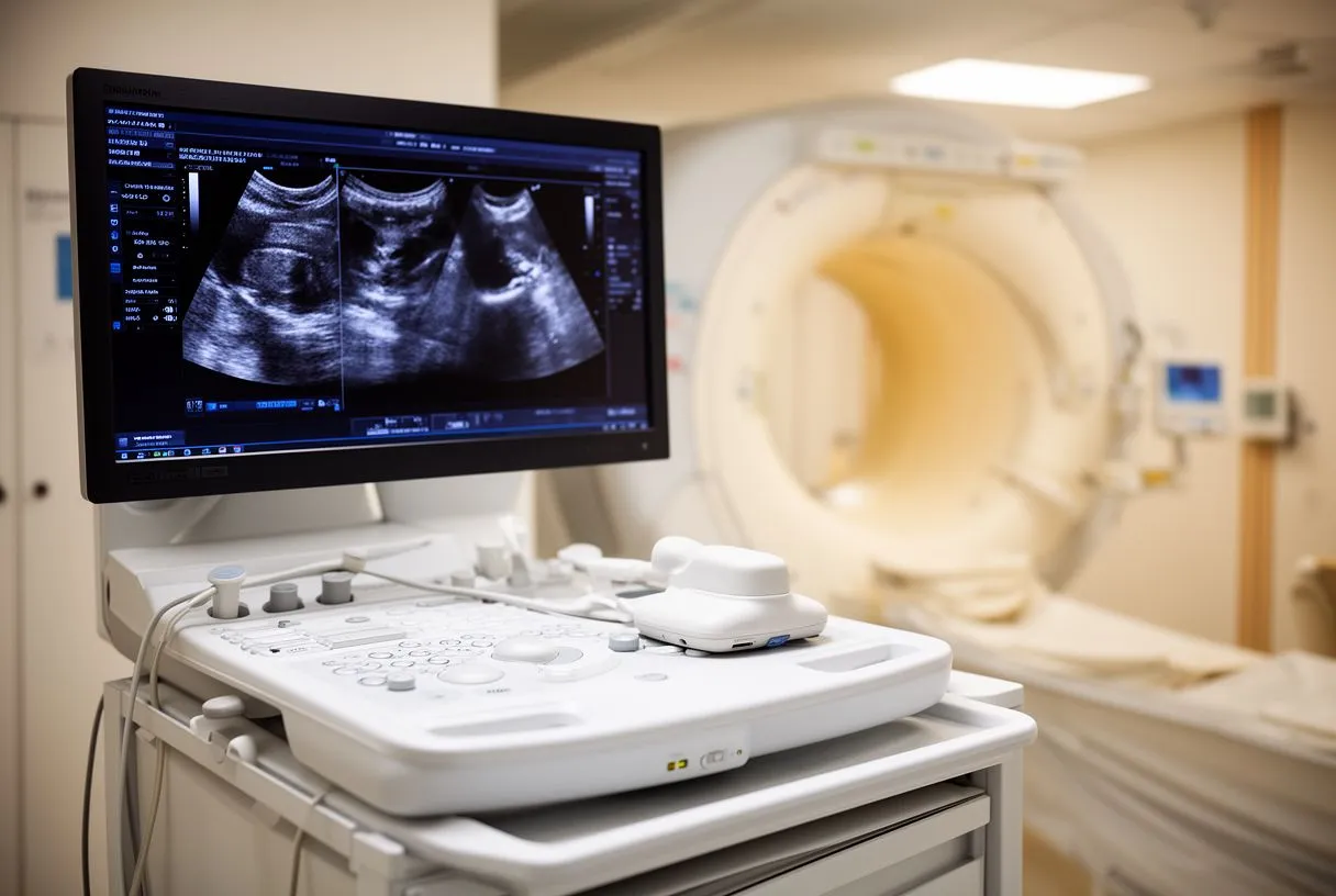
The ultrasound that sees your heart working. Echocardiography uses sound waves to create a live moving picture of your heart. It shows the four chambers, the four valves (aortic, mitral, tricuspid and pulmonary), and how well the heart squeezes. An echocardiogram interpretation doctor measures the opening of a narrowed valve, the leak of a leaking one, and the pumping strength of the heart muscle — a number called the ejection fraction.
Why the reader matters so much. Echocardiography is operator-dependent: the images and the measurements depend on the skill of the person acquiring and reading them. Subtle findings — a mild valve thickening, a small pericardial effusion, early signs of heart muscle stiffening — can be missed or over-called by a less experienced eye. This is precisely where the cardiac imaging specialist role earns its place in your care.
What the report should tell you. A good report states the findings in plain terms: which valve is affected, how severely, and what the pumping function is. It should end with a conclusion that answers the question the referring doctor asked. If your report is full of numbers without explanation, ask for a conversation — a good team will walk you through it. In our consultations we take the time to explain what the studies actually show, in plain words, and to answer every question before any decision is made.
When Is a Cardiac MRI or Heart CT Scan Needed?
Cardiac MRI: the tissue specialist. Cardiac MRI uses a powerful magnet and no radiation to produce highly detailed images of the heart muscle. It is the test of choice when doctors need to know the condition of the muscle itself — whether there is scar from a previous heart attack, inflammation from myocarditis, or the fibrosis of certain inherited conditions. A cardiac MRI specialist near you may also be involved in assessing complex congenital heart disease, where the anatomy was abnormal from birth. Many patients searching for a pediatric cardiac imaging specialist are looking for exactly this kind of expertise, because children with congenital heart disease need imaging tailored to their unique anatomy.
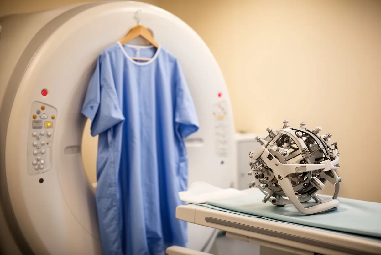
Cardiac CT: the map of the arteries. Cardiac CT is fast and excellent for two things: measuring calcium in the coronary arteries and visualizing the arteries themselves with a CT angiogram. It can rule out a blockage with high confidence when symptoms are ambiguous. It has also become essential for planning transcatheter procedures — for TAVR, the catheter-based aortic valve replacement, a CT scan defines the size of the valve, the access route and the anatomy of the surrounding structures. Standardized scanning protocols and reporting for these procedures have been established by international consensus documents, such as the one published by the European Society of Cardiovascular Radiology (European Society of Cardiovascular Radiology), which set out precisely which measurements must be taken before valve implantation.
Which one do you need? The choice depends on the question. If your doctor wants to know "is there a blockage in the arteries?", CT is usually the answer. If the question is "what is the state of the heart muscle?", MRI is usually the answer. If the question is about valve function, an echocardiogram usually comes first. The cardiac imaging specialist role includes knowing which test to use, and when — and, just as importantly, when not to order more.
Who Reads a Nuclear Stress Test and What It Shows?
The specialist behind the report. If you are wondering who reads a nuclear stress test, the answer is usually a nuclear cardiologist — a cardiac imaging specialist with specific training in nuclear techniques. The test involves a tiny, safe amount of radioactive tracer, injected into a vein, which is taken up by the heart muscle. Images taken under stress (usually with exercise or medication) and at rest are compared. If an area of the muscle takes up the tracer at rest but not under stress, that indicates a blockage that only limits blood flow when the heart works harder.
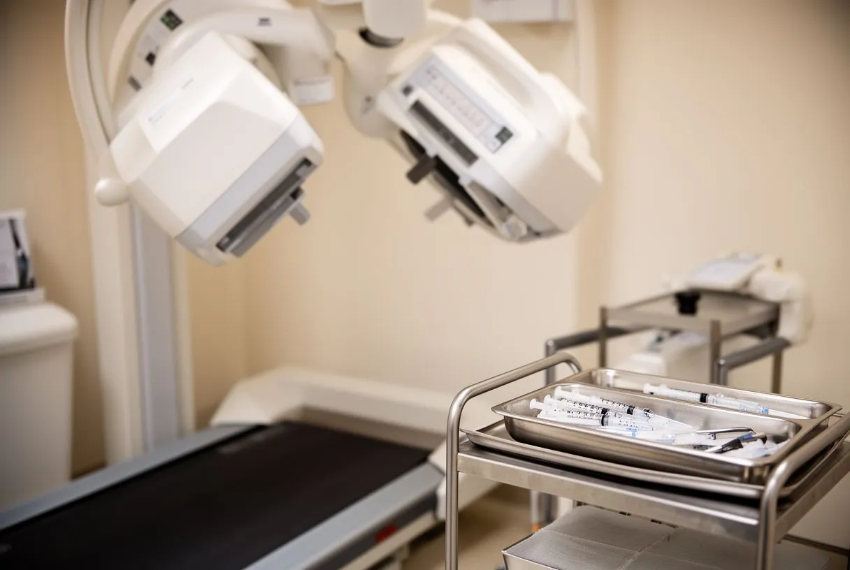
What a nuclear perfusion scan can tell you. A nuclear perfusion scan answers a different question than an echocardiogram: it shows the blood supply to the heart muscle, not just the structure. It is particularly useful when chest pain has not been clearly explained, or when a previous angioplasty or bypass graft needs follow-up. The specialist also assesses how well the heart pumps under stress — a measure called the stress ejection fraction, which adds prognostic information beyond the pictures themselves.
Radiation and safety. Nuclear tests involve a small dose of radiation, similar in scale to other established medical imaging. Modern scanners and protocols keep doses as low as reasonably possible. If you are pregnant or think you may be, tell your team before the test — alternatives exist.
Cardiac Imaging for Surgery Planning: What Your Team Looks For
Imaging decides the approach. Before any heart operation — valve repair or replacement, coronary bypass, aortic surgery, or a transcatheter procedure — the surgical team relies on imaging to plan the operation. A cardiac imaging specialist for surgery planning will map the valve anatomy, measure the aorta (the main artery leaving the heart, where a diameter above 5.5 cm generally triggers a discussion about repair), and assess the heart's function. This is exactly what an echocardiogram interpretation doctor and a cardiac CT specialist do together before recommending a procedure.
What we look for in our team. Every case is reviewed by our multidisciplinary team against international practice guidelines before a recommendation is made. Our work covers valve disease, coronary disease, the aorta and structural heart conditions, and we perform conventional, minimally invasive, robotic and transcatheter procedures — we recommend the less invasive route only where the evidence supports it for that patient. The imaging findings are central to that discussion: the same aortic valve narrowing may point to a surgical replacement in one patient and a TAVR in another, depending on anatomy, age and other conditions.
The patient receives the plan, not just the images. After the team review, the patient receives a clear proposal with the alternatives, and the final decision is always theirs. Imaging findings are explained in everyday language: what the pictures show, what the options are, and what the trade-offs between them look like for your specific situation. That conversation — not the scan alone — is what leads to a good decision.
Heart Scan Second Opinion: When to Ask for One
You are allowed to ask. A heart scan second opinion is a normal, sensible step when a major decision looms — valve surgery, a procedure on the aorta, or any intervention with meaningful trade-offs. Specialists expect it, and most welcome it. A fresh reading of your scans can confirm the plan, refine it, or occasionally reveal an option nobody had mentioned. In our practice we encourage second opinions and treat seeking one as a normal part of deciding about heart surgery.
What you need to bring. For a second opinion to be useful, the specialist needs the original images, not just the written report. Most hospitals can export images to disc — a service often called imaging on disc — which you can then send to the reviewing team. Bring the written report as well, along with any other recent tests. The reviewing specialist will then compare their own reading with the original.
How the second opinion works here. The first contact is a free video consultation: we get to know the patient, talk through the condition, and say which studies are still needed so no unnecessary ones are ordered. A face-to-face visit at the consulting rooms follows when the patient decides to go ahead. We see patients with complex valve and structural heart disease from Guadalajara, Querétaro and Hermosillo, and we review every case as a multidisciplinary team before recommending anything.
How to Prepare for Your Cardiac Imaging Specialist Appointment
Before the scan. Preparation depends on the test. For an echocardiogram, no preparation is needed. For a cardiac CT, you may be asked to avoid caffeine and to take a beta-blocker to slow your heart rate for clearer images. For cardiac MRI, remove all metal objects and tell the team about any implanted devices — pacemakers and defibrillators require specific protocols. For nuclear tests, avoid caffeine on the day of the stress portion. Always follow your facility's specific instructions.
What to bring and what to ask. Bring your previous reports, your medication list, and any questions you have written down. Good questions to ask the person reading your scan include:
- "What exactly does this scan show?" — in your own words, not just numbers.
- "What does this change about my treatment?" — a test that changes nothing may not be needed.
- "What happens if we do nothing?" — understanding the natural course of your condition.
- "Do you need any more images, or is this enough?" — to avoid unnecessary testing.
After the results. When your results are ready, book a proper conversation — not a rushed hallway summary. The cardiac imaging specialist role includes explaining what the images show, what they mean for your life, and what the realistic options are. If a procedure is being considered, ask about the risks, the recovery time, and the less invasive alternatives. If you are unsure, seek a second opinion. And above all, do not try to interpret complex scans on your own — discuss your situation with a qualified cardiac team, and book a consultation with a specialist who can look at your images and talk them through with you, face to face or by video.