

Connective Tissue Disorders and Aortic Aneurysm Risk
Connective tissue disorder aorta: learn early warning signs, MRI/CT screening schedules, and protective treatments.
When someone is diagnosed with a connective tissue disorder aorta, it means the main artery of the body may be weakened by an underlying genetic condition that affects the tissue holding it together. Connective tissue is the body's scaffolding — it gives strength and flexibility to the skin, joints, blood vessels, and the walls of the aorta. When that scaffolding is faulty, the aorta can slowly stretch and enlarge over time, a condition called an aortic aneurysm. The good news is that, with the right monitoring and timely treatment, most people with these conditions live full lives and dramatically reduce their risk of a serious aortic event.
What Is a Connective Tissue Disorder Aorta?
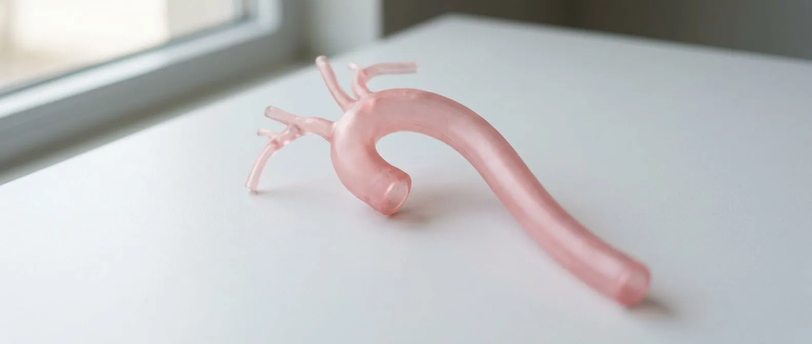
Connective tissue disease aorta changes begin at the microscopic level. The aorta is the largest artery in the body, carrying oxygen-rich blood from the heart through the chest and into the abdomen. Its wall is built from layers of collagen and elastin — proteins that let the artery stretch with each heartbeat and then spring back. In inherited connective tissue conditions, one of those building blocks is defective, so the wall is weaker than it should be.
How weakening happens over time. Under the constant pressure of thousands of heartbeats a day, a structurally weak aortic wall gradually stretches. This process is called dilation. Over years, dilation can progress to a PubMed-indexed body of research confirming what clinicians see in practice: that aortic enlargement in connective tissue disorders follows a predictable, monitorable pattern rather than appearing without warning. Research also shows that aging and cellular stress accelerate this vascular remodeling, and studies of abdominal aneurysm formation have documented how mitochondrial damage in vessel cells drives this process — findings that apply to the weakening mechanisms seen in genetic aortic disease.
Why diagnosis matters so early. Knowing you have a genetic connective tissue condition transforms a hidden threat into a manageable one. Once identified, the aorta can be measured regularly, and treatment can begin long before the artery reaches a dangerous size. Many patients we meet at our consulting rooms in Guadalajara, Querétaro and Hermosillo arrive after a relative was diagnosed, and that family history is often the first clue.
How Do Connective Tissue Disorder Aorta Symptoms Appear?
The quiet phase. Most connective tissue disorder aorta symptoms are absent until the aorta is significantly enlarged. This is the most important thing to understand: an aneurysm can grow for years without a single warning sign. That is why people with a known genetic condition should never rely on how they feel.
When symptoms do appear. As the aorta widens, some people notice a deep, steady chest or upper back pain, a hoarse voice, or difficulty swallowing if the enlarged vessel presses on nearby structures. Others may feel shortness of breath during activity. These signs usually mean the aneurysm has already grown large enough to need urgent evaluation.
Emergency signs. A sudden, tearing or ripping pain in the chest or back, pain that moves to the abdomen, fainting, or signs of stroke are red flags for AHA Journals-covered disease mechanisms aside — an acute event such as a dissection or rupture, which is a medical emergency requiring an immediate call to emergency services.
Marfan Syndrome and the Aorta: What Should You Know?
The most common genetic cause. Marfan syndrome is the best-known connective tissue disorder aorta condition, caused by a fault in the fibrillin-1 gene. It affects roughly one in several thousand people, and it tends to show up in a recognizable pattern: tall, slender build, long limbs and fingers, eye lens dislocation, and joint flexibility.
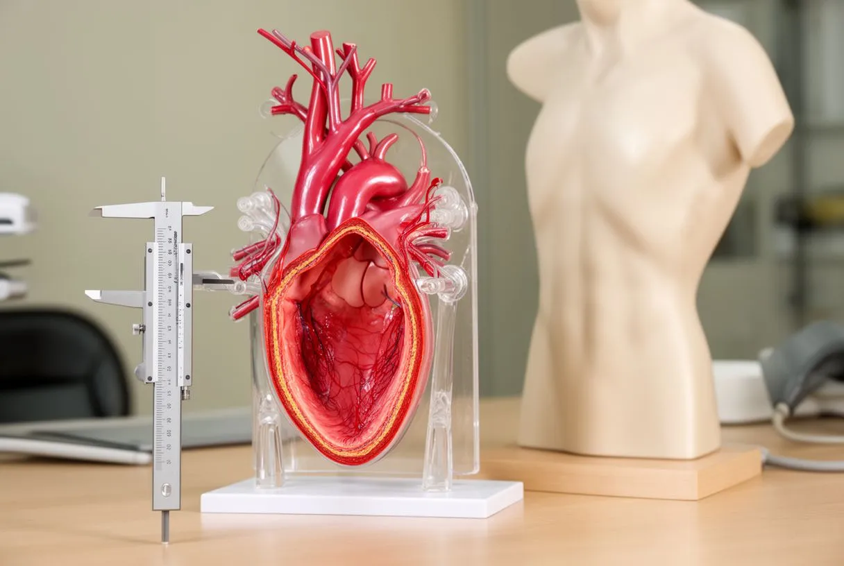
How it affects the heart. In Marfan syndrome, the problem usually starts at the aortic root — the section where the aorta leaves the heart. The root can dilate and stretch the valve between the heart and the aorta, causing leaking. This is why cardiac imaging, not just joint and eye checks, is central to Marfan care from childhood onward.
What treatment looks like. Medications that lower blood pressure and reduce the force of each heartbeat are the first line of protection, because they slow the rate of dilation. When the aortic root approaches a surgical threshold — typically around 5.0 to 5.5 cm in diameter, with lower thresholds for certain patients — elective aortic aneurysm repair offers excellent long-term protection. In our practice, every case like this is reviewed by the full multidisciplinary team against international practice guidelines before a recommendation is made, and the patient receives a clear proposal with the alternatives.
What Is the Ehlers-Danlos Aorta Risk?
Why the vascular type matters. Ehlers-Danlos syndrome is a family of collagen disorders, and its vascular type is the one that concerns the heart team most. The Ehlers-Danlos aorta risk is different from Marfan's: instead of gradual dilation, the artery can rupture or dissect at a relatively normal size. That means patients may have less warning and require earlier, more frequent monitoring.
Recognizing the broader picture. People with vascular Ehlers-Danlos often have very thin, translucent skin, easy bruising, and distinctive facial features. Frequent bowel or uterine ruptures can also occur. Because arteries throughout the body are affected — not just the aorta — imaging sometimes extends beyond the heart to other arterial beds.
How care differs. Surgical thresholds may be set at smaller diameters, and the choice of surgical technique matters because fragile tissue tears easily. When we see a patient with suspected vascular Ehlers-Danlos, we take extra time in the consultation to explain what the studies actually show, in plain words, before any decision is made. Genetic testing to confirm the type is also a key step, because management is different for each subtype.
What Is Loeys-Dietz Aorta Syndrome?
A newer, recognized condition. Loeys-Dietz aorta syndrome was identified in the mid-2000s. It is caused by changes in genes involved in the TGF-beta signaling pathway, and it shares features with both Marfan and Ehlers-Danlos — plus a few of its own, such as widely spaced eyes, a cleft palate or bifid uvula, and a characteristic twist of the arteries.
Why the surgical threshold is lower. In Loeys-Dietz, the aorta can dissect at smaller diameters than in Marfan syndrome. Many teams therefore recommend elective aortic aneurysm repair earlier — often around 4.5 to 5.0 cm — and monitor the entire aorta, including the branches, with regular imaging. Some patients also need surgery on the arteries supplying the head and brain.
Family screening is essential. Loeys-Dietz is passed from parent to child about half the time, and new mutations also occur. If one family member is diagnosed, genetic testing and imaging for relatives can catch silent aneurysms before they become emergencies.
How Does Connective Tissue Disorder Aorta Screening Work?
Screening starts with a family history. If you or a relative has been diagnosed with a genetic connective tissue condition — or if there is a history of sudden death from aortic disease — screening is the first step. A connective tissue disorder aorta diagnosis is confirmed through imaging plus, when appropriate, genetic testing.
What a typical screening schedule looks like. The exact intervals depend on your condition, the current size of your aorta, and how fast it has grown. Your team will personalize this, but general patterns include:
- Baseline echocardiogram: an ultrasound of the heart that measures the aortic root and ascending aorta, often repeated every 6 to 12 months when changes are active or surgery is being considered.
- Genetic testing: a blood or saliva test that can identify the specific gene change, guide surveillance intensity, and inform decisions for family members.
- Full aortic imaging: CT or MRI of the chest, abdomen and pelvis to look beyond the root, especially in conditions where the whole aorta is at risk.
- Eye and skeletal evaluation: important in Marfan syndrome, where lens dislocation and spinal changes help confirm the diagnosis.
Why you should not wait for symptoms. Screening is designed to find problems while they are still silent and easy to manage. An aneurysm discovered at 4 cm is a planning conversation; one discovered at 8 cm with a rupture is an emergency. The difference between those two outcomes is often simply a scheduled scan.
Which Connective Tissue Disorder Aorta Imaging (MRI or CT) Is Right for You?
Both are excellent tools. A connective tissue disorder aorta imaging plan usually combines echocardiography with either CT or MRI. CT is fast, widely available, and ideal for emergencies; it uses X-rays and radiation. MRI avoids radiation, shows the aortic wall and valve in exceptional detail, and is well suited to the decades of repeat scans that connective tissue patients need.
Making the choice. For young patients who will face dozens of scans across a lifetime, minimizing radiation exposure is a real consideration, and many teams favor MRI for routine surveillance. CT takes priority when a dissection is suspected, when surgical planning needs precise detail quickly, or when MRI is not possible due to a pacemaker, implanted devices, or claustrophobia.
What is checked at each scan. The measurements that matter most are the maximum diameter at the aortic root, the ascending aorta, the arch, and the descending aorta — plus the growth rate between scans. A growth rate above about 0.5 cm per year is a signal to intensify surveillance or consider repair sooner. In our consultations we walk through each image with the patient so they understand exactly what their own aorta is doing.

What Are the Connective Tissue Disorder Aorta Treatment Options?
Medication comes first. Connective tissue disorder aorta treatment usually begins with medication. Beta-blockers reduce the force of each heartbeat, slowing dilation. Losartan and related drugs have shown benefit in Marfan syndrome as well. These are long-term, preventive medicines — they protect the artery while it is still small enough to protect.
When surgery is recommended. Elective aortic aneurysm repair is planned when the aorta reaches a size or growth rate where the risk of waiting outweighs the risk of surgery. For patients with connective tissue disorders, the standard approach is aortic root replacement with a valve-sparing or composite graft technique, and lifelong follow-up afterward. These decisions are guided by risk stratification — a structured way of weighing each patient's anatomy, condition, and overall health.
Less invasive options where they fit. For patients with a functioning aortic valve but a narrowed or diseased valve later in life, Transcatheter Aortic Valve Implantation approaches such as TAVR — a valve delivered through a catheter in the groin — can offer a lower-risk route for suitable candidates. Similarly, for the abdominal aorta, endovascular repair with a stent-graft can sometimes be used. In our practice we perform conventional, minimally invasive, robotic and transcatheter procedures, and we recommend the less invasive route only where the evidence supports it for that patient. One of the most careful decisions in connective tissue patients involves the location of the aortic cross-clamp during surgery — supra-renal versus infra-renal clamping — and studies of elective open aortic aneurysm repair have compared outcomes between these clamping positions to help protect the kidneys and organs during the operation.
When to See an Aorta Specialist for a Connective Tissue Disorder?
Recognizing the right moment. Knowing when to see an aorta specialist about connective tissue conditions comes down to a few clear triggers. You should request an evaluation if you have a diagnosed genetic connective tissue condition and no cardiac follow-up yet; if a close relative has an aortic aneurysm or died suddenly of aortic disease; if you notice physical features of Marfan, Loeys-Dietz or Ehlers-Danlos alongside chest pain; or if any scan has shown an aorta above normal size.
What an aortic center offers. Aortic disease in connective tissue disorders is specialized care. The surgical technique, the timing, the graft choice and the follow-up plan all differ from ordinary aneurysm care, and outcomes are best at teams that see these cases regularly. Asking how many connective tissue cases a team handles each year is a fair and reasonable question.
Getting a second opinion is normal. If you have been told you need surgery — or told you do not — a second opinion can confirm the plan or open alternatives. We encourage second opinions and treat seeking one as a normal part of deciding about heart surgery. The first contact is a free video consultation: we get to know the patient, talk through the condition, and say which studies are still needed so no unnecessary ones are ordered; a face-to-face visit at the consulting rooms follows when the patient decides to go ahead.
How Can You Live Well With a Connective Tissue Disorder Aorta?
Protective habits. Living well with a connective tissue disorder aorta means combining medical care with sensible daily choices. None of these replace your treatment plan, but together they lower day-to-day stress on the aorta:
- Control blood pressure: keep it at the target your cardiologist sets, since high pressure directly stretches the aortic wall.
- Never smoke: tobacco damages the aortic wall and accelerates aneurysm growth more than almost any other habit.
- Choose appropriate exercise: steady, moderate activity is good; heavy straining, maximal weight lifting, and contact sports are usually avoided because they spike blood pressure sharply.
- Ask about anticoagulation: some connective tissue patients carry a higher thrombotic risk alongside the bleeding risk of thin skin and fragile vessels, so any blood-thinner decision should be made with a specialist who knows both sides.
Planning pregnancy and procedures safely. Pregnancy raises blood pressure and cardiac output, which increases aortic risk, so women with these conditions should have their aorta assessed before conception and monitored throughout. Dental work and surgery may also need antibiotic or anticoagulation planning, depending on your specific condition and medications.
Building a lifelong partnership. The single most protective step is simply staying connected to a team that knows your aorta. Regular scans, honest conversations, and early planning turn what was once a feared diagnosis into a well-managed one. If you or a family member may have a connective tissue disorder affecting the aorta, do not self-diagnose — book a consultation with a qualified cardiac team, bring your questions, and let the experts map out your plan. Knowledge and monitoring, applied early, are your strongest protection.






