

Deep Hypothermic Circulatory Arrest in Aortic Surgery
Learn how deep hypothermic circulatory arrest aortic surgery works, why cooling protects the brain, and what recovery looks like.
What is Deep Hypothermic Circulatory Arrest Aortic Surgery?
Deep hypothermic circulatory arrest aortic surgery is a highly specialized technique used by cardiac surgeons to repair complex problems in the aorta, the largest artery in the human body. The aorta carries oxygen-rich blood directly from the heart to the rest of the body. When disease affects the aortic arch or the ascending aorta—the vital section of the vessel that rises directly from the heart—surgeons often need to temporarily stop blood flow to perform delicate repairs safely. Stopping the circulation would normally deprive the brain and vital organs of oxygen, causing irreversible cellular damage within minutes. To prevent this catastrophic outcome, the surgical team deliberately cools the patient's body to a profoundly low temperature, usually between 18 and 20 degrees Celsius (64 to 68 degrees Fahrenheit).
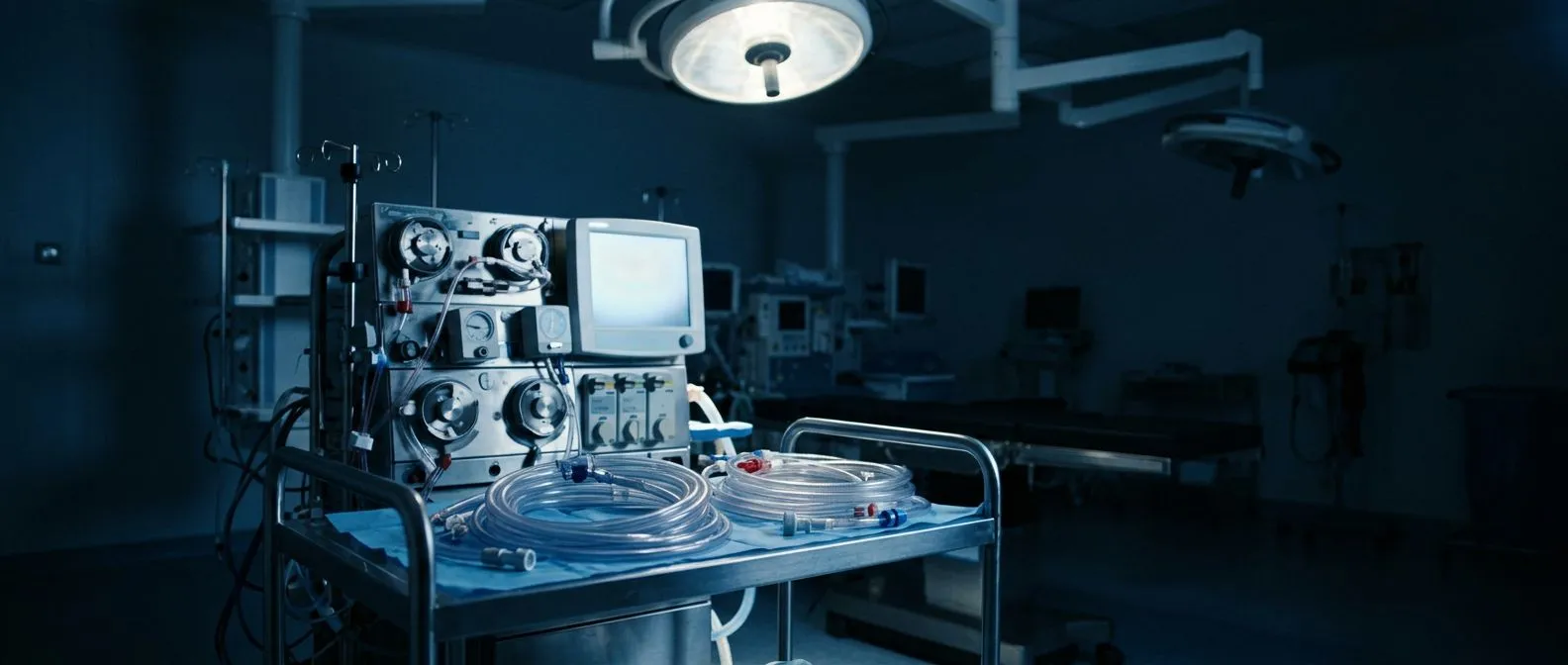
This profound state of hypothermia dramatically reduces the metabolic rate of the body's tissues, allowing them to survive without a continuous supply of oxygenated blood for a limited, carefully monitored period. This approach is typically reserved for the most challenging cardiac conditions, such as complex arch aneurysms or acute aortic dissections, where traditional surgical clamps cannot be used safely. By lowering the body's temperature, the brain's oxygen demand drops to a fraction of its normal requirement, buying the surgeon precious time to perform precise, life-saving repairs. The process relies entirely on the use of a cardiopulmonary bypass machine, which mechanically takes over the function of the heart and lungs during the cooling and warming phases.
Understanding this complex procedure can be overwhelming for patients and families, but the underlying principle is essentially a natural protective mechanism. Just as cold water preserves living tissue, cooling the body preserves brain and organ function while the surgical team addresses the most critical structures in the chest. The technique represents a remarkable testament to modern medicine's ability to safely push the boundaries of human physiology to repair life-threatening damage to the cardiovascular system.
How Does Circulatory Arrest Work?
The intricate process begins once the patient is under general anesthesia and completely unresponsive. The surgical team inserts an arterial cannula, a specialized tube, into a major blood vessel, connecting the patient to a cardiopulmonary bypass machine, commonly known as a heart-lung machine. This machine pumps blood out of the body, removes carbon dioxide, adds oxygen, and precisely regulates the temperature of the blood before returning it to the patient. To initiate cooling, the machine runs a built-in heat exchanger that progressively lowers the blood temperature, cooling the body efficiently from the inside out rather than relying on external ice packs alone.
As the core temperature steadily drops, the heart eventually stops beating on its own, a process often assisted by medical solutions that protect the heart muscle. According to Verywell Health, this profound cooling slows the electrical activity and metabolic processes of all cells in the body. Once the target temperature is reached and sophisticated monitoring confirms that the brain's electrical activity is sufficiently suppressed, the bypass machine is temporarily turned off. At this crucial point, there is no blood actively circulating through the body, creating a completely bloodless and still surgical field.
With the circulation legally arrested, the surgeon can safely open the diseased aorta and meticulously repair it, whether by excising an aneurysm, sewing in a synthetic graft, or carefully reattaching crucial branch arteries that supply the head and neck. The complete absence of blood flow and the absolute stillness of the tissues allow for extremely precise suturing, which would be virtually impossible if blood were actively pulsing through the fragile vessels. Once the structural repair is complete, the bypass machine is carefully restarted, and the patient is slowly warmed back to a normal physiological body temperature.
Why Cooling the Brain Protects the Body
The brain is the most sensitive organ in the human body when it comes to oxygen deprivation. Under normal physiological conditions, brain cells begin to die within four to six minutes without oxygen. Deep hypothermia fundamentally alters this strict timeline by reducing the body's overall metabolic rate. For every 7 degrees Celsius drop in body temperature, the metabolic rate of the brain decreases by approximately 50 percent. By cooling the patient to 18 degrees Celsius, the brain's oxygen requirement drops so remarkably low that it can tolerate the complete cessation of blood flow for up to 30 to 45 minutes, depending on the specific circumstances and baseline patient health.
Brain protection strategies: During this critical time, the surgical team continuously monitors the brain's electrical activity using electroencephalography (EEG). The ultimate goal is to achieve a flatline EEG, indicating that the brain has reached a deep state of metabolic suppression where it is maximally protected from ischemic injury. In addition to whole-body cooling, surgeons may use selective cerebral perfusion, where cold blood is directed specifically to the brain through the arteries of the neck, providing an extra layer of safety and extending the available surgical time for highly complex repairs.
The success of the entire procedure relies heavily on this delicate physiological balance. The surgical team must execute the physical repair efficiently while strictly respecting the physiological limits of the cooling process. By understanding these protective mechanisms, patients can better appreciate why this surgery takes significantly longer than standard cardiac operations and why the meticulous preparation phase is so critical to the final outcome.
When Do Surgeons Need Circulatory Arrest?
Surgeons typically employ deep hypothermic circulatory arrest when operating on the aortic arch, the curved top portion of the aorta that gives rise to the vital arteries supplying the brain and arms. Conditions that specifically affect this area include aortic arch aneurysms, where the vessel wall weakens and balloons outward, and acute aortic dissections, where the inner layer of the aorta tears, allowing blood to split the vessel wall. These conditions are immediately life-threatening and require urgent intervention to prevent fatal rupture, stroke, or severely compromised blood flow to vital organs.
Aortic dissection urgency: In cases of acute dissection, the sudden tear in the aorta can disrupt blood flow to the brain, kidneys, or legs, leading to rapid organ failure. Surgeons use circulatory arrest to safely open the arch, meticulously inspect the full extent of the tear, and repair the damage without the extreme risk of massive bleeding or air entering the bloodstream and traveling to the brain. The completely bloodless field is absolutely essential for visualizing the fragile intimal flap and ensuring that the repaired aorta will hold securely under high pressure.
Another critical scenario involves complex re-operations, where dense scar tissue from previous surgeries makes standard surgical approaches incredibly dangerous. The lack of bleeding provided by circulatory arrest allows the surgeon to carefully dissect through tough scar tissue without risking catastrophic injury to the heart, lungs, or other major blood vessels. In these incredibly high-stakes situations, the profound cooling provides a necessary safety net for both the patient's survival and the surgical team's ability to perform the required work.
Types of Aortic Aneurysms Requiring Body Cooling
Aneurysms are permanent, localized enlargements of an artery caused by a progressive weakening of the vessel wall. When they occur in the aortic arch, they are often completely asymptomatic until they become very large or begin to dangerously leak. Patients may eventually experience noticeable symptoms like chest pain, persistent back pain, or arm pain if the expanding aneurysm presses on nearby nerves or anatomical structures. The decision to operate is typically based on the absolute size of the aneurysm, with established guideline thresholds often suggesting surgery when the aortic diameter exceeds 5.5 centimeters, or earlier if the patient has a genetic connective tissue condition like Marfan syndrome.
Managing complex arch aneurysms: The presence of an atheromatous plaque—a dangerous buildup of cholesterol and other cellular substances in the artery wall—can severely complicate these aneurysms. The plaque can become fragile and easily break off during surgical manipulation, causing a devastating stroke if it travels to the brain. Deep hypothermic circulatory arrest allows surgeons to open the aorta and carefully remove these dangerous plaques before they can dislodge. For more detailed clinical insights into aortic conditions like intramural haematoma, which can closely mimic an aneurysm and requires careful coordination, research published in the Journal of Cardiothoracic Surgery highlights the critical decision-making process between cardiologists and cardiac surgeons.
The surgical repair involves completely replacing the diseased segment of the aorta with a durable synthetic tube, most often made of a woven polyester fabric. If the aneurysm involves the critical arteries that branch off to supply the head and neck, the surgeon may use a specially designed branched graft or meticulously reattach the native blood vessels to the new graft. This intricate, high-precision work demands the still, bloodless environment that circulatory arrest provides, ensuring the surgeon can create secure, leak-proof connections that will function reliably for the rest of the patient's life.
Risks of Deep Hypothermic Circulatory Arrest
While deep hypothermic circulatory arrest is a remarkable and life-saving technique, it inherently carries significant risks that patients must fully understand before consenting to surgery. The profound cooling and subsequent rapid warming process can stress the body's individual cells and delicate blood clotting systems. Patients may experience temporary confusion, delirium, or memory loss after waking up from anesthesia, a condition medically known as postoperative cognitive dysfunction, which typically resolves over several weeks. More serious neurological risks include stroke, which can occur if microscopic particles of calcified plaque or trapped air enter the bloodstream during the procedure.
Bleeding and clotting concerns: The prolonged use of the heart-lung machine and the extreme temperature changes can severely affect how blood naturally clots, leading to a higher risk of bleeding during and immediately after surgery. To combat this, the surgical team utilizes autologous blood—blood that is safely collected from the patient before bypass and returned to them—to minimize the need for donor transfusions and reduce associated infectious risks. However, despite these measures, patients may still require additional blood products, and the surgical team prepares extensively for this possibility, having screened blood readily available in the operating room at all times.
- Neurological risks: Stroke or temporary cognitive changes due to the delicate nature of brain blood flow and the temporary cessation of circulation.
- Bleeding: Increased risk of postoperative bleeding requiring blood transfusions due to temperature-related changes in the body's clotting cascade.
- Organ strain: Prolonged heart-lung bypass time can temporarily affect kidney and lung function, requiring temporary dialysis or respiratory support in rare cases.
Other potential complications include acute kidney injury, lung inflammation, and hospital-acquired infection. The extensive length of the surgery and the necessary use of prolonged heart-lung bypass can temporarily strain these vital organs. Patients with pre-existing conditions, such as advanced age, chronic kidney disease, or severe lung disease, face a statistically higher risk of these complications. Surgeons carefully weigh these risks against the immediately life-threatening nature of the aortic disease to determine the absolute lowest risk approach for your specific situation.
What to Expect During Aortic Surgery
On the day of surgery, the patient arrives at the hospital early to complete final medical preparations. The anesthesiology team places multiple critical monitoring lines, including an arterial line to constantly and accurately measure blood pressure, and a central venous catheter to administer powerful medications and fluids. The patient is then given general anesthesia, and a specialized breathing tube is inserted to manage respiration throughout the lengthy procedure. The surgery itself can take anywhere from six to ten hours, heavily depending on the complexity of the structural repair and the unique anatomy of the individual patient.
Once the patient is safely connected to the heart-lung machine and cooled to the exact target temperature, the physical repair begins. The surgical team works meticulously, often utilizing advanced intraoperative imaging and specialized graft materials. In some complex cases, surgeons may employ modern hybrid techniques, combining traditional open surgery with minimally invasive endovascular approaches to repair the aorta from the inside out. For example, the use of a physician-modified endograft for symptomatic zone 2 penetrating ulcer of the aortic arch without bridging stenting of the left subclavian artery for vertebral preservation, as detailed in a recent study from ScienceDirect, demonstrates how custom-made stent grafts can treat complex arch pathology while preserving critical blood flow to the brain.
Throughout the entire operation, the highly specialized team—including the lead surgeon, anesthesiologist, perfusionist who runs the heart-lung machine, and surgical nurses—communicates constantly and clearly. They meticulously monitor the time spent in circulatory arrest down to the exact second, ensuring the brain's protective physiological limits are strictly observed. Once the physical repair is fully finished, the delicate rewarming phase begins, slowly and carefully bringing the patient's body temperature back to normal before attempting to safely restart the heart.
Recovery Timeline for Aortic Surgery
Recovery from deep hypothermic circulatory arrest aortic surgery is a marathon, not a sprint, requiring immense physical and mental patience. After the procedure is completed, the patient is immediately transferred to the intensive care unit (ICU) for close, continuous monitoring. They will remain on a mechanical ventilator to help them breathe for the first few hours or overnight, depending entirely on their neurological and respiratory progress. The medical team closely watches blood pressure, heart function, and neurological status, frequently checking for any signs of confusion or weakness that might indicate a postoperative complication. Some patients may even experience temporary atrial flutter, an irregular heartbeat that is managed with medication as the heart recovers from the stress of surgery.
Once the breathing tube is safely removed and the patient is hemodynamically stable, the focus shifts to actively regaining physical strength. Patients are highly encouraged to sit up, stand, and eventually walk with physical therapy assistance within the first few days. Pain management is carefully balanced to keep the patient comfortable enough to participate fully in physical therapy without over-sedating them. The profound cooling the body endured means the patient often feels deeply fatigued and weak, which is a completely normal part of the physiological recovery process that requires patience, adequate rest, and proper nutrition.
Most patients spend one to two full weeks in the hospital before being safely discharged home or to a specialized cardiac rehabilitation facility. Complete recovery, including returning to normal daily activities and potentially work, typically takes six to eight weeks, though some older patients or those with extensive procedures may need up to three months to feel fully recovered. Regular follow-up appointments with the cardiac surgery team are absolutely essential to monitor the healing process and ensure the repaired aorta remains structurally healthy over time.

Are You a Candidate for This Procedure?
Determining candidacy for deep hypothermic circulatory arrest aortic surgery is a highly individualized and complex process. Surgeons evaluate multiple critical factors, including the specific anatomical location of the aortic disease, the patient's biological age, and the presence of other serious medical conditions. A thorough preoperative workup involves advanced imaging, such as high-resolution CT scans or MRIs, to accurately map the aorta in three dimensions, as well as a complete functional assessment of the heart, lungs, and kidneys to ensure they can withstand the surgery.
Assessing surgical risk: The surgical team uses standardized scoring systems, such as EuroSCORE II, to mathematically estimate the potential risk of mortality and complications based on the patient's comprehensive health profile. If the calculated risks of the open surgery outweigh the natural risks of the underlying aortic disease, the team may explore alternative treatments, such as endovascular stenting or aggressive medical management. However, for complex arch aneurysms or severe dissections where endovascular options are anatomically limited, circulatory arrest often remains the safest and most effective path to a definitive long-term cure.
Patients facing this major surgery should have an open, honest, and detailed discussion with their cardiac surgeon about exactly what to expect. It is crucial to understand not only the technical aspects of the procedure but also the realistic expectations for recovery time and quality of life afterward. Making an informed medical decision requires carefully weighing the significant surgical risks against the potentially life-saving benefits of repairing a severely diseased aorta before it ruptures.

Long-Term Outcomes and Follow-Up Care
The long-term clinical outcomes for patients who successfully undergo this complex surgery are generally excellent, provided they strictly adhere to their medical regimen and maintain regular, lifelong follow-up care. The synthetic grafts used to replace the diseased aorta are incredibly durable and rarely fail, meaning the specific repaired section of the vessel is essentially permanently cured of the aneurysm or dissection. However, the rest of the natural aorta remains at risk over time, especially in patients with underlying genetic connective tissue disorders or widespread atherosclerosis.
- Imaging surveillance: Annual CT or MRI scans are absolutely critical to monitor the entire length of the aorta for new aneurysms or gradual enlargement over time.
- Medication adherence: Strict blood pressure control is essential to reduce physical stress on the remaining aortic wall; patients typically take medications like beta-blockers for life, and sometimes Aspirin to prevent clotting.
- Lifestyle adjustments: Avoiding heavy lifting and adopting a strict heart-healthy diet support long-term vascular health and protect the surgical repair from excessive internal pressure.
Living well after deep hypothermic circulatory arrest aortic surgery requires a dedicated partnership between the patient and their medical team. Routine visits to the cardiologist and cardiac surgeon ensure that any new vascular issues are caught early and managed effectively before they become dangerous. If you or a loved one are facing an aortic condition that may require this advanced surgical technique, we highly encourage you to discuss your specific situation with a qualified cardiac team and book a consultation to explore your options rather than self-diagnosing.






