

Descending Aorta Aneurysm: Monitoring and Repair
Descending aorta aneurysm monitoring and repair options explained by Guadalajara heart specialists.
Receiving a diagnosis of a descending aorta aneurysm can feel overwhelming, especially when you are navigating healthcare systems from another country. The aorta is the main pipeline carrying oxygen-rich blood from your heart to the rest of your body. When a section of this vessel in the chest or abdomen weakens and expands like a balloon, it requires careful monitoring and, sometimes, surgical repair. Our goal at Cirugía de Corazón is to bridge complex cardiology with clear, calm explanations so you can make informed decisions about your care alongside Dr Alejandro Molina.
What is a descending aorta aneurysm and how does it form?
Anatomy of the aortic arch and descending segment: The aorta curves out of the heart, forming the ascending aorta and the arch, before traveling downward through the chest and abdomen. The descending aorta aneurysm specifically occurs in this downward section, after the blood has passed the major vessels supplying the head and arms. Over time, the muscular and elastic walls of this vessel can degrade. This degeneration is often driven by chronic high blood pressure, smoking, or genetic conditions that affect connective tissue.
The role of atherosclerosis: In many patients, the underlying mechanism is the buildup of an atheromatous plaque—a deposit of cholesterol and cellular debris—within the artery wall. This plaque causes chronic inflammation, weakening the tissue and causing it to stretch. As the vessel wall thins, the diameter of the aorta expands. If the enlargement reaches a dangerous threshold, the risk of tearing (dissection) or rupturing increases significantly, making proactive management essential.
Distinguishing from other cardiac conditions: It is important to differentiate a structural aneurysm from other heart issues. For example, while an arrhythmia like atrial flutter affects the electrical rhythm of the upper chambers, an aneurysm is a physical enlargement of a blood vessel. General resources like MedlinePlus provide helpful overviews, but your specific anatomy requires evaluation by a specialized cardiac team to determine the exact size and growth rate of the aneurysm.
Descending aorta aneurysm symptoms and warning signs
The challenge of silent enlargement: One of the most difficult aspects of a descending aorta aneurysm is that it often produces no symptoms until it becomes critically large or begins to tear. Many aneurysms are discovered incidentally during a routine chest X-ray or an ultrasound performed for an entirely different reason. This silent progression is why screening is vital for patients with risk factors like a family history of aortic disease or long-standing hypertension.
When the aneurysm makes itself known: If the expanding vessel begins to press against surrounding structures, you might experience localized discomfort. Patients sometimes report a deep, constant pain in the chest, back, or abdomen. In some cases, the pain may radiate, causing distinct arm pain if the enlargement compresses nearby nerves. This discomfort should never be ignored, as it can indicate that the vessel wall is under significant physical stress.
Signs of a tear or rupture: A sudden, severe, tearing pain in the chest or back is the classic sign of an aortic dissection, a life-threatening emergency. Other symptoms might include a sudden drop in blood pressure, difficulty breathing, or signs of decreased blood flow to the lower body. If you experience these acute symptoms, immediate emergency medical attention is required, as the vessel wall has failed and blood is escaping its normal pathway.
How is a descending aorta aneurysm diagnosed?
Initial clinical evaluation: Diagnosis begins with a thorough physical examination. During this exam, your physician will listen to your heart and blood vessels using auscultation, a method of listening to internal body sounds. While a stethoscope cannot measure the aneurysm, it can reveal murmurs or abnormal blood flow sounds that prompt further investigation. Your doctor will also review your blood pressure history and lipid profiles, looking for markers like elevated apolipoprotein B, which indicates higher cardiovascular risk.
Advanced imaging techniques: To confirm the presence and measure the exact dimensions of a descending aorta aneurysm, imaging is mandatory. A computed tomography (CT) angiogram is the gold standard, providing highly detailed cross-sectional images of the aorta. This scan allows the surgical team to map the aneurysm’s location, its length, and its relationship to branching arteries, which is critical for planning any potential intervention.
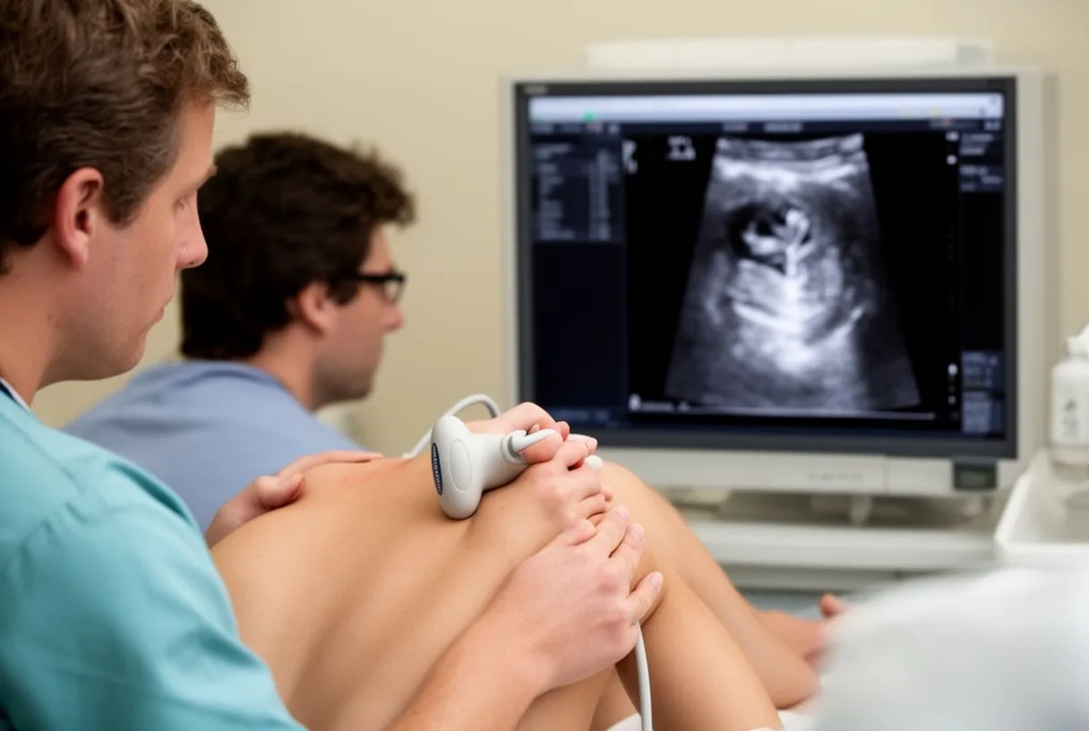
Incidental findings during imaging: Because CT scans and echocardiograms provide detailed views of the entire chest, they occasionally reveal other unrelated cardiac anomalies that must be addressed before surgery. For instance, preoperative imaging might detect a cardiac tumor, such as a papillary fibroelastoma. Evaluating these incidental masses is crucial, as detailed in studies like Left Atrium Papillary Fibroelastomas: A Cause of Cerebral Emboli, ensuring the surgical plan accounts for all potential risks to the patient.
Descending aortic aneurysm size criteria for surgery
Understanding the threshold for intervention: Not every enlarged aorta requires immediate surgery. The decision to operate is heavily based on descending aortic aneurysm size criteria established by major cardiovascular guidelines. For the descending thoracic aorta, the threshold for surgical intervention is generally set at an external diameter of 5.5 centimeters or greater. This measurement is chosen because the risk of rupture increases significantly once the vessel reaches this size.
- Diameter: Surgery is typically recommended when the descending aorta reaches 5.5 cm or larger in patients without connective tissue disorders.
- Growth rate: Rapid expansion, often defined as growing more than 0.5 cm in a six-month period, is a strong indicator for repair even if the absolute diameter is below the standard threshold.
- Symptom onset: The development of new pain, vocal cord paralysis, or other pressure symptoms warrants surgical evaluation regardless of the exact measurement.
Personalized risk assessment: Size criteria are guidelines, not absolute rules. A patient’s overall health, age, and surgical risk profile (often calculated using tools like EuroSCORE II) heavily influence the timing of surgery. For a smaller, frail patient, the risk of surgery might outweigh the risk of the aneurysm at a slightly smaller diameter, whereas a younger, healthier patient with a strong family history of rupture might be advised to intervene earlier.
Aortic aneurysm watchful waiting and surveillance guidelines
What active surveillance looks like: If your aneurysm is below the surgical threshold, aortic aneurysm watchful waiting is the standard approach. This does not mean ignoring the condition. It involves a strict schedule of regular imaging—usually a CT scan or MRI every six to twelve months—to meticulously track the aneurysm’s growth rate. This monitoring ensures that if the vessel begins to expand rapidly, the surgical team can intervene before a rupture occurs.

Medical management during waiting: While you wait, medical therapy is essential to reduce the stress on the aortic wall. This primarily involves strict blood pressure control, often using medications like beta-blockers. Patients are typically advised to take daily aspirin to mitigate cardiovascular risks, though your specific prescription will depend on your overall cardiac health. The goal is to keep the blood pressure in a safe, controlled range to prevent the aneurysm from stretching further.
Lifestyle modifications: Watchful waiting also requires lifestyle adjustments. Patients must stop smoking entirely, as tobacco use drastically accelerates aortic degeneration. Heavy lifting and strenuous activities that cause sudden spikes in blood pressure should be avoided. Your cardiac team will help you understand which exercises are safe and which activities might put undue stress on the weakened vessel wall during the surveillance period.
Open descending aorta surgery vs endovascular repair
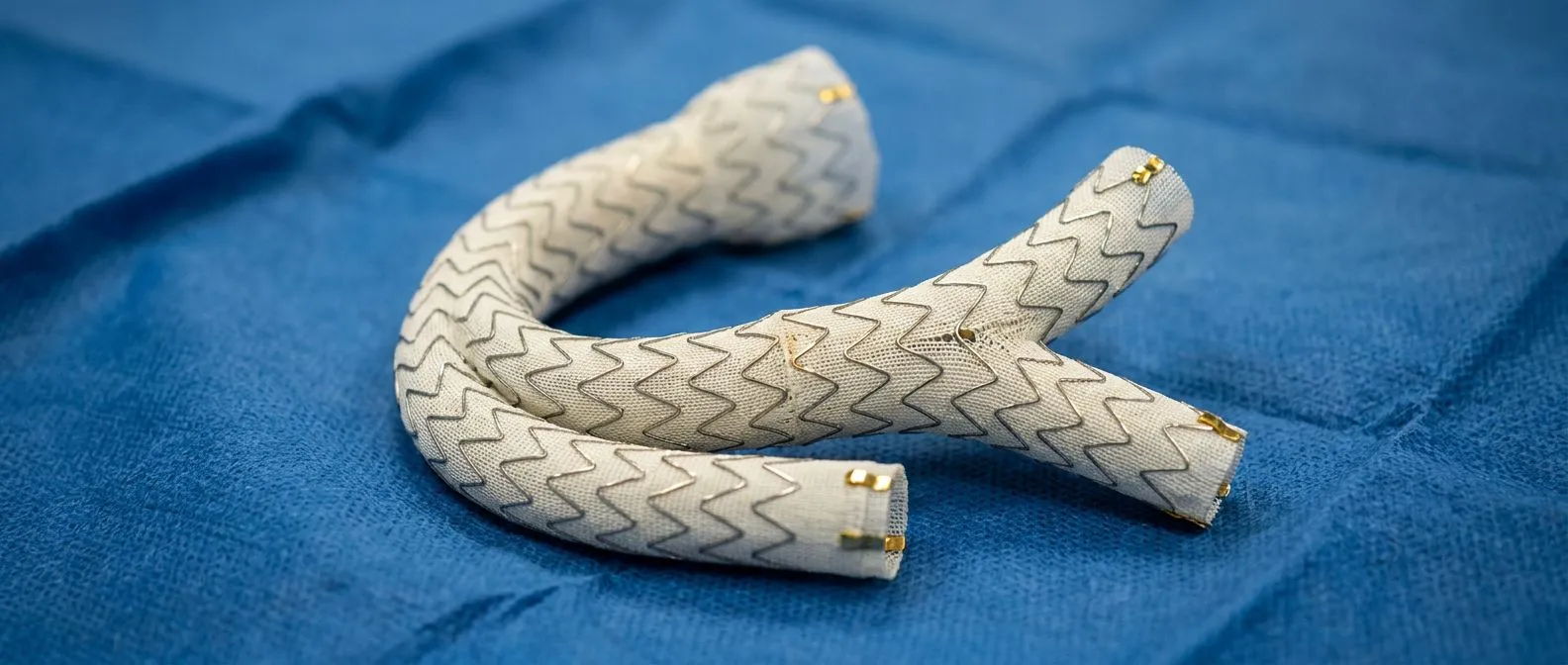
The traditional open approach: Open descending aorta surgery is a major operation where the surgeon makes an incision in the chest or side to directly access the aorta. The weakened section is removed and replaced with a synthetic arterial graft. This approach provides a highly durable repair and is often necessary if the aneurysm has a complex shape or involves branches that supply blood to the spinal cord or abdominal organs.
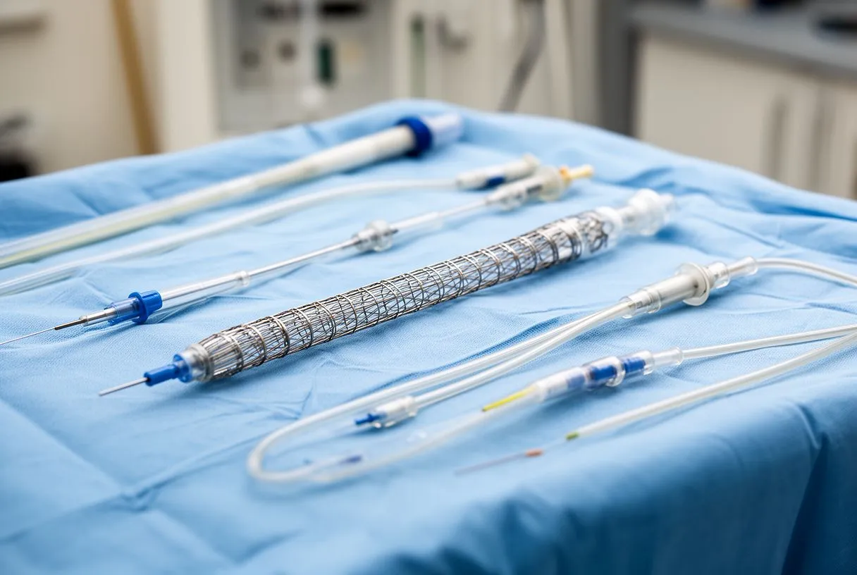
The minimally invasive alternative: Endovascular descending aorta repair, known as TEVAR (Thoracic Endovascular Aortic Repair), is a less invasive option. Instead of a large chest incision, the surgeon makes small incisions in the groin to access the femoral arteries. A stent graft is guided up into the descending aorta using X-ray imaging and deployed inside the vessel, creating a new, strong inner channel for blood to flow, effectively bypassing the aneurysm.
Weighing the trade-offs: The choice between open surgery and endovascular repair depends on the patient's anatomy and overall health. TEVAR offers a faster initial recovery and is often preferred for older patients or those with significant comorbidities who might not tolerate open surgery well. However, TEVAR requires specific anatomical features, like adequate "landing zones" for the stent, and may require future monitoring for stent leaks or migration.
What to expect during endovascular descending aorta repair
Preoperative preparation: Before endovascular descending aorta repair, you will undergo a detailed CT scan to map the exact dimensions of your aorta. If the preoperative workup includes an echocardiogram, it might reveal other conditions, such as aortic valve tumors, as noted in case reports like Asymptomatic papillary fibroelastoma of the Aortic valve in a young woman - a case report. Your anesthesiologist will review whether the procedure will be done under general or regional anesthesia, depending on the surgical plan.
The stent deployment process: During the procedure, you will have an arterial line placed to monitor your blood pressure continuously. The surgical team advances a catheter through the blood vessels to the descending aorta. Once the stent graft is correctly positioned over the aneurysm, it is expanded, sealing off the weakened aortic wall from the high-pressure blood flow. In some cases, techniques similar to balloon angioplasty are used to ensure the stent graft fits snugly against the aortic walls.
Immediate post-operative care: Because TEVAR avoids a large chest incision, the immediate recovery is much smoother. Patients are typically monitored in the intensive care unit for a short time to ensure the stent is functioning correctly and that blood flow to the lower body is stable. Pain is usually minimal and managed with standard medications, allowing patients to begin walking and eating within a day or two of the procedure.
Recovery timeline after descending aorta aneurysm repair
Immediate hospital stay: The length of your hospital stay depends on the type of repair. For endovascular repair, patients are often discharged within two to four days. Open descending aorta surgery requires a longer stay, typically a week or more, including time in the intensive care unit where the medical team monitors your heart rate, spinal cord function, and organ perfusion closely. In complex cases, techniques like awake ECMO might be discussed for respiratory support, though this is rare for straightforward repairs.
- Weeks 1-2: Focus on wound healing and managing post-operative discomfort. Walking is encouraged, but heavy lifting is strictly prohibited.
- Weeks 3-6: Gradual return to light daily activities. Many patients undergoing endovascular repair return to desk work during this period.
- Months 2-3: Full recovery from open surgery is usually achieved, allowing a return to normal exercise routines, though contact sports should still be avoided.
Long-term follow-up: Regardless of the surgical approach, descending aorta aneurysm repair requires lifelong follow-up. You will need periodic CT scans or MRIs to ensure the repair remains intact. If you received a stent graft, the medical team will check for "endoleaks"—a condition where blood continues to seep into the aneurysm sac—and monitor the stent's position. If you required other cardiac interventions, like TAVR (transcatheter aortic valve replacement) for a coexisting valve issue, your follow-up will also include echocardiograms to assess valve function.
Long-term prognosis and lifestyle adjustments
Life expectancy and outcomes: The descending aorta aneurysm prognosis is generally very positive following successful repair and consistent medical management. Once the aneurysm is excluded from the bloodstream, the risk of rupture is drastically reduced. Patients can often return to a full, active life. The key to long-term survival is preventing future aneurysms in other parts of the aorta, which requires strict blood pressure control and regular imaging.
Cardiovascular risk management: Because aortic disease is often linked to systemic vascular issues, managing your overall cardiovascular risk is paramount. Your physician will monitor your cholesterol and may prescribe medications to prevent the formation of new atheromatous plaque. If you had an open repair, the use of your own blood for transfusions (autologous blood) might have been utilized to minimize risks, but ongoing blood health is still monitored. Maintaining a heart-healthy diet and regular, moderate exercise are crucial components of your long-term strategy.

Emotional and psychological recovery: Recovering from a major cardiac event or surgery involves more than just physical healing. Many patients experience anxiety about their heart health or fear of the aneurysm returning. It is entirely normal to feel this way. We encourage patients to discuss these feelings with their healthcare team, as psychological well-being is a critical part of returning to a high quality of life. Support groups or counseling can be highly beneficial during this transition.
Preparing for your consultation in Mexico
Gathering your medical records: For our international patients traveling to Guadalajara, Querétaro, or Hermosillo, organizing your medical history beforehand streamlines your consultation. Please bring copies of any CT scans, MRIs, or echocardiograms performed in your home country, along with a list of your current medications. Having this information available allows Dr Alejandro Molina to provide a precise assessment of your aneurysm without repeating unnecessary tests.
Understanding logistics and costs: We understand that traveling for medical care involves significant planning.
Taking the next step: An aneurysm diagnosis is a call to action, but it is not a journey you have to take alone. We encourage you to discuss your specific situation with a qualified cardiac team to understand your options fully. Rather than self-diagnosing or letting anxiety dictate your choices, book a consultation with us. Together, we can review your imaging, discuss whether watchful waiting or surgical repair is the safest path for your anatomy, and create a personalized plan for your heart health.






