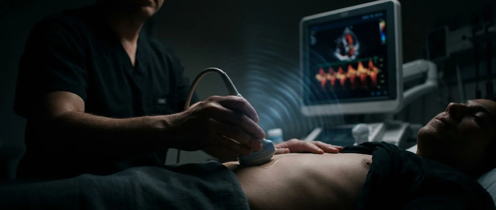
Doppler Echocardiography: Measuring Blood Flow in the Heart
Doppler echocardiography blood flow testing measures how blood moves through heart valves and chambers.
If your cardiologist has mentioned a Doppler echocardiography blood flow study, you may be wondering what it actually measures and why it matters. In simple terms, a Doppler echocardiogram is an ultrasound scan of the heart that does not just show the heart's structure — it measures the speed and direction of blood moving through your chambers and valves. That motion information is what turns a still picture into a working diagnosis, and it is often the study that decides whether a leaky valve needs watching, medication, or surgery. This guide walks you through what the test is, what it detects, how to prepare, what results mean, and what to expect in terms of comfort, time and cost.
What Is Doppler Echocardiography?
The basic principle. Doppler echocardiography applies the Doppler effect — the same physics that makes a passing ambulance siren change pitch — to ultrasound waves bounced off moving red blood cells. When blood flows toward the probe, the reflected sound waves come back at a higher frequency; when it flows away, the frequency drops. A computer converts those shifts into precise velocity measurements, usually expressed in metres per second.
Why velocity matters. Blood speed tells a cardiologist about pressure. When blood squeezes through a narrowed (stenotic) valve, it accelerates, much like water through a pinched hose. The faster the jet, the higher the pressure gradient across the valve, and from that the team can calculate how severe the narrowing is — without ever putting a catheter inside your heart.
What it looks like on screen. Most studies combine 2D greyscale imaging — the moving anatomical picture of your heart — with Doppler displays shown as coloured overlays or velocity waveforms. Together they answer two questions at once: what does the heart look like, and how is blood actually moving through it?
What Is Doppler Echocardiography Used For?
Valve disease. This is the classic indication. Doppler quantifies how narrowed a stenotic valve is and how much a leaking (regurgitant) valve lets blood flow backwards. It also estimates pressures inside the heart, including pulmonary artery pressure, which matters greatly in planning treatment.
Investigating symptoms and murmurs. Doctors frequently order a Doppler echo to explain a heart murmur heard with a stethoscope, shortness of breath, chest discomfort, palpitations, or unexplained dizziness. Many murmurs turn out to be harmless — Doppler is how that distinction gets made.
Congenital heart disease and monitoring. In our practice, which covers valve disease, coronary disease, the aorta and structural heart conditions, nearly every surgical decision begins with a Doppler echocardiogram. It is equally central in paediatric cardiology, where it maps blood flow in children born with heart defects and tracks how repairs are holding up over the years.
Echo vs Doppler: Are They the Same Test?
The short answer. An echocardiogram is the overall ultrasound study of the heart; Doppler is one of the tools built into it. So "echo and Doppler" are not two separate appointments — a complete modern echocardiogram almost always includes Doppler measurements by default.
What "2D echo with Doppler" means. When a report says "2D echocardiogram with Doppler study," it means the sonographer captured the standard moving images of your heart's chambers and valves, then added Doppler measurements of blood flow velocities and pressure gradients. The two parts are interpreted together: the 2D images show structure, the Doppler shows function.
When only one part is enough. Occasionally a quick screening echo may be limited to 2D imaging, and occasionally a focused Doppler study is repeated just to track one valve. But for a new patient or a pre-surgical assessment, expect the full combined study.
Putting it in context at our practice. At our consulting rooms in Guadalajara, Querétaro and Hermosillo, we take the time in consultation to explain in plain words what the echo images and Doppler numbers actually show, and to answer every question before any decision is made.
What Does Doppler Show in the Heart?
Blood flow direction. The colour display paints flow moving toward the probe in one colour and flow moving away in another. A jet of colour spraying backwards through a closed valve instantly reveals regurgitation that a still image could miss.
Flow velocity and pressure gradients. Continuous wave and pulsed wave Doppler trace the speed of blood across valves. From the peak velocity across the aortic valve, for example, the machine calculates the valve's opening area and the pressure drop across it — numbers that directly drive treatment decisions such as when a patient becomes a candidate for TAVR or surgical valve replacement.
Chamber pressures and heart function. Doppler also estimates pressure in the lung arteries, detects diastolic dysfunction (a stiffening of the heart's filling phase), and — combined with 2D measurements — contributes to calculating the ejection fraction, the percentage of blood the heart pumps out with each beat. Cardiac ultrasound technology has advanced to the point where this non-invasive study guides most heart valve decisions; an overview in the medical literature describes it as a cornerstone of contemporary cardiology practice (Frontiers in Cardiovascular Medicine).
Types of Doppler Echocardiography
Pulsed wave Doppler. This mode measures blood velocity at one specific, small location — a chosen point inside the heart. It is precise about where the measurement comes from, though it cannot record very high velocities. It is used for normal flows, such as blood entering the left ventricle.
Continuous wave Doppler. Here the probe sends and receives sound continuously along an entire line, sacrificing location precision to capture very fast jets — exactly what is needed across a severely narrowed or leaking valve. The two modes complement each other.
Other variants worth knowing:
- Colour Doppler: paints flow direction and relative speed across a whole region, ideal for spotting leaky valves at a glance.
- Tissue Doppler: measures the motion of the heart muscle itself rather than blood, helping assess filling pressures and coordination of contraction.
- Transesophageal Doppler echo (TEE): a probe swallowed into the oesophagus gives far clearer images, especially of valves at the back of the heart, infections, and the aorta — a systematic review notes it detects acute aortic dissection with high accuracy where transthoracic images are limited (Frontiers in Cardiovascular Medicine).
Stress Doppler echocardiography. This study takes images and Doppler measurements before and immediately after exercise or a medication that makes the heart work harder. It reveals valve problems or blocked coronary arteries that only appear under load, and it is often the missing piece when a resting echo looks normal but symptoms persist.
How to Prepare for a Doppler Echocardiography Test
For a standard transthoracic study. Preparation is genuinely simple: eat and take your medications as usual, wear a top that is easy to remove from the waist up, and avoid body lotion on the chest, which can interfere with the ultrasound gel and image quality. There is no fasting, no dye and no needle.
If a transesophageal study is planned. Because the throat is numbed and a sedative is given, you will fast for several hours beforehand, arrange a driver home, and discuss any swallowing problems or loose teeth with the team in advance.
If a stress study is planned. Wear comfortable shoes and clothing you can exercise in, and ask beforehand whether you should hold any beta-blockers or other medications, since some drugs blunt the heart rate response the test needs. When patients come to us, we tell them exactly which studies are still needed so no unnecessary ones are ordered — often the Doppler echo they already have is enough to start the conversation.
What Happens During the Test and How Long It Takes
The procedure itself. You lie on your left side while a sonographer presses a small handheld probe (transducer) against your chest, moving it to different positions to capture the heart from several angles. Cool gel is applied to help the sound waves travel. You may be asked to breathe slowly or hold your breath briefly for a few measurements.
How long does an echo Doppler test take? A complete transthoracic echocardiogram with full Doppler typically takes 30 to 60 minutes. A transesophageal study takes about the same plus recovery time from sedation; a stress study adds the exercise or infusion period. It is an outpatient test — you go home the same day.
Comfort and what you feel. Aside from the firm pressure of the probe and the cool gel, you feel nothing. There is no radiation, no injection and no discomfort during a standard study. Our team reviews every echocardiogram together against international practice guidelines before recommending anything, so the numbers on the report are never read in isolation.
Understanding Abnormal Doppler Echocardiography Results
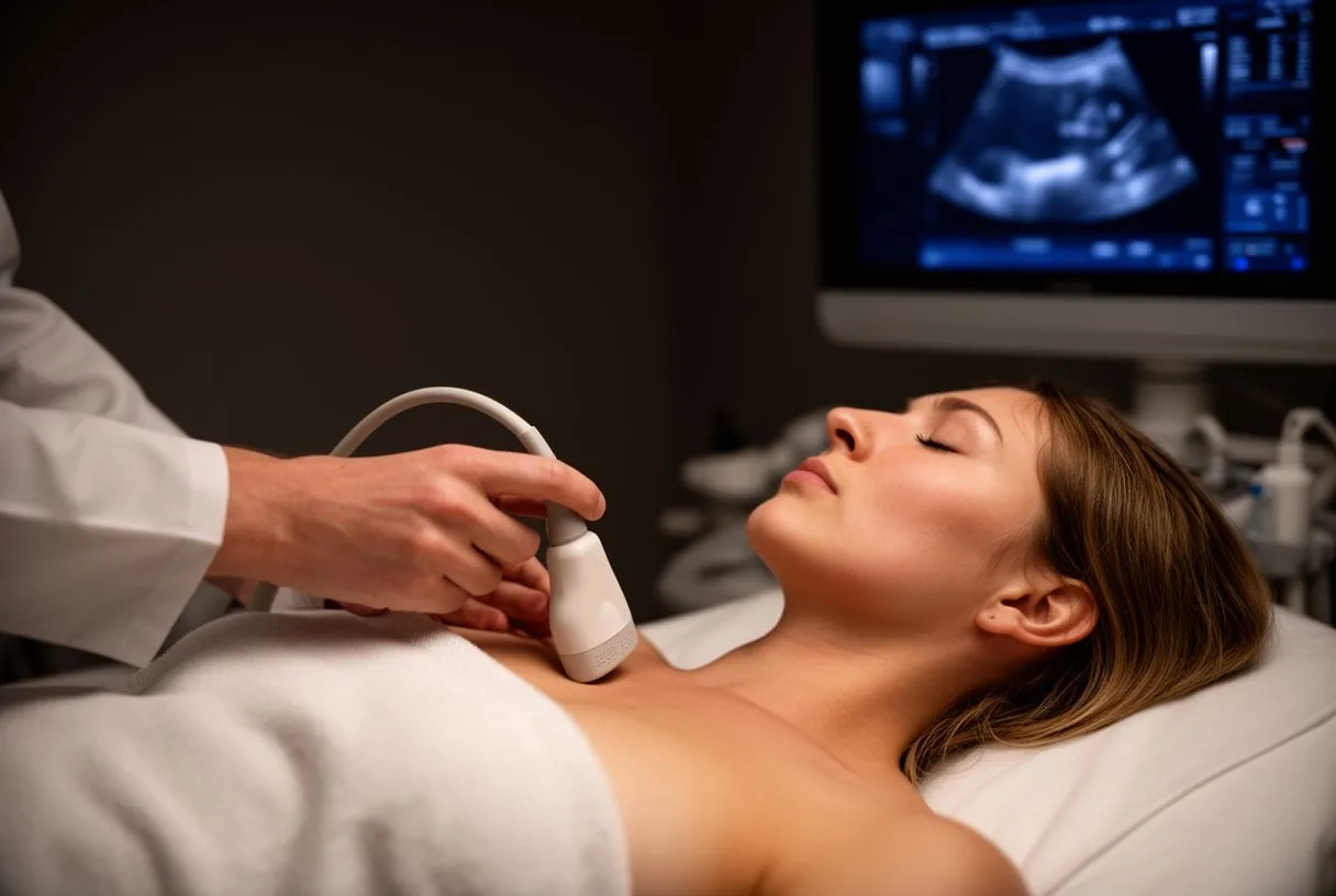
What the report contains. A Doppler echo report lists chamber sizes, valve areas, pressure gradients, regurgitation grades (usually trivial, mild, moderate or severe), estimated lung artery pressures and the ejection fraction. Each number has a normal range, and severity is graded against international thresholds.
Common abnormal findings. These include aortic stenosis (a narrowed aortic valve), mitral regurgitation (a leaking mitral valve), pulmonary hypertension (raised pressure in the lung arteries), and reduced pumping function after a heart attack. An abnormal result is a starting point for a conversation, not a verdict: mild findings often only need periodic monitoring.
What happens next. Severity decides the pathway. Mild valve disease is usually watched with repeat echoes every few years; severe, symptomatic disease typically leads to a discussion of valve repair, surgical replacement, or a transcatheter option such as TAVR for suitable patients with aortic stenosis. Major bodies such as the Mayo Clinic emphasise that treatment timing depends on symptoms and severity together, not on the scan alone.
Getting a second reading. We encourage second opinions and treat seeking one as a normal part of deciding about heart surgery — bringing your echo images and report to another qualified team is always reasonable, and we are glad to review studies done elsewhere.
Are There Downsides or Risks to a Doppler Echo?
The standard study is very low risk. A transthoracic Doppler echocardiogram uses sound waves, not radiation, and has no known harmful effects. It can be repeated as often as needed to track a condition over time — a major advantage over scans that carry radiation exposure.
Practical limitations. The main downside is image quality. In patients with obesity, lung disease, or certain chest shapes, ribs and air in the lungs can block the sound waves, degrading the images. Contrast agents injected through a small vein can help, or a transesophageal study can bypass the problem entirely.
Risks of the specialised versions. A transesophageal echo involves sedation and a small risk of throat irritation or, rarely, oesophageal injury. A stress echo carries the usual risks of exercise in heart patients and is performed with monitoring. Each is ordered only when the extra information justifies it.
Cost considerations. The cost of a Doppler echocardiography study in Mexico varies widely by city, facility and whether it is part of a larger work-up. Many international patients combine the test with a surgical consultation, which can reduce total travel costs compared with paying for studies piecemeal.
What to ask before booking. Not all echo reports are equal — completeness matters, especially if surgery is on the table:
- Full Doppler panel: confirm the study will include continuous and colour Doppler across all valves, not just a limited screening view.
- Raw images available: a surgical team needs to review the actual stored images, not only a written report.
- Time to explain results: a report nobody walks you through is only half a test.

How we work with international patients. The first contact with our team is a free video consultation: we get to know the patient, talk through the condition, and say which studies are still needed so no unnecessary ones are ordered; a face-to-face visit at the consulting rooms follows when the patient decides to go ahead. Every case is then reviewed by our multidisciplinary team — led by Dr Alejandro Molina, trained in cardiothoracic surgery at the Universidad de Guadalajara and in cardiac, minimally invasive, robotic and structural surgery at the University of Montreal — before a recommendation is given, and the final decision is always the patient's.
Key Takeaways on Doppler Echocardiography
Doppler echocardiography is the study that turns pictures of your heart into measurements of how it actually works: how fast blood moves, how much pressure builds across a narrowed valve, and how badly a leaking valve lets blood escape backwards. It is painless, radiation-free, quick to perform, and — for most valve and structural heart conditions — the test that starts and steers every treatment decision, from watchful waiting to minimally invasive repair or TAVR. In our experience, patients who understand what their doppler echocardiography blood flow numbers mean make calmer, more confident decisions about their care.
If you have been told you have a murmur, a leaky or narrowed valve, or unexplained breathlessness, the sensible next step is not to wait and worry — it is to sit down with a qualified cardiac team and have the results explained to you in plain language. Book a consultation with Dr Alejandro Molina's team in Guadalajara, Querétaro or Hermosillo, bring any echo you already have, and let a specialist walk you through what it shows and what, if anything, needs to be done about it.