

Endocardium Infection and Inflammation: Endocarditis Basics
Endocardium endocarditis explained: causes, symptoms, diagnosis and treatment from heart specialists.
What Is Endocardium Endocarditis and Why It Matters
Endocardium endocarditis is an infection and inflammation of the endocardium, the thin, smooth layer of tissue that lines the inside of your heart's chambers and covers the heart valves. When bacteria or, far less often, fungi settle on that lining, they form clusters of debris called vegetations that cling to the valve surface. For many patients the first hint that something is wrong is a fever that simply will not settle, a new feeling of exhaustion, or a heart murmur their doctor hears for the first time. Understanding the endocardium endocarditis basics matters because the infection can damage valve tissue quickly and, left untreated, can send fragments of infected material through the bloodstream to other organs.
The endocardium itself is not a passive surface. It is the innermost layer of the heart wall, and where it folds over the valves it creates the hinge points that open and close with every beat. An infection that begins on a valve disrupts that hinge mechanism, so the valve may leak or fail to open fully. The result is the same mechanical problem a worn valve would cause: the heart must pump harder to move blood, and downstream chambers like the atrium can enlarge over time. Unlike wear-and-tear valve disease, however, endocarditis can progress over days to weeks rather than years, which is why cardiologists treat even a suspected case as urgent.
Who gets it and how common is it? Endocardium endocarditis is uncommon in the general population, but it is not rare among people with certain heart conditions, prosthetic valves, or indwelling hardware that gives bacteria a route into the blood. Internationally, the patients most often affected are those who already have a structurally abnormal valve, a replaced valve, or a long-term intravenous line. The reassuring point is that the condition is well understood, the diagnosis pathway is established, and treatment works when it is started promptly. For a calm, evidence-based overview of how major heart centres approach this condition, you can read Stanford Health Care.
Endocarditis Causes: How Infection Reaches the Heart
Bacterial endocarditis almost always begins with bacteria entering the bloodstream — a process doctors call bacteraemia — and then settling on a valve or area of damaged endocardium. The bacteria do not arrive because the heart is weak; they arrive because everyday events briefly push bacteria from the skin, mouth, gut, or urinary tract into the blood. In a healthy person the immune system clears that small bacterial load within minutes. In someone with an abnormal valve, or with a prosthetic surface that bacteria can stick to, a few organisms survive, multiply, and build a protective layer called a vegetation.
Common entry routes for bacteria Understanding where the bacteria come from helps patients make sense of prevention advice. The mouth is the most discussed source, because dental procedures that cause bleeding can push oral bacteria into the blood, but the same can happen with routine brushing or gum disease. Other recognised routes include skin infections, indwelling venous catheters, intravenous drug use, and procedures on the gut or urinary tract. After cardiac surgery, an arterial cannula or arterial line used for monitoring, or an arterial graft placed during bypass, can rarely become a source if sterile technique is breached.
Not every organism that reaches the blood can cause endocarditis. The bacteria most often responsible are streptococci from the mouth, staphylococci from the skin, and enterococci from the gut. A category called the HACEK group of fastidious bacteria accounts for a smaller share of cases. Fungi such as Candida can also cause endocarditis, usually in patients with long intravenous lines or severely weakened immunity, and fungal cases tend to produce larger, more fragile vegetations. Knowing the likely organism guides the choice of endocarditis antibiotics before lab results are back, which is why blood cultures are drawn before treatment starts.
Types of Endocardium Endocarditis: Acute vs Subacute
Clinicians describe endocardium endocarditis as either acute or subacute, based on how quickly it progresses. Acute endocarditis is caused by highly aggressive organisms — most often Staphylococcus aureus — and can destroy a normal valve within days, producing high fever, rapid deterioration, and early heart failure. Subacute endocarditis, sometimes called the classic "endocarditis lenta," develops over weeks to months with lower-grade fever, weight loss, and vague fatigue; the patient may feel unwell for a long time before the diagnosis is made. The distinction is not purely academic: acute disease demands same-day hospital admission and intravenous antibiotics, while subacute disease still needs urgent treatment but may allow a little more time for work-up.
Native valve versus prosthetic valve infection The other key split is whether the infected valve is the patient's own (native valve endocarditis) or a surgically placed replacement (prosthetic valve endocarditis). Native valve infections are more common and often involve previously damaged valves. Prosthetic valve endocarditis is rarer but more serious, because bacteria stick more easily to foreign material and the infection can loosen the sewing ring that holds the valve in place. Early prosthetic endocarditis, within the first year of surgery, may even be acquired in the operating room; late cases behave more like native valve disease.

The border between acute and subacute is not always sharp, and a subacute case can suddenly worsen if a vegetation breaks off and blocks a vessel. For that reason cardiologists do not rely on the label alone; they use the patient's overall stability, blood cultures, and echocardiogram findings to decide how aggressively to act. If you are being evaluated for a valve replacement, your team will explain that the long-term risk of infection on a prosthetic valve is low but lifelong, and that certain dental and medical procedures afterwards warrant preventive antibiotics in selected patients.
Endocarditis Symptoms: What Patients Actually Feel
Endocarditis symptoms are notoriously non-specific, which is part of why diagnosis can be delayed. Many patients describe a flu-like illness that lingers: low-grade fever, chills, sweating at night, loss of appetite, and a tiredness that does not improve with rest. Because these symptoms overlap with common viral illnesses, people often wait before seeking care, and the infection can advance during that window. When fever is combined with a known heart murmur, a prosthetic valve, or recent dental or medical procedures, it should never be dismissed as "just a virus."
Symptoms tied to valve damage As the infected valve begins to leak or stiffen, patients may notice new shortness of breath, especially on exertion or when lying flat, and a sensation of rapid or irregular heartbeat. Some develop swelling in the legs or abdomen as the heart struggles to keep up. These are signs that the infection has begun to affect heart function, and they move the situation from "needs investigation" to "needs the same-day attention of a cardiac team."
Some symptoms come not from the heart itself but from fragments of vegetation travelling in the bloodstream. A small embolus reaching the arm can cause sudden arm pain or a cold, pale hand; one reaching the brain can cause a stroke-like event; one in the kidney can cause flank pain and blood in the urine. Less common but classic findings include small, tender nodules on the fingertips, painless red spots on the palms and soles, and tiny red splinters under the nails. None of these alone confirms endocarditis, but together they prompt the tests described in the next sections.
Endocarditis Risk Factors and High-Risk Groups
Endocarditis risk factors fall into two broad groups: those that make a valve easier for bacteria to colonise, and those that give bacteria more opportunities to enter the blood. Structural valve problems are the most important of the first group. A bicuspid aortic valve, a valve with only two leaflets instead of three, is a well-recognised example, as are rheumatic valve damage, floppy mitral valves (mitral valve prolapse with regurgitation), and any prosthetic valve. Congenital heart disease with unrepaired shunts also raises risk, because abnormal blood flow patterns can roughen the endocardium where bacteria settle.
Procedures and hardware that increase risk The second group is about access. Long-term intravenous lines, dialysis catheters, and implanted devices all create a direct pathway from skin to bloodstream. Among patients who inject drugs, the risk is high because non-sterile injections repeatedly introduce skin bacteria. Certain medical procedures — dental work that causes bleeding, procedures on infected skin, and some urinary or bowel investigations — can briefly seed the blood. Your cardiologist weighs both groups together to decide whether you specifically need preventive antibiotics.
- Dental procedures: cleaning with bleeding, extractions, and gum surgery are the classic triggers; good daily oral hygiene lowers the background bacteraemia that happens between procedures.
- Prosthetic valves and repairs: the first year after replacement carries the highest added risk, so many teams treat these patients as higher-risk for life.
- Indwelling lines and devices: meticulous care of catheters and prompt removal when no longer needed is one of the most effective practical measures.
- Unrepaired congenital shunts: residual abnormal flow keeps the endocardium roughened until the lesion is corrected, sustaining risk even with good hygiene.

It is worth being clear about who is not in the high-risk group. People with a simple, previously repaired septal defect, a coronary stent, or a pacemaker without valve involvement are generally not considered to need preventive antibiotics for dental work. Over-prescribing carries its own harms, including allergic reactions and antibiotic resistance. This is why guideline-based prevention is selective, and why the decision is made by the cardiac team that knows your anatomy, not by a blanket rule.
Endocarditis Diagnosis: Tests Your Cardiologist Uses
Endocarditis diagnosis rests on a combination of clinical findings, blood tests, and imaging, combined using the internationally accepted Duke criteria. The first practical step is taking several sets of blood cultures from different veins before antibiotics are started, because identifying the organism and its antibiotic sensitivities shapes the entire treatment plan. If antibiotics have already been given, blood cultures may be negative, which makes diagnosis harder; in that situation your team may pause antibiotics briefly to repeat cultures, balancing speed against the need to identify the bacteria.
What the echocardiogram shows The key imaging test is the echocardiogram, an ultrasound of the heart. A transthoracic echo, done through the chest wall, is safe and quick and may show large vegetations or valve leakage. A transoesophageal echo, done with the probe passed into the swallowing tube, sits millimetres from the heart and gives much finer detail; it is the preferred test for small vegetations, for prosthetic valves, and for complications such as abscesses around the valve. Neither test is painful in the usual sense, though the transoesophageal study uses light sedation.
Supporting tests round out the picture. Blood counts often show anaemia and raised inflammatory markers such as C-reactive protein. A chest X-ray may reveal heart enlargement or fluid in the lungs if the valve is failing. An electrocardiogram checks for rhythm disturbance; atrial flutter or other arrhythmias can complicate severe valve regurgitation. In some centres a CT or PET scan helps detect infected material on prosthetic material or in unusual sites. The combination of positive blood cultures, imaging evidence, and clinical signs is what allows a confident diagnosis rather than any single test alone.
Endocarditis Heart Murmur and Other Physical Signs
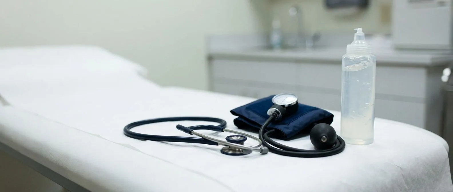
The endocarditis heart murmur is one of the most recognised physical signs, heard by auscultation — the careful listening with a stethoscope that forms the core of the bedside cardiac exam. A new murmur, or a change in a murmur that was already known, suggests that the infected valve has begun to leak or stiffen. Not every patient with endocarditis has a murmur, however; right-sided infections and some early cases can be silent on exam. The absence of a murmur therefore does not rule the diagnosis out, which is why imaging is always required.
Other classic but variable findings Beyond the murmur, clinicians look for signs that small infected clots have broken off and lodged elsewhere. Tiny red-brown spots in the whites of the eyes, painless haemorrhages on the palms and soles, and tender nodules on the fingertips are the textbook "stigmata" of endocarditis. They are useful when present, but they are absent in many modern cases, partly because patients present earlier and partly because acute staphylococcal disease can outpace these slow-developing signs. Splenomegaly — an enlarged spleen felt in the left upper abdomen — is another older sign still mentioned in teaching.

Because physical signs are inconsistent, the real diagnostic value lies in combining them with the history and tests. A patient with a prosthetic valve, three weeks of night sweats, and a newly audible murmur is very different from a young athlete with a fever after a viral illness and a flow murmur that has been present for years. Your cardiac team interprets the whole pattern, and if there is genuine suspicion, they proceed to blood cultures and echocardiography rather than waiting for the full set of classic signs to appear.
Bacterial Endocarditis Antibiotics and Treatment Plan
Endocarditis treatment centres on high-dose, bactericidal antibiotics given intravenously for several weeks, most commonly four to six, guided by the organism found in blood cultures. The choice of endocarditis antibiotics is not standard; it depends on the bacterium, whether the valve is native or prosthetic, and whether the patient has allergies or kidney function that limits certain drugs. A typical regimen starts with broad cover until cultures return, then narrows to a targeted agent once sensitivities are known. Throughout treatment, blood is re-cultured to confirm the bacteria have cleared, and inflammatory markers are tracked to confirm the infection is responding.
When surgery is needed alongside antibiotics Antibiotics alone cure many cases, but surgery becomes necessary in a sizeable minority. The main reasons are heart failure from a failing valve, uncontrolled infection despite appropriate antibiotics, large or mobile vegetations at risk of breaking off, and infection with organisms that antibiotics cannot reliably eradicate, such as some fungi or highly resistant bacteria. Surgery removes the infected tissue and replaces the damaged valve with a mechanical or biological prosthesis. Where the infection involves the ascending aorta or has produced an abscess, the operation is more complex and is undertaken at a centre experienced in such reconstructions.
Patients sometimes ask why treatment takes so long when they feel better within days. The reason is that vegetations protect bacteria from both the immune system and antibiotics, so stopping early risks a relapse with a resistant organism. Clinical studies and registries have refined how long and how intensively to treat; for an example of a prospective observational study that documents real-world effectiveness of targeted therapy in a related cardiac condition, see the COLLIGO-HCM multinational observational study. The same principle — following defined patients carefully over time to see what treatment actually achieves — underpins how endocarditis outcomes are measured today.
Endocarditis Complications and When to Seek Help
Endocarditis complications fall into three categories: heart damage, embolic events, and immune-mediated effects. Heart damage includes worsening valve leakage, abscesses that burrow into the surrounding muscle and conducting tissue, and, in severe cases, rupture of a valve component or fistula formation between chambers. Embolic events happen when pieces of vegetation break off and block arteries, most dangerously in the brain, where they cause stroke, and in the coronary arteries, where they can produce an atypical infarction that is not caused by the usual atheromatous plaque. Immune-mediated effects include kidney inflammation and arthritic-type pains that resolve as the infection is treated.
Signs that should prompt urgent attention During treatment, and for some weeks afterwards, patients are told to watch for specific warning signs. A return of fever, new breathlessness, sudden weakness or numbness on one side, severe headache, or chest pain should never be "waited out" at home. Some patients may already be taking long-term medications such as aspirin for other cardiac reasons, or an anticoagulant such as those studied in the ARISTOTLE trial; any change in their rhythm, such as new atrial flutter, or any sign of bleeding needs urgent review because infection can alter how these drugs behave.
The risk of complications is why endocarditis is never managed as an outpatient at the start, even when the patient looks well. The first phase of treatment is usually in hospital so that fever, inflammatory markers, and valve function can be watched closely and surgery arranged quickly if needed. After discharge, close follow-up continues, often with a repeat echocardiogram to confirm the valve has stabilised. For an example of how observational data help clinicians understand complications and outcomes in complex cardiovascular patients, see PubMed study 24547542.
Endocarditis Prevention and Living with Heart Valve Infection
Endocarditis prevention is built on three practical pillars: good daily oral and skin hygiene, selective antibiotic prophylaxis before high-risk procedures for high-risk patients, and careful management of any indwelling lines or implanted devices. The single most effective everyday measure is excellent dental care, because the mouth is the most common source of the bacteria involved. Regular cleaning, treating gum disease, and avoiding elective dental work during active infections all lower the background level of bacteria entering the blood. Selective prophylaxis — a single dose of antibiotic before certain dental procedures — is reserved for patients with the highest-risk heart conditions, not for everyone.
Living well after treatment Most patients who complete treatment recover well and return to their usual lives, though some carry a new prosthetic valve and the lifelong considerations that come with it. Long-term follow-up usually involves periodic echocardiograms to check valve function, attention to oral health, and awareness of the warning signs described above. Patients who have had endocarditis once are at higher risk of a second episode, so prevention advice is given more firmly. A cardiac rehabilitation-style approach — gradual return to activity, managing other risk factors such as high blood pressure, and staying up to date with vaccinations — supports overall cardiovascular health without directly "preventing" endocarditis.
In summary, endocardium endocarditis is a serious but treatable infection of the heart's inner lining and valves, recognised by persistent fever in someone with valve disease or a prosthetic valve, confirmed by blood cultures and echocardiography, and treated with weeks of intravenous antibiotics, with surgery reserved for cases of valve failure or uncontrolled infection. The diagnosis can be frightening, but the pathway is well established and outcomes are good when treatment begins promptly. If you or a family member have a heart murmur, a replaced valve, or symptoms that worry you, the right next step is not to search for reassurance online but to discuss your specific situation with a qualified cardiac team. Dr Alejandro Molina and the team at Cirugía de Corazón see patients in Guadalajara, Querétaro and Hermosillo, and you can book a consultation to have your case reviewed properly rather than self-diagnosing.






