

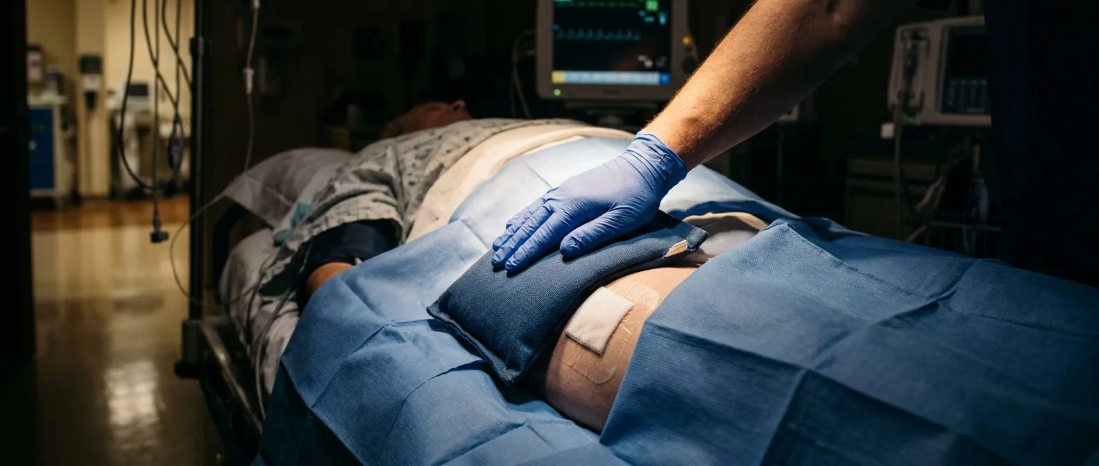
Femoral Artery Access for Catheter Heart Procedures
Femoral artery access is how most catheter heart procedures begin.
If you or a family member has been told you need a heart catheterization, a stent, or a transcatheter valve procedure, someone will have mentioned where the catheter will enter your body. For many years, that entry point has most often been the femoral artery access site in the groin. Understanding how femoral artery access works, what it feels like, and what recovery involves can take much of the fear out of the day itself. This guide walks through the whole journey, from the puncture in the groin to going home.
What Is Femoral Artery Access?
Femoral artery access is the technique of reaching the arteries of the heart and the rest of the body through a single small puncture in the groin. The femoral artery is the large artery that carries blood down each leg. Because it sits close to the skin and is fairly wide, doctors can place a thin tube, called a catheter, into it and guide the catheter upward to the heart using X-ray imaging.
Once the catheter is in place, the team can measure pressures inside the heart, inject contrast dye to film the coronary arteries, open blocked arteries with balloons and stents, or replace a narrowed aortic valve through a TAVR procedure. Almost everything a heart team needs to diagnose or treat can be done through this one doorway.
Why the groin, of all places? The artery there is easy to feel, easy to compress by hand if bleeding occurs, and large enough to accept the widest tubes used in structural heart procedures. In our consultations we take the time to explain in plain words why the femoral route is proposed for a particular patient, and what the alternatives would be.
Where Is the Femoral Artery Located in the Leg?
The femoral artery is in both legs — you have one in each thigh. It begins where the external iliac artery passes under the inguinal ligament, the band of tissue that runs between your hip bone and your pubic bone, roughly at the crease where your leg meets your trunk. From there it travels down the front-inner thigh, becoming the popliteal artery behind the knee.
You can feel it yourself: press gently just below the crease of your groin, slightly toward the middle of your thigh, and you will feel a firm pulse. That pulse is exactly what the doctor uses. During the procedure, the puncture is made in the common femoral artery, the segment just below the ligament, because that part sits over the femoral head, the ball of the hip joint. Puncturing over bone makes compressing the artery afterwards much safer and more effective.
What about the veins next to it? The groin also holds the femoral vein, which runs just inside the artery. For some procedures — right heart catheterization or placement of a temporary pacing wire — the vein rather than the artery is used. The team identifies which vessel they are in with ultrasound before puncturing, which is now standard practice.
Why Is Femoral Artery Access Used for Cardiac Catheterization?
Femoral artery access for cardiac catheterization remains the workhorse route in heart catheter laboratories worldwide, and for good reasons. The artery is large, so it accepts introducer sheaths of many sizes. Small diagnostic catheters, medium stent tubes, and the very large tubes used for TAVR can all go through the same route, depending on the patient.
From the groin, the catheter follows a smooth, natural curve up the aorta, the body's main artery, and into the openings of the coronary arteries. This stable route makes precise manoeuvres easier, which matters when a blockage is complex or when a valve must be positioned millimetre by millimetre.
Some patients have no other good option. If the arteries of the wrist are too small, calcified, or blocked, or if both arms have already been used or injured, the femoral route may be the safest one available. Every case we treat is reviewed by our multidisciplinary team, where we weigh the access route against each patient's own anatomy before recommending one.
How Is Femoral Artery Access Performed, Step by Step?
The femoral artery access procedure follows a well-practised sequence. Knowing the steps in advance makes the day far less mysterious. Here is the typical flow of the puncture itself:
- Preparation: the groin is shaved and cleaned with antiseptic, and sterile sheets cover everything but the puncture area.
- Ultrasound mapping: the doctor scans the artery to see its size, locate the correct segment, and avoid the vein behind it.
- Local anaesthetic: the skin and deeper tissues are numbed with lidocaine, much like at the dentist.
- Needle puncture: a hollow needle is advanced into the artery at a deliberate angle, and pulsing blood confirms entry.
- Wire and sheath: a soft guidewire is passed into the artery, the needle is removed, and a short plastic tube — the sheath — is slid over the wire into the vessel.
From that moment the sheath is the doorway. Every catheter for the rest of the procedure passes through it. Good technique matters at each step, and the puncture location is confirmed with X-ray before the larger tubes go in. Ultrasound-guided puncture is widely used, because a precise first puncture is one of the best ways to prevent complications later.
Does the whole thing hurt? Most patients feel only the sting of the numbing injection. The puncture itself, and the passage of catheters inside the arteries, are usually not painful — some people feel brief warm flushes when contrast dye is injected.
Femoral Artery Access vs Radial Artery Access: Which Is Right for You?
Radial access means entering through the radial artery at the wrist. In recent years many centres have shifted routine diagnostic work and simple stents to the wrist, because the artery there is near the skin surface and can be pressed firmly against bone for a long time, and bleeding from the wrist rarely becomes dangerous. Patients also sit up and walk sooner.
But the comparison is not one-sided, and the choice depends on the specific procedure and the specific patient:
- Size of equipment: TAVR and other structural heart procedures need large tubes that the wrist artery usually cannot accommodate, so femoral access remains standard for them.
- Complexity of the case: chronic total blockages, complicated anatomy, or multi-vessel stenting may be easier and safer from the groin.
- Patient factors: peripheral artery disease in the leg, prior groin surgery, obesity, or severe calcification can all tip the decision the other way.
We recommend the less invasive route only where the evidence supports it for that patient — sometimes that is the wrist, sometimes the groin, and the reasoning is always explained before the procedure is scheduled.
Femoral Artery Access for an Angiogram: What Does It Feel Like?
Femoral artery access for angiogram — the dye study that films your coronary arteries — is a calm, well-tolerated experience for most people. You stay awake, lying on a padded table in a cool, dimly lit room. Screens above you show the X-ray images in real time, and a monitor displays your heart rhythm throughout.
The sequence of sensations is predictable. First comes the cold swipe of antiseptic. Then the numbing injection stings for a few seconds. After that, most patients feel pressure and a sense of the doctor working, but not sharp pain. When contrast dye enters the coronary arteries, a wave of warmth may spread through your chest — it lasts seconds and is harmless. Some people feel as though they need to urinate briefly during dye injections.
How long does it take? A diagnostic angiogram typically runs thirty to sixty minutes. A stent or other intervention takes longer, depending on what is found. Ask before the procedure how long your particular study is expected to take, so the time on the table holds no surprises.
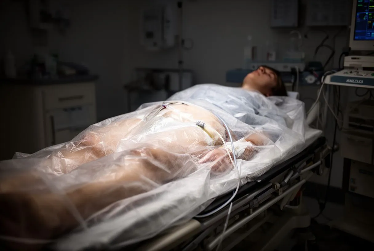
What if I need to move or cough? Just tell the team — everyone in the room understands, and the doctor will pause until you are comfortable. Speaking up during a catheterisation is normal and expected, never a bother.
What Are the Complications of Femoral Artery Access?
Femoral artery access complications are uncommon with modern technique, but they are honest to discuss, and knowing the signs helps you recover safely. The most frequent problem is bleeding at the puncture site, which can form a collection of blood under the skin called a haematoma. Most haematomas are small and settle on their own with pressure and time.
More serious, but rarer, problems include injury to the artery wall itself. These are the main ones a heart team watches for:
- Bleeding and haematoma: swelling and bruising at the groin; occasionally blood tracks up or down the thigh.
- Pseudoaneurysm: a small pocket where blood escapes the artery wall but is contained by surrounding tissue; it causes a pulsating lump and is usually treated with an ultrasound-guided injection or compression.
- Blocked leg artery: clot or damage at the puncture can slow blood flow to the leg, causing coldness, pain, or colour change — this needs urgent attention.
- Retroperitoneal bleeding: bleeding that tracks backward into the pelvis instead of outward; it is rare, but it can cause a drop in blood pressure and hidden symptoms.
What happens if you damage your femoral artery? In most cases the damage is found quickly — either during the procedure on X-ray or afterwards on ultrasound — and it can be repaired, often without open surgery. Before you leave the recovery area, the groin is usually checked carefully, and you should receive written instructions on which warning signs to watch for at home.
It helps to put the risk in proportion: serious vascular complications are a minority of cases in contemporary registries, and the technique — ultrasound, correct puncture site, and proper closure — is designed precisely to keep them rare.
Can a Femoral Artery Be Fixed? How the Access Site Is Closed
Yes — a femoral artery can be fixed, and in the ordinary case it heals by itself. The artery has a tough three-layer wall, and a small puncture closes when steady pressure is applied for fifteen to twenty minutes, or when a closure device is used. If a larger problem does arise — a pseudoaneurysm, a dissection flap, or a blocked segment — there are established treatments, from ultrasound-guided compression to a covered stent placed through the same or another artery.
Closure devices deserve a word. These are small collagen plugs or suture-based devices deployed at the puncture site at the end of the procedure. They seal the hole immediately, let the patient sit up sooner, and are standard for larger sheaths such as those used in TAVR. For small diagnostic punctures, manual pressure alone works well.
The choice of closure method is usually planned before the procedure starts, matched to the sheath size and the patient's artery. The key point for patients is this: the overwhelming majority of femoral punctures heal cleanly, and even the uncommon complications have recognised, effective treatments.
What Does Femoral Artery Access Recovery Look Like?
Femoral artery access recovery is usually quicker than patients expect. After the sheath is removed, you lie flat for a few hours — often two to four for a diagnostic case — while pressure or a closure device seals the artery. The nursing team checks the groin, the pulses in the foot, and your blood pressure at regular intervals during that time.
The first evening and next day: once you are cleared, you can sit up, walk to the bathroom, and usually go home the same day after a diagnostic angiogram, or the next day after a stent. For the first twenty-four to forty-eight hours the instructions are simple:
- Keep it gentle: short walks are fine; avoid bending sharply at the hip, heavy lifting, stairs in a hurry, and straining.
- Rest the leg: do not lift anything heavier than a few kilograms for the first few days.
- Keep the site clean and dry: a small dressing covers the puncture; showering is usually allowed after a day or two, but no baths or pools for about a week.
- Arrange the ride home: driving is typically resumed after a few days, once your doctor confirms it is safe — sooner after a simple diagnostic test, later after an intervention.
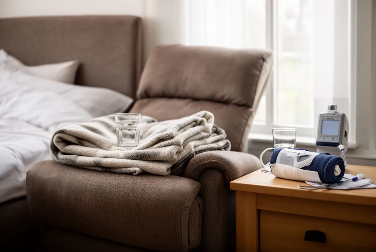
A small bruise is normal. Many patients go home with a palm-sized bruise and a pea-sized firm lump at the puncture. Both fade over one to two weeks. The puncture site is usually reviewed at follow-up, and a carefully done femoral puncture rarely slows anyone down for more than a few days.
When Should You Call Your Doctor After Femoral Artery Access?
Most recoveries are uneventful, but a few warning signs should prompt a phone call the same day, or a trip to the emergency department if they are severe. Knowing them is part of going home safely. Contact your medical team promptly if you notice:
- New or growing swelling at the groin that is firm, pulsating, or rapidly enlarging.
- Active bleeding that soaks the dressing and does not stop with ten minutes of firm hand pressure.
- A cold, pale, numb, or painful leg or foot on the side of the puncture.
- Fever, spreading redness, or discharge from the puncture site.
- Dizziness, fainting, or sudden severe pain in the abdomen, back, or thigh.
What are symptoms of femoral artery blockage? Away from the puncture itself, a blocked femoral artery in general circulation problems causes cramping pain in the calf or thigh when walking that eases with rest, coldness of the lower leg, slow-healing wounds, and in advanced cases pain at rest. If a catheter procedure has narrowed the artery where it was punctured, walking may become harder on that side — a change worth reporting rather than tolerating.
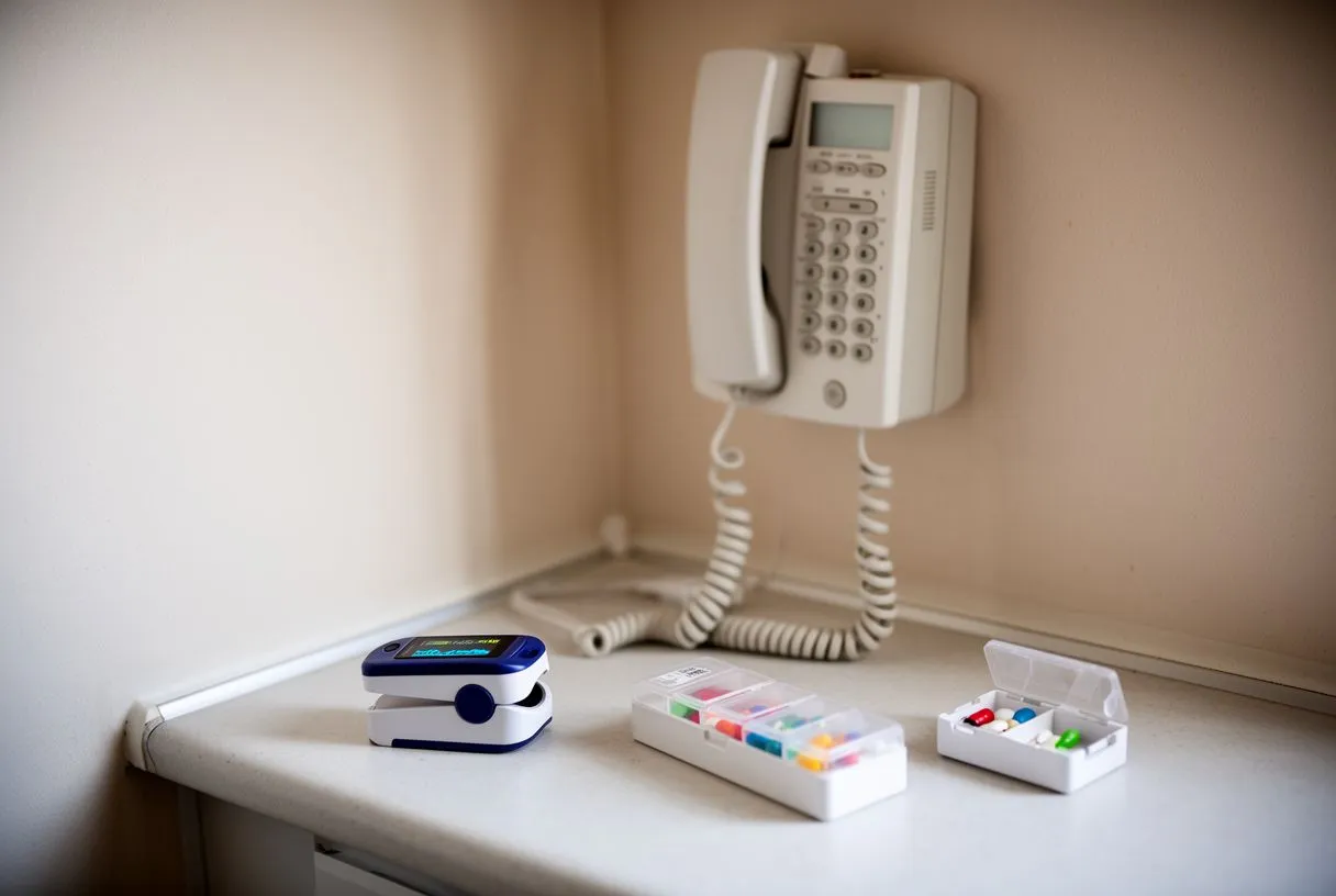
Ask for these warning signs in writing before discharge, because recognising a problem early is the best way to keep it minor. Never wait out a worrying symptom overnight — call, and let the team decide.
A Final Word on Femoral Artery Access
Femoral artery access has carried cardiac medicine for more than half a century, and it remains, for many procedures, the most dependable doorway to the heart. The puncture is small, the discomfort modest, and the recovery measured in days rather than weeks. The risks are real but uncommon, they have clear warning signs, and the treatments for them are well established.
The most important decision is not the puncture itself, but choosing the right route and the right procedure for your particular heart, and that choice deserves a specialist team. Our multidisciplinary cardiac team, led by Dr Alejandro Molina with consulting rooms in Guadalajara, Querétaro and Hermosillo, reviews every case together against international guidelines, encourages second opinions, and always leaves the final decision with the patient — starting with a free video consultation to talk through your situation and exactly which studies are needed. If a heart catheterisation, stent, or TAVR has been suggested for you or someone you love, bring your questions to a qualified cardiac team and discuss your own case in person rather than deciding from a screen.






