Coronary Ostia Narrowing: Diagnosis and Repair
Coronary ostia stenosis diagnosis and repair from Guadalajara heart specialists.
Deep inside the chest, where the aorta leaves the heart, two tiny openings feed the arteries that keep the heart muscle itself alive. When those openings — the coronary ostia — become narrowed, the whole heart can be quietly starved of blood. Coronary ostia stenosis is less common than blockages further along the coronary arteries, but it is often missed precisely because it sits in a place standard tests do not always examine closely. This guide explains what the ostia are, why they narrow, which symptoms to watch for, how the diagnosis is made, and what repair options exist, so you can walk into a consultation knowing the right questions to ask.
What Are the Coronary Ostia?
Ostia in medical terms simply means openings or mouths. In the heart, the coronary ostia are the two points where the left and right coronary arteries arise from the very first stretch of the aorta, just above the aortic valve. The left coronary artery quickly divides into vessels that supply the front and side of the heart; the right coronary artery wraps around to supply the bottom and back.
These openings are small — a few millimetres across — yet every drop of blood reaching the heart muscle passes through them. In most people the ostia sit within the sinuses of Valsalva, the small bulges just above the aortic valve, positioned so the arteries can fill during the moment the heart relaxes between beats. Cadaveric and imaging studies have documented considerable natural variation in their exact height, angle and position, which matters when surgeons plan any work near the aortic root.PubMed
Anatomy also explains why ostial disease behaves differently from disease further down the artery. A narrowing at the ostium blocks an entire coronary tree at its source, rather than one branch, so the territory at risk is larger. That is why a seemingly small ostial lesion can produce symptoms out of proportion to its appearance on a standard angiogram.
What Is Coronary Ostia Stenosis?
Stenosis means narrowing. Coronary ostia stenosis is a constriction at the mouth of a coronary artery — usually where it leaves the aorta — that limits blood flow into the vessel. The narrowing may be caused by plaque, by scar tissue, or by disease of the aortic wall itself pressing in on the opening.
It helps to picture the plumbing. If a garden hose is pinched at the tap, no amount of widening the hose further along will restore full flow. The same logic applies here: an ostial lesion controls the supply to the whole artery downstream. Ostial lesions account for a meaningful share of significant coronary artery disease, and they carry particular importance because stents placed at an ostium can shift position or be flattened, which influences the choice between stenting and surgery.
Some narrowing is also congenital — present from birth. Unusual origin or course of a coronary artery (for example, an anomalous right coronary artery arising from the left side and running between the great vessels) can squeeze the artery with every heartbeat. In rare cases this pattern is linked to sudden cardiac events in young athletes, which is why congenital repair is considered in selected patients.
What Causes Ostial Coronary Artery Disease?
Atherosclerosis is the most common cause. The same plaque process that narrows arteries elsewhere in the body can build up right at the coronary openings, where the turbulent flow at the aortic wall encourages plaque formation. Risk factors are familiar: smoking, high blood pressure, diabetes, high cholesterol and family history. Management of ostial coronary artery disease therefore always includes treating these underlying drivers, whatever procedure is chosen.
Aortic valve surgery is another well-recognised cause. Narrowing can appear months or years after an aortic valve replacement, when the graft or valve sewing ring sits close to the coronary openings, or when calcification from the diseased valve extends upward into the ostia. Radiation therapy to the chest, and inflammatory conditions affecting the aorta such as certain large-vessel vasculitides, can also narrow the ostia by thickening the aortic wall itself.
- Common drivers of ostial narrowing: atherosclerotic plaque at the aortic origin, post-surgical changes after valve replacement, radiation injury, aortic wall inflammation, and congenital variants of the coronary origin.
- Why the cause matters: plaque in the aortic wall may need patch or graft surgery, while a stent-friendly lesion from simple atherosclerosis may suit a catheter-based approach.
Identifying the cause is not academic — it determines the treatment. A lesion caused by calcified aortic disease responds poorly to a coronary stent, whereas a soft atherosclerotic plaque may be treated from the inside. This is why imaging that shows the aortic root, not just the artery, is essential.
What Are the Symptoms of Heart Blockage at the Ostium?
The classic symptom is angina. Chest tightness or pressure, typically brought on by exertion and relieved by rest, is the hallmark of any significant coronary narrowing, including ostial disease. Because an ostial lesion restricts an entire artery, the discomfort may come on more quickly and with less exertion than expected — a pattern worth reporting in detail to your doctor.
Other warning signs include: shortness of breath during activity, unusual fatigue, and breathlessness lying flat. In some patients — particularly women, older adults, and people with diabetes — the presentation is atypical: jaw or arm discomfort, nausea, or simply a sudden decline in exercise tolerance. If the narrowing triggers an unstable pattern (symptoms at rest or escalating rapidly), that is an emergency and requires immediate care.
Ischaemic heart disease — the medical term for the heart muscle not receiving enough oxygenated blood — can also announce itself as a heart rhythm disturbance or, in advanced cases, as heart failure symptoms such as leg swelling and breathlessness at rest. Ischaemic heart disease treatment always begins with confirming where and how severely the blood supply is compromised, which brings us to diagnosis.
How Is Coronary Ostia Stenosis Diagnosed?
Coronary CT angiography has changed the game. A CT scan of the coronary arteries shows the aortic root, the ostia and the arteries in a single three-dimensional picture, and is particularly good at depicting calcified ostial lesions that a catheter angiogram can under-call or even miss when the catheter cannot engage the opening properly. A coronary calcium score, obtained from the same scanner, quantifies the plaque burden and helps interpret borderline findings.
The echocardiogram plays a supporting role. An echocardiogram (an ultrasound of the heart) cannot reliably visualise the ostia themselves, but it shows the consequences: areas of the heart muscle that move abnormally, valve disease that may be the underlying cause, and the function of the heart overall. Stress testing, whether on a treadmill or with imaging, demonstrates how the narrowing affects the heart under effort.
Cardiac catheterisation confirms the picture. In an invasive angiogram, a thin tube is advanced to the coronary openings and dye is injected. Ostial lesions require specific technique — the catheter can itself occlude a critical ostium — so an experienced team interprets the pressure traces as well as the images. In our practice, every imaging study is reviewed jointly, and we take the time in consultation to explain in plain words what the studies actually show before any recommendation is made.Every case is then reviewed by our multidisciplinary team against international practice guidelines before a proposal is given, and the final decision always rests with the patient.
How Is Ostial Narrowing Treated? Interventional Options
Medication remains the foundation. Antiplatelet therapy, statins, beta blockers and nitrates reduce symptoms and slow plaque progression, and for lesions that are not severely obstructive, medication alone may be entirely appropriate. No procedure removes the need for aggressive risk-factor control.
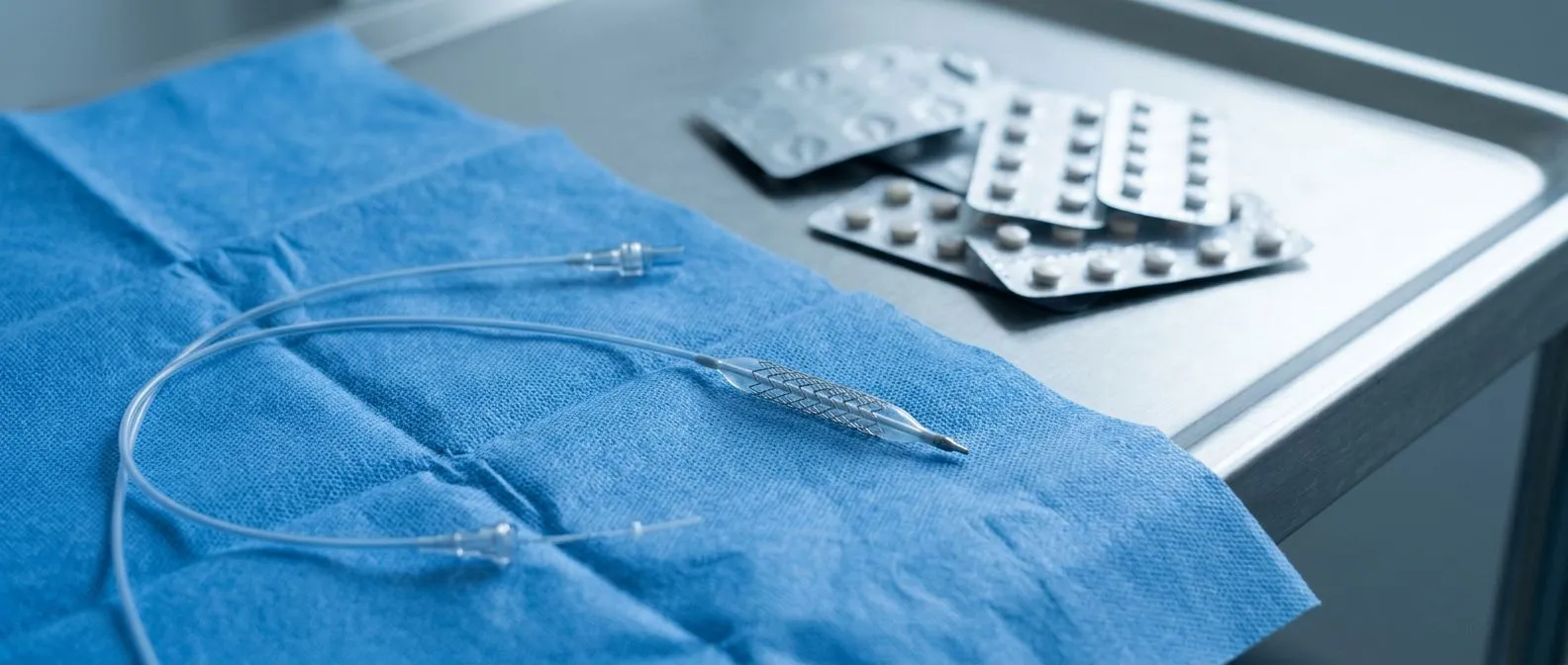
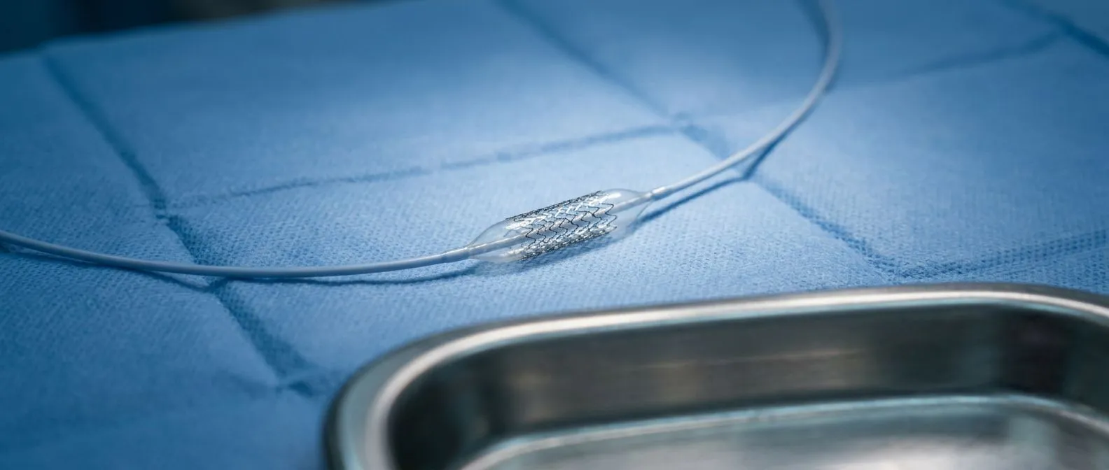
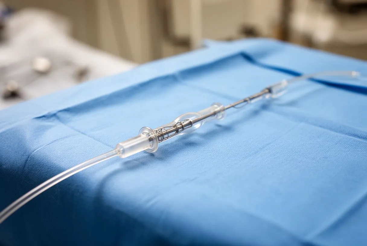
Percutaneous coronary intervention — opening the artery from the inside with a balloon and a coronary stent — is a widely used option for ostial disease. However, ostial lesions are among the more technically demanding for stenting: the opening must be precisely covered, the stent must not protrude too far into the aorta, and calcified or aortic-wall disease raises the risk of the stent compromising either the ostium or the valve. Specialised techniques, including dedicated ostial stenting positions and intravascular imaging, are used to improve results.
Selection is everything. Not every ostial stenosis needs a procedure, and not every stenosis is suitable for a stent. The decision weighs the severity of the narrowing, the presence of symptoms despite medication, the anatomy of the aortic root, and the patient's overall health. In our team's experience, the less invasive route is recommended only where the evidence supports it for that specific patient — and we actively encourage patients to seek a second opinion, treating it as a normal part of deciding about heart surgery.When intervention is chosen, Dr Molina and the team perform conventional, minimally invasive, robotic and transcatheter procedures, so the recommendation is shaped by the anatomy rather than by the technique we happen to offer.
When Is Surgery Needed for Coronary Ostia Narrowing Repair?
Surgery is considered when the anatomy demands it. Coronary ostia narrowing repair through the classic surgical route — coronary artery bypass grafting, where a vessel from the chest wall or leg is used to carry blood around the blockage — offers a durable solution that does not depend on the diseased ostium staying open. Bypass is often preferred when the ostial lesion sits alongside other disease: severe left main involvement, multivessel coronary artery disease, or a badly calcified aortic root.
Aortic root surgery is a different situation. When the narrowing is caused by disease of the aortic wall or by a previously implanted aortic valve, the repair may involve replacing or patching the aortic root and re-implanting the coronary arteries with their small attached buttons of aortic wall — the so-called coronary buttons — into the new graft. This is precise, open-heart work, and it is usually combined with treatment of the valve or an aortic aneurysm repair in the same operation. Endovascular aneurysm repair, delivered through catheters, is generally not suitable when the coronary ostia themselves must be reconstructed, because they arise in exactly the segment the graft must cover.
Outcomes depend on patient selection. For isolated ostial disease in a suitable patient, both stenting and bypass can offer excellent relief of symptoms; the choice turns on anatomy, calcification, and the patient's other conditions such as chronic kidney disease, which influences both the contrast burden of catheter procedures and the surgical risk. Aortic root surgery carries more upfront risk than a stent but addresses the underlying cause when that cause is the aorta itself. In our practice, we have seen that the honest discussion of these trade-offs — including the risks we would rather not have — is what allows patients to choose with confidence.Dr Molina trained in cardiac, minimally invasive, robotic and structural surgery at the University of Montreal and in structural cardiology and TAVR at CHU Bordeaux, and he brings that combined surgical-transcatheter perspective to every root-and-ostium decision.
Recovery After Ostial Coronary Artery Disease Management
Recovery after a stent is measured in days. Most patients who undergo percutaneous intervention go home the next day, with a groin or wrist puncture that heals within a week. Normal activities resume gradually over one to two weeks, and cardiac rehabilitation — a supervised programme of exercise and education — is strongly advised.
Recovery after bypass or root surgery is measured in weeks. Hospital stay is typically around five to seven days, with the first two often in intensive care. Sternal precautions (no lifting, driving or pushing for six to eight weeks while the breastbone heals) apply after conventional surgery; minimally invasive and robotic approaches use smaller incisions and generally allow a faster return to daily life, though the underlying healing timeline is similar. Full recovery and a return to work commonly take six to twelve weeks, depending on the operation and the patient's baseline fitness.
Lifelong follow-up is part of the treatment. Whatever the procedure, patients continue medical therapy, attend surveillance imaging where indicated, and work on the risk factors that caused the disease. Follow-up visits should focus on how the patient is actually living, not just how the images look.
Costs vary with the procedure chosen. A stent procedure and an open surgical repair are different operations with very different resource needs, so a single "price" for coronary ostia stenosis treatment is not meaningful until the diagnosis and recommended approach are clear. What patients can reasonably expect is a detailed, itemised proposal after the studies are reviewed — covering hospital, surgeon, anaesthesia, perfusion, and device or graft costs — rather than a vague package figure.
International patients often find Mexico accessible. Guadalajara, Querétaro and Hermosillo are all served by major airports, and private hospitals in these cities routinely care for international patients. Travel logistics should be planned around the recovery: for open surgery, patients should plan to remain locally for several weeks before flying home, and should confirm with their insurer, in writing, what is covered for treatment abroad.
- Before travelling for surgery: obtain all imaging on disc or electronically, confirm insurance coverage for out-of-country care, and arrange a remote review before booking flights.
- What a good proposal includes: the recommended operation and why, the alternatives considered and rejected, expected hospital stay, recovery milestones, and total itemised costs.
The first contact with our team is a free video consultation: we get to know the patient, talk through the condition, and identify which studies are still needed so no unnecessary ones are ordered. A face-to-face visit at the consulting rooms follows only when the patient decides to go ahead.This structure exists so that cost and travel planning rest on a firm diagnosis rather than on assumptions.

When to Seek Help for Suspected Ostial Narrowing
Certain symptoms should never wait for an appointment. Chest pressure at rest lasting more than a few minutes, pain radiating to the jaw, arm or back with sweating or nausea, sudden severe breathlessness, or fainting all warrant calling emergency services immediately. Ostial lesions, because they can cut off an entire coronary artery, deserve the same urgency as any acute coronary syndrome.
Planned evaluation is appropriate for exertional symptoms. New chest tightness on climbing stairs, a steady loss of exercise tolerance, or breathlessness that has crept up over months deserves a scheduled cardiology assessment within days to weeks. Bring a list of symptoms, when they occur, and any prior imaging — this history guides which test is ordered first and can save weeks of uncertainty.
Reducing arterial plaque is a lifelong project, and it starts with the basics: stop smoking, control blood pressure and diabetes, take prescribed statin therapy, exercise as tolerated, and attend follow-up. These measures do not dissolve established ostial stenosis, but they slow its progression and protect the rest of the circulation. Calcified arteries are a marker of accumulated risk, and while no single number predicts an individual's life expectancy, controlling the drivers of plaque is the most reliable way to protect the years ahead.If you are reading this because symptoms have already begun, our team offers a free initial video consultation to help you understand your studies and decide on the next step — and we will always tell you honestly when the best plan is the one you already have.
Coronary ostia stenosis is an uncommon but important form of coronary artery disease: a narrowing at the very source of the heart's own blood supply, with symptoms like any other blockage but anatomy that changes the treatment calculus. Accurate diagnosis usually depends on imaging that looks at the aortic root — coronary CT angiography in particular — and treatment spans medication, coronary stents, and surgical repair including bypass and aortic root reconstruction. The right answer is specific to each patient's anatomy and circumstances, which is why it should be reached with a qualified cardiac team, never by self-diagnosis. If any of what you have read matches your situation, book a consultation and bring your questions; a good team will welcome every one of them.