

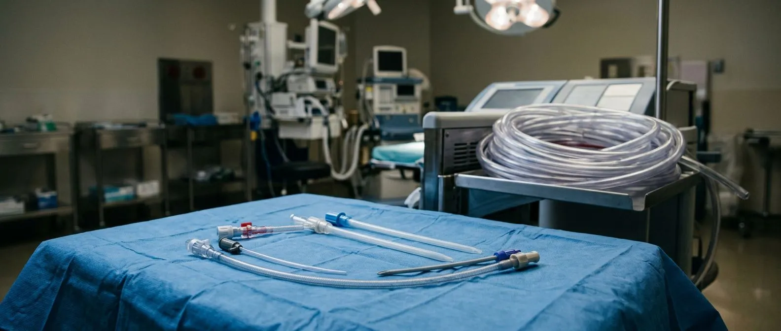
Femoral Cannulation in Minimally Invasive Heart Surgery
Femoral cannulation heart surgery enables minimally invasive procedures through the groin instead of open chest.
Femoral cannulation heart surgery is one of the reasons a growing number of patients can have a heart operation without a full open-chest incision. If your surgeon has mentioned "going through the groin" or "peripheral access," they were most likely describing this technique. In simple terms, femoral cannulation means placing special tubes, called cannulas, into the large blood vessels at the top of the thigh — the femoral artery and the femoral vein — so that a heart-lung machine can take over the work of the heart and lungs during the operation. It is a well-established approach used in minimally invasive valve surgery, robotic procedures, and some catheter-based treatments. This guide explains what happens during the procedure, why surgeons choose it, what the risks are, and what recovery actually looks like, so you can ask better questions and feel less anxious about the part of the operation you will never see.
What Is Femoral Cannulation?
Femoral cannulation is the technique of inserting cannulas into the blood vessels of the groin to connect a patient to a cardiopulmonary bypass machine, often called the heart-lung machine. The femoral artery is the vessel that carries oxygen-rich blood from the heart down the leg; the femoral vein carries blood back up toward the heart. During minimally invasive cardiac surgery, the surgeon does not open the chest through the breastbone, so the tubes that normally connect the heart-lung machine to the heart itself have to be placed somewhere else.
Why the groin? The femoral vessels sit close to the surface of the skin, are large enough to carry the blood flow the bypass machine needs, and can be reached through a small incision of a few centimeters. This is what makes minimally invasive approaches possible: the surgeon can work through small incisions between the ribs or a small chest incision, while the bypass circuit is connected through the groin.
Open or percutaneous. There are two main ways to place the cannulas. In an open technique, the surgeon makes a small incision and directly exposes the vessel before tying it in. In a percutaneous technique — meaning "through the skin" — the cannula is guided in with a needle and wire, much like placing a large intravenous line, and the entry hole is closed with a special suture device. Both are standard, well-practiced methods in modern cardiac surgery.
Femoral Cannulation vs Sternotomy: How Do They Compare?
A sternotomy is the traditional approach to heart surgery: the breastbone is divided down the middle so the surgeon can see the heart directly and connect the bypass machine to the aorta and the venous chambers of the heart. It is the reference point against which every newer technique is measured, and it remains the right choice for many operations.
The trade-offs. Femoral cannulation minimally invasive cardiac surgery avoids dividing the breastbone entirely. That usually means smaller incisions, less bleeding, and a faster return to daily activity. It can also reduce the risk of wound infection at the chest, which matters especially for patients with diabetes, lung disease, or previous chest surgery. The trade-off is that femoral access carries its own risks, particularly at the groin site, which we will cover in detail below.
Neither approach is universally better. The right choice depends on the operation, the anatomy of the patient's vessels, and the overall clinical picture. Guidelines from international cardiac societies support both approaches, and responsible surgeons recommend the less invasive route only where the evidence supports it for that specific patient. Every case is reviewed by our multidisciplinary team against international practice guidelines before a recommendation is given, and the patient receives a clear proposal with the alternatives — the final decision is always theirs.
How Does Femoral Cannulation Heart Surgery Work?
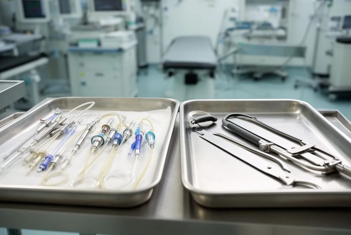
During femoral cannulation for cardiopulmonary bypass, the system works as a two-way circuit. The venous cannula, placed in the femoral vein, drains blood away from the body by gravity and pump suction. That blood is routed through the heart-lung machine, which removes carbon dioxide, adds oxygen, and returns the oxygenated blood through the arterial cannula in the femoral artery. From there, the fresh blood travels backward up the aorta to the rest of the body.
What the surgeon does at the same time. Once the heart-lung machine is running, the heart can be quieted and held still, giving the surgeon a calm, bloodless field to repair a valve or a vessel through a small incision. This is the principle behind robotic heart surgery femoral access and behind minimally invasive valve repair through the ribs. In robotic procedures, the surgeon works entirely with instruments controlled from a console, while the patient's circulation is managed through the femoral circuit.
Who watches the circuit? A dedicated perfusionist — the specialist who operates the heart-lung machine — monitors flow, pressure, temperature, and oxygen delivery throughout the operation. The team can also run checks during the case, such as pressure measurements in the arterial line, to confirm the cannula is positioned correctly and blood is flowing the right way.
How Do You Put a Femoral Arterial Line?
This question is often asked by patients preparing for surgery, and the answer helps demystify the process. A femoral arterial line — the catheter femoral artery heart bypass uses — is placed in a sequence of careful steps, either in the operating room or in a hybrid setting with imaging support.
Step-by-step placement. The sequence is deliberately methodical:
- Ultrasound first: the surgeon or anesthesiologist maps the femoral artery and vein with ultrasound, checking the vessel's size, the quality of its wall, and the presence of any plaque or calcification.
- Needle and wire: a small needle enters the artery, a guidewire is passed carefully into the vessel, and the tract is dilated in stages so the artery is never stretched abruptly.
- Cannula insertion: the bypass cannula is passed over the wire to the right depth, and its position is confirmed before the bypass machine is connected.
Local anesthesia, general anesthesia. The patient is usually under general anesthesia for the operation itself, though some femoral lines — for example, those used for certain catheter procedures — can be placed with local numbing and light sedation. The groin incision is small, often three to five centimeters, and is closed in layers at the end of the procedure.
What Are the Different Techniques Used for Arterial Cannulation?
Arterial cannulation is not one-size-fits-all. The technique used for arterial cannulation depends on the operation, the anatomy, and the surgeon's judgment. Understanding the options helps you see why your team may have chosen one approach over another.
The main techniques include:
- Femoral artery cannulation: the standard peripheral access for minimally invasive valve surgery, robotic procedures, and endovascular heart surgery cannulation.
- Aortic cannulation: direct cannulation of the aorta during a sternotomy, still the most common route in open surgery.
- Axillary cannulation: cannulation of the artery under the collarbone, often chosen when the aorta is diseased or when femoral access is not suitable.
- Central cannulation: direct cannulation of the heart's own vessels, used in specific situations where peripheral access is inadequate.
Why technique matters. The choice of cannulation route affects the direction of blood flow, the risk of complications at the access site, and the surgeon's ability to protect the brain from particles or air during the operation. This is why the assessment is individualized — the same operation may be done with different cannulation strategies in different patients. Our team performs conventional, minimally invasive, robotic and transcatheter procedures, and we recommend the less invasive route only where the evidence supports it for that patient.
What Is Femoral Cannulation for Aortic Surgery?
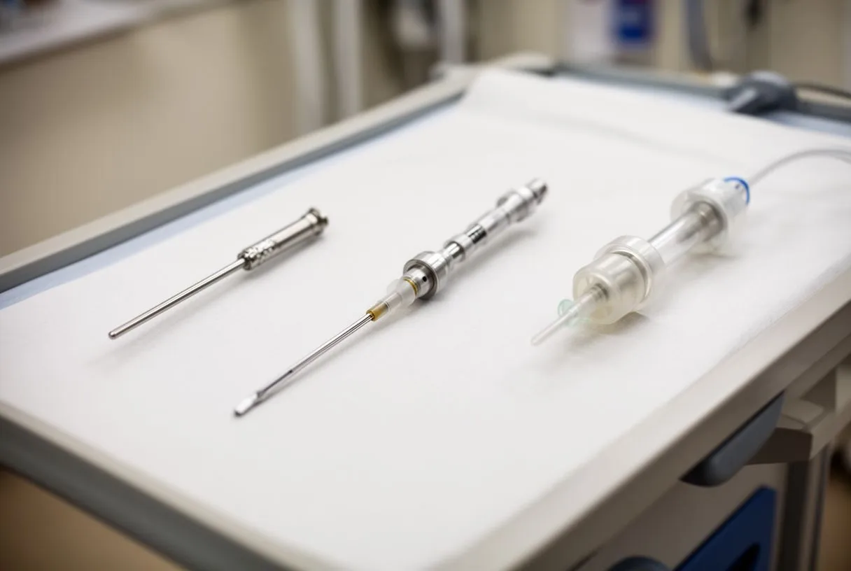
Femoral cannulation for aortic surgery deserves its own discussion, because the aorta — the body's main artery — presents particular challenges. In operations on the aortic valve or the ascending aorta, blood must flow in the correct direction through the cannula, and the surgeon must manage how the aorta is perfused during the procedure.
Reverse flow and its implications. When blood is returned through the femoral artery, it travels up the aorta in the opposite direction from normal flow — this is called retrograde perfusion. It is generally well tolerated, but it requires attention to how the aorta is clamped and how the brain is protected. Surgeons manage this with careful monitoring, including pressure measurements and, in some cases, imaging of the aorta before choosing the route.
When the aorta is diseased. If the aorta carries significant plaque or calcification, passing a cannula through it can dislodge particles. In these situations, surgeons may prefer axillary or central cannulation instead. Preoperative imaging — usually a CT scan of the aorta — is the standard way to make this call. Published experience with minimally invasive aortic valve surgery, including center series such as the one reported in Cirugía y Cirujanos, shows that with careful patient selection this technique can be performed safely.
What Are the Potential Complications of Femoral Artery Catheterization?
Femoral cannulation complications are real but well understood, and most are preventable with careful technique and patient selection. You should know about them honestly, because an informed patient asks better questions.
The most common issues. Potential complications of femoral artery catheterization include bleeding or hematoma at the groin site, injury to the arterial wall, narrowing of the artery afterward (dissection or stenosis), and reduced blood flow to the leg during the bypass period. Infection at the incision site is uncommon but possible. The reported rates of these complications vary across studies, and the risk depends heavily on the state of the patient's vessels.
How the team reduces them. Prevention starts before the incision. Ultrasound mapping of the femoral artery and vein identifies vessels that are too small or too diseased. Sizing the cannula correctly, using anticoagulation management, checking leg perfusion during the case, and closing the artery properly all reduce the risk. Specialized closure devices are often used to seal the puncture site immediately.
Warning signs after surgery. Patients should watch for:
- Leg changes: coolness, numbness, or a pale or bluish color in the leg on the cannula side.
- Groin swelling or pain: a growing lump, bleeding, or increasing pain at the incision site.
- Fever: any fever after surgery, particularly with groin redness, should be reported promptly.
In our consultations we take the time to explain what the studies actually show, in plain words, and to answer every question before any decision is made.
How Do You Know If You Hit the Femoral Artery?
This question usually comes from the world of training and simulation — students learning vascular access — but it matters to patients too, because it explains how the team confirms the right vessel is being used.
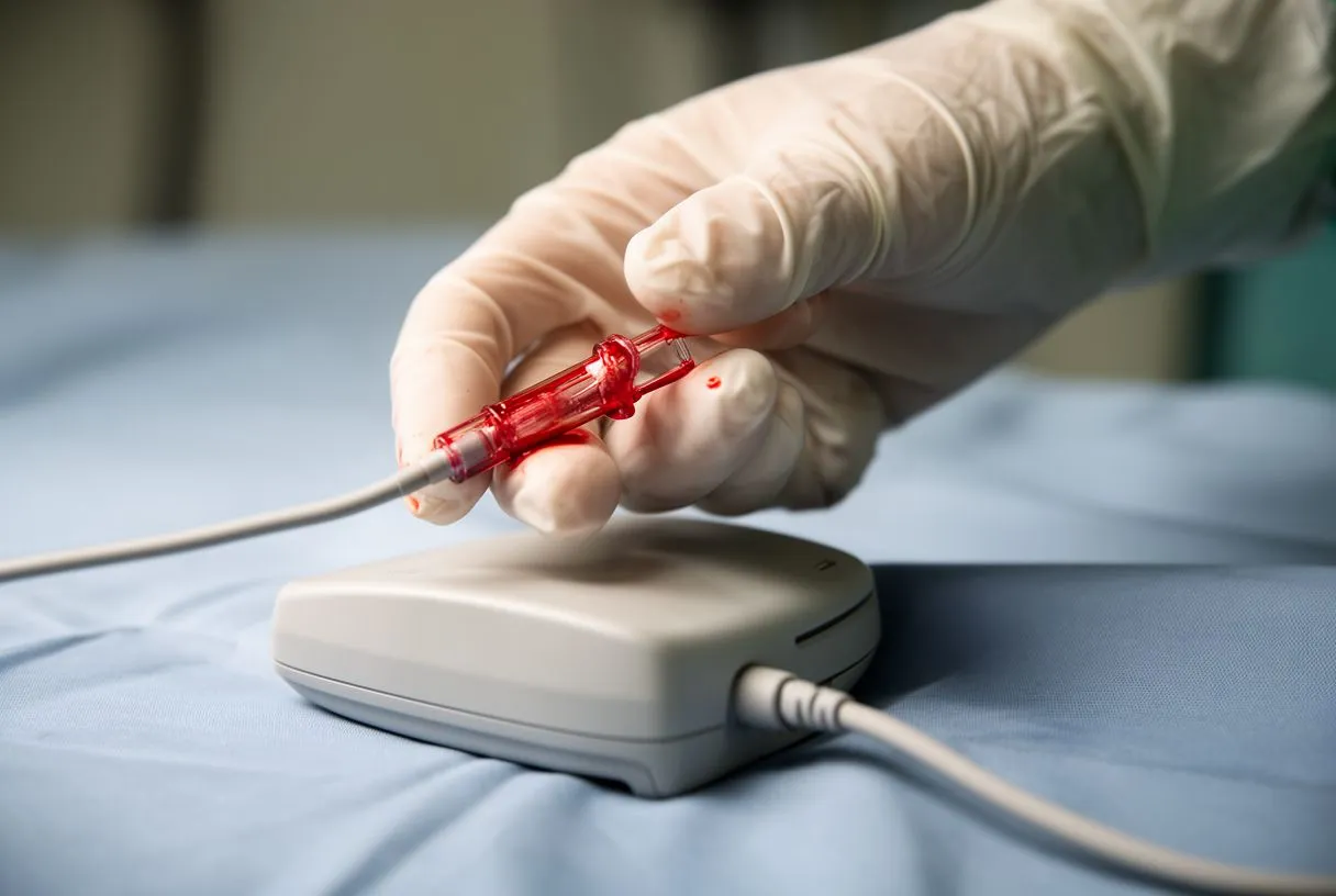
The clinical signs. When a needle enters the femoral artery, the telltale sign is bright red, pulsatile blood returning through the needle, often in rhythm with the heartbeat. Venous blood is darker and flows continuously rather than pulsing. A small pressure transducer connected to the line can confirm the arterial pressure waveform, which is the most reliable bedside confirmation.
Ultrasound confirmation. In modern practice, ultrasound is used in real time, so the team sees the needle tip entering the vessel before it goes in. This has largely replaced the older "blind" technique and has made the procedure safer and more predictable. Ultrasound-guided access has evolved from a novelty into a standard step in preparing for cannulation.
What Is Femoral Cannulation Recovery Time Like?
Femoral cannulation recovery time is usually not the limiting factor in a patient's overall recovery — the heart operation itself is. But the groin site does have its own healing timeline worth understanding.
The first days. The groin incision typically heals within one to two weeks. Patients are usually asked to avoid heavy lifting, deep bending, or strenuous leg activity for the first week or two to let the artery wall and the skin layers heal. Bruising at the site is common and fades over a couple of weeks.
Compared to sternotomy. This is where the difference shows. After a sternotomy, the breastbone takes six to eight weeks to knit, with restrictions on lifting and driving during that period. After minimally invasive surgery with femoral access, those bone-healing restrictions largely disappear, and patients often describe returning to normal life sooner. The minimally invasive experience has been described in detail in studies of early recovery after these procedures, including work published in Reanimatology on the specific features of the early period after minimally invasive cardiac surgery.
What to expect at follow-up. Your team will check the groin at follow-up visits, feeling for the pulse in the leg and looking at the incision. Most patients leave with nothing more than a small scar. If any narrowing of the artery occurred, it is usually detected at these visits through symptoms or a simple ultrasound check.
Who Is a Candidate for Femoral Cannulation?
Femoral cannulation candidate criteria center on the health of the groin vessels and the type of operation planned. Not everyone is a good candidate, and that is a strength of the process, not a limitation.
Favorable factors. Good candidates generally have healthy femoral arteries on imaging, no severe peripheral artery disease, an aorta that can tolerate retrograde flow, and an operation suited to a minimally invasive approach — such as mitral valve repair, isolated aortic valve work, or robotic procedures. Dr Molina trained in cardiothoracic surgery at the Universidad de Guadalajara, in cardiac, minimally invasive, robotic and structural surgery at the University of Montreal, and in structural cardiology and TAVR at CHU Bordeaux — training that shapes how we match each patient to the right access route.
Factors that steer the team elsewhere. Significant plaque in the femoral arteries, a very small vessel diameter, a diseased abdominal aorta, severe peripheral vascular disease, or prior femoral surgery can all make femoral access less suitable. In those cases, axillary or central cannulation offers a safer path. Preoperative CT imaging of the aorta and leg vessels is the standard tool for this decision.
The role of TAVR and catheter procedures. Transcatheter aortic valve replacement, or TAVR, uses femoral artery access routinely — the catheter is threaded from the groin up to the heart, and no bypass machine is needed at all in the transfemoral approach. For patients who need less invasive heart surgery options, this family of techniques — percutaneous cannulation for minimally invasive surgery, TAVR, robotic access — means "heart surgery without open chest" is a genuine possibility rather than an idea. The first contact with our team is a free video consultation: we get to know the patient, talk through the condition, and say which studies are still needed so no unnecessary ones are ordered. If you are weighing these options, the most useful next step is to discuss your own situation with a qualified cardiac team — start with a consultation, bring your imaging, and ask directly whether femoral cannulation heart surgery is the lowest-risk route for you. We also encourage second opinions and treat seeking one as a normal part of deciding about heart surgery.






