Haemodynamic Study: Measuring Pressures Inside the Heart
A haemodynamic study heart test measures pressures inside chambers and vessels to guide treatment.
When doctors suspect a problem with the pressures inside your heart, an ultrasound picture alone may not be enough to decide on treatment. A haemodynamic study heart test measures those pressures directly, using a thin tube threaded into the heart itself. It sounds intimidating, but it is one of the most routine and informative tests in modern cardiology. In this guide we walk through what the test is, how it works, what it feels like, what the numbers mean, and how the results shape decisions about surgery or other treatment.
What Is a Haemodynamic Study?
The definition in plain words. A haemodynamic study is a test that measures blood flow and pressure inside the heart and the large vessels connected to it. It is performed during cardiac catheterisation, a procedure in which a long, flexible tube called a catheter is guided through a blood vessel into the heart. Sensors at the catheter tip record pressures in each chamber and artery.
Why direct measurement matters. Every heartbeat depends on pressure differences: blood moves from one chamber to the next only when the pressure in front of it is lower. When a valve is narrowed, a lung artery is strained, or the heart muscle weakens, those pressure patterns change. Measuring them directly gives the medical team numbers no scan can fully replicate.
What it is not. It is important to understand what a haemodynamic study does not do. It is not a diagnostic scan that produces pictures of your heart, and it is not usually a treatment in itself — although it can be combined with procedures like stent placement or valve replacement. Think of it as the surveyor's measurement before any building work is planned.
What Does Haemodynamic Mean in Medical Terms?
Haemodynamics in a simple way. "Haemodynamic" simply means "relating to blood flow." In everyday terms, haemodynamics is the plumbing of the heart: how hard the blood pushes, how much flows through with each beat, and how much resistance the vessels put up. If you imagine the heart as a pump and the arteries as pipes, a haemodynamic study is the pressure gauge attached to the pipes.
The seven variables. Doctors usually describe haemodynamics through a handful of variables: heart rate, blood pressure, cardiac output, blood volume, chamber pressures, vascular resistance and flow. Cardiac output — the amount of blood the heart pumps each minute — is one of the most important. When it is too low, tissues and organs stop receiving enough oxygen.
How is cardiac output measured? There are several methods, including thermodilution (a cold saline injection measured by the catheter), the Fick method (using oxygen consumption), and non-invasive options such as pulse-contour analysis, bioimpedance, and cardiopulmonary exercise testing. During a catheter study, both thermodilution and the Fick method are commonly used together, which is why the two results are often compared against each other for accuracy.
Why Do I Need a Haemodynamic Study Heart Test?
Heart valve problems. A narrowed aortic valve (aortic stenosis) or a leaking mitral valve changes the pressure gradients inside the heart. The study measures exactly how severe the blockage or leak is, which is central to deciding whether valve replacement — including TAVR, a transcatheter aortic valve replacement — is appropriate.
Heart failure and lung pressures. In patients with breathlessness or suspected heart failure, a heart failure haemodynamic study measures pressure in the pulmonary artery, the vessel carrying blood to the lungs. Raised pulmonary artery pressure tells the team whether the lungs are being "backed up" by the left side of the heart, which changes the treatment plan considerably.
Before surgery and other treatments. A preoperative cardiac haemodynamic assessment is often requested before major non-cardiac surgery or before cardiac surgery itself, to confirm the heart can tolerate the operation. It also guides treatment in congenital heart disease in adults, where a congenital heart disease haemodynamic study maps pressures and shunts that may have existed since birth. In our team, every proposed study and every proposed procedure is reviewed together against international practice guidelines before we recommend it, and you receive a clear proposal with the alternatives explained.
How Does Cardiac Catheterisation Measure Pressures?
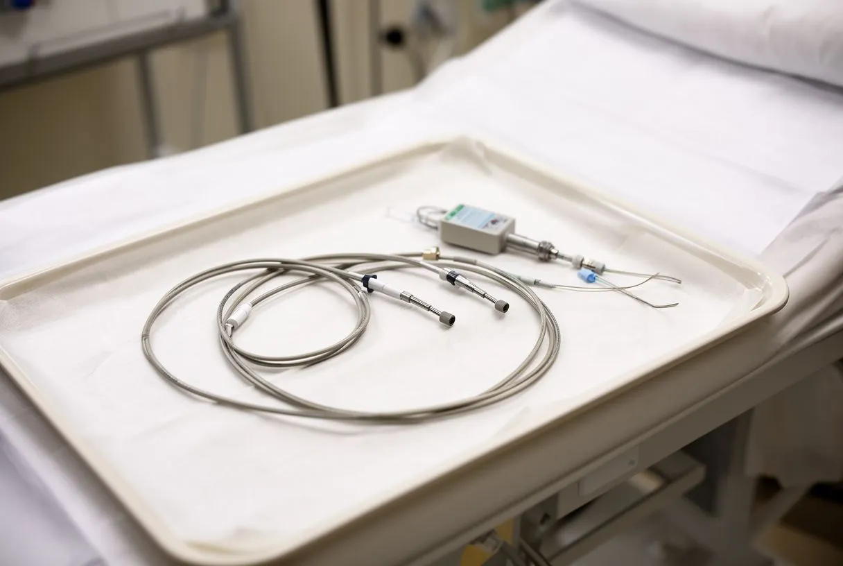
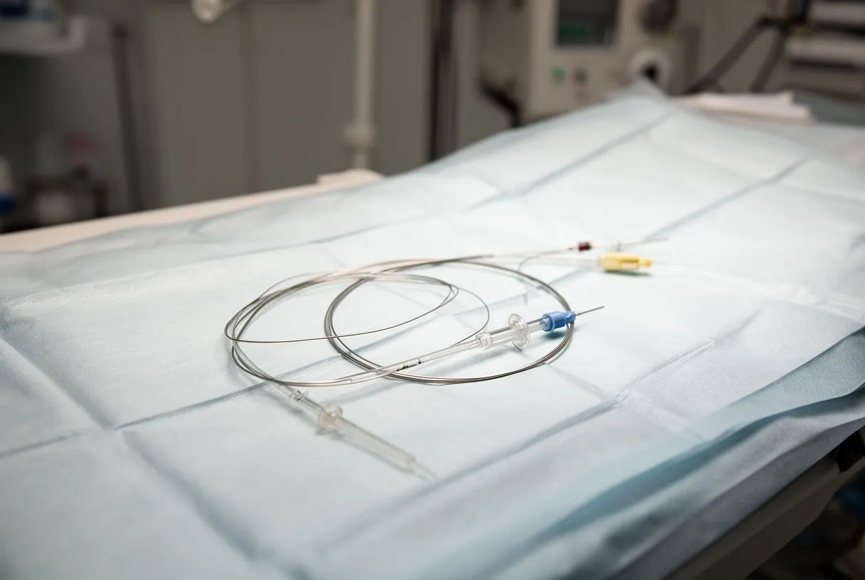
The catheter itself. Cardiac catheterisation uses a catheter roughly the width of a pencil lead, with pressure sensors at its tip. It is introduced through an artery or vein — usually at the wrist or the groin — and guided under X-ray imaging to the right side or left side of the heart. Because the sensor sits inside the chamber itself, the readings are direct rather than estimated.
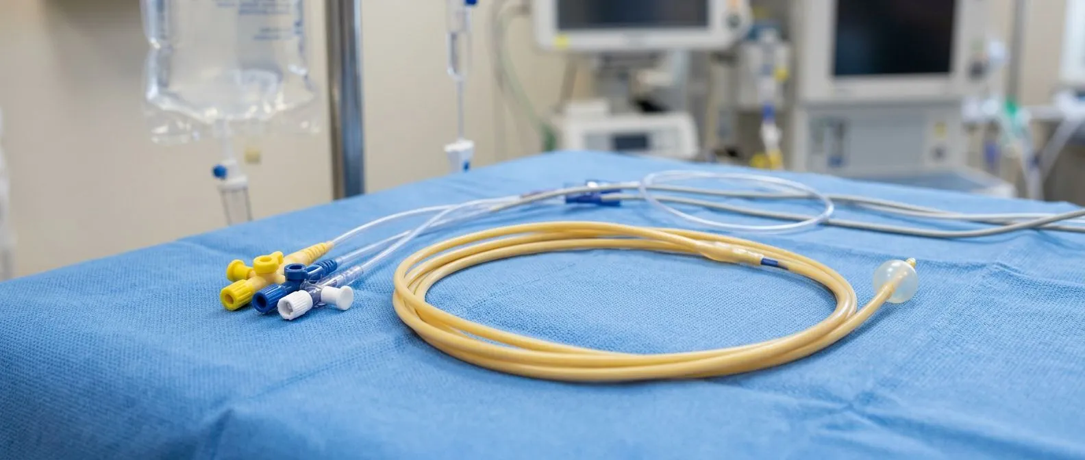
Right-heart catheterisation and the Swan-Ganz catheter. A Swan-Ganz catheter heart test is the classic right-sided study. This soft, balloon-tipped catheter travels through the right atrium, the right ventricle and into the pulmonary artery, recording pressures along the entire path. This pulmonary artery pressure test is the standard way to confirm or rule out pulmonary hypertension — abnormally high pressure in the lung circulation.
Left-heart catheterisation. A left-sided study passes through the artery into the aorta and left ventricle. It measures the pressure the left ventricle generates with each beat and, when the aortic valve is narrowed, compares the pressure above and below the valve to calculate the severity of the narrowing. This is the interventional cardiology pressure study that most often precedes a valve decision. We find that when we explain these pressure curves to patients in plain words, the whole decision about a valve becomes far less frightening.
What Is the Procedure for a Haemodynamic Study?
Step by step. The heart pressure measurement procedure follows a predictable sequence. Knowing the order of events removes much of the anxiety patients bring to the day.
- Access: the wrist or groin is numbed with local anaesthetic, and a thin sheath (a short plastic tube) is placed into the vessel.
- Guidance: the catheter is advanced toward the heart under X-ray fluoroscopy; you cannot feel it travelling.
- Measurement: pressures are recorded in each chamber and vessel; blood samples may be taken for oxygen levels.
- Optional extras: contrast dye may be injected (coronary angiography with haemodynamics), or medications may be given to test the heart's response.
- Removal: the catheter and sheath are removed and pressure is applied, or a small closure device is used.
What you feel. You are awake but mildly sedated for most of the test. The only sensation most patients report is the local anaesthetic sting, a moment of warmth if dye is injected, and a fluttering or "skipped beat" feeling when the catheter touches the ventricle. That flutter is normal and brief.
Combining tests. Often the same catheter session includes coronary angiography (dye pictures of the heart arteries), since the access route is already in place. This avoids a second procedure and gives the team a complete picture in one sitting — one reason a combined study is frequently the most efficient option for patients.
Haemodynamic Study Preparation: What to Do Before
Medications. Haemodynamic study preparation is usually straightforward. You will be told whether to take your usual blood thinners, diabetes tablets or insulin on the day — instructions vary, so follow your own team's written plan rather than general advice. Bring a full list of every medication and supplement you use.
Fasting and logistics. You will typically be asked not to eat for six to eight hours beforehand, though small sips of water are usually allowed. Arrange for someone to drive you home, since the sedation makes driving unsafe for the rest of the day. Wear loose, comfortable clothing and leave jewellery at home.
Questions to ask in advance. A good team will welcome your questions before the day. Useful ones include: will my study include coronary angiography? Will I need sedation and how will I feel afterwards? Who will explain the results to me, and when? Our first contact is a free video consultation: we get to know the patient, talk through the condition, and say which studies are still needed so no unnecessary ones are ordered.
Haemodynamic Study vs Echocardiogram: What's the Difference?
Estimate versus measurement. The haemodynamic study vs echocardiogram question comes up constantly, and the distinction is simple: an echocardiogram is an ultrasound scan that estimates pressures from blood flow patterns, while a catheter study measures them directly. Echo is painless, quick and free of risk; catheterisation is invasive but definitive.
When echo is enough. For most screening and follow-up, echo is genuinely sufficient. It is the right first test for murmurs, breathlessness and valve surveillance. Many patients never need a catheter study at all because their echo results are clear enough to guide treatment.
When the catheter adds something echo cannot. The invasive study is requested when echo and clinical findings disagree, when pressures are borderline and the treatment decision hinges on precision, when pulmonary hypertension needs confirmation, or when a procedure such as TAVR or stenting is planned. In those situations, the measured numbers — not estimates — are what the whole decision rests on.
Haemodynamic Parameters Normal Values Explained
The numbers your report shows. Haemodynamic parameters normal values are broadly stable across adults, though exact reference ranges vary slightly between laboratories. Your report will list measured pressures alongside these reference values, and the interpretation is your cardiologist's job — the numbers below are simply to help you read the report with understanding.
What is a bad cardiac output number? A normal cardiac output is roughly 4 to 8 litres per minute for an adult at rest, and it is often adjusted for body size as a "cardiac index" of about 2.5 to 4 litres per minute per square metre. A cardiac index persistently below about 2.2 is generally considered low and is one marker of cardiogenic shock — a situation where the heart cannot pump enough to sustain the organs.
What is the normal range for systemic vascular resistance? Systemic vascular resistance (SVR) describes how tightly the body's arteries are squeezed. A typical normal range is roughly 800 to 1,200 dynes·sec/cm⁵. A very high SVR means the heart is working against stiff pipes; a very low SVR, as in sepsis or certain shock states, means the pipes are too relaxed and blood pressure collapses.
Chamber pressures. In rough terms: right atrial pressure sits around 2 to 6 mmHg, pulmonary artery pressure around 15 to 30 mmHg at its peak, and the left ventricle reaches about 100 to 140 mmHg with each contraction while relaxing to a low single-digit or low-teens diastolic pressure. Consistently elevated filling pressures point toward heart failure or stiffening of the heart muscle. A detailed review of haemodynamic principles and their clinical use can be found in the medical literature, for example through Europe PMC.
Heart Catheter Test Risks: What Could Go Wrong?
The honest risk picture. Heart catheter test risks are real but small when the test is done for a clear indication. The most common issues are minor: bruising or bleeding at the access site, temporary soreness, or an allergic reaction to the contrast dye. These almost always settle with simple care.
Less common complications. Serious complications — heart rhythm disturbance during catheter passage, vessel injury, stroke, kidney injury from contrast, or heart attack — are rare and occur far more often in patients who are already severely ill or have advanced kidney disease. This is precisely why the test is ordered only when the information it provides changes the treatment plan. Our team reviews every case against international practice guidelines before recommending an invasive study, so that no patient is exposed to catheter risk without a clear reason.
Telling your team what matters. Before the test, mention any previous reactions to contrast dye, kidney problems, bleeding disorders, or allergies to latex or adhesives. Also mention if you might be pregnant. These details let the team adjust the plan — different dye, different access site, or additional monitoring — and keep the risk as low as possible for your situation.
How Long Does a Haemodynamic Study Take, and What Is Recovery Like?
The duration. How long does a haemodynamic study take? A straightforward right- or left-heart study typically lasts 30 to 60 minutes. When coronary angiography or a combined right-and-left study is included, allow 60 to 90 minutes. Pre-procedure preparation and post-procedure monitoring extend the total hospital visit to most of a day.
Going home. Most patients go home the same day. If the wrist was used, a few hours of rest and a light dressing are usually enough. If the groin was used, you will lie flat for a few hours so the puncture seals, and you should avoid heavy lifting, climbing stairs quickly, and driving for about 24 to 48 hours.
Getting the results. Many teams discuss the preliminary findings with you before you leave, and a full written report follows within days. The results feed directly into your treatment decision — whether that is medication, TAVR, conventional surgery, or simply continued monitoring. Research on cardiovascular testing has shown how central these studies remain to decision-making; for instance, the INCAPS COVID 2 study documented the recovery of cardiovascular testing in Asia after the pandemic, and work published in the Journal of the American Heart Association has used advanced cardiopulmonary exercise testing to investigate lingering exercise intolerance after COVID-19.
What drives the price. The haemodynamic study cost in Mexico varies mainly with whether the study is combined with coronary angiography or an intervention, which hospital and catheterisation suite is used, and whether you are an international patient requiring additional coordination. The catheter lab time, the contrast, the closure device and the anaesthesia support all form part of the total. Always ask for a written, itemised estimate before the day — reputable teams provide one without hesitation.
For patients travelling from abroad. Many international patients combine their study and their treatment consultation into a single trip. To do that well, send your previous reports — echo results, ECGs, blood tests including Liver Function Tests if surgery is being considered, and any earlier catheter reports — before you travel. This lets the team confirm which studies are genuinely still needed rather than repeating everything.
Second opinions are normal. If you are facing a decision about valve surgery or another intervention, a second opinion is not a lack of trust — it is a normal part of deciding. A good team will tell you exactly which studies were done, what they showed, and what the alternatives are. We actively encourage second opinions and treat seeking one as a normal part of deciding about heart surgery; the final decision is always the patient's.

Our experience. Dr Molina trained in cardiothoracic surgery at the Universidad de Guadalajara, in cardiac, minimally invasive, robotic and structural surgery at the University of Montreal, and in structural cardiology and TAVR at CHU Bordeaux, and the team also trained at Jena, Bad Berka and Buffalo — experience we bring to interpreting every haemodynamic study our patients undergo. That background matters less as a credential than as a habit: explaining each pressure curve in plain words before anything else is proposed.
When Pressure Numbers Change the Plan
Small numbers, big decisions. It can be surprising how much of cardiac medicine turns on a handful of pressure readings. Whether a valve is repaired or replaced, whether a patient is a candidate for TAVR inside a previous TAVR (a TAVI Inside TAVI procedure, sometimes needed when a first transcatheter valve fails), or whether a patient is managed with medication alone — all of these decisions lean on measured pressures and gradients.
Other tests that fit around it. A haemodynamic study rarely stands alone. It is usually combined with an echocardiogram, an exercise or Stress Test such as a Treadmill Test, breathing studies like Pulmonary Function Tests when lung disease is suspected, and in selected cases a Viability Study or Haemodynamic Study follow-up after treatment. In intensive care, continuous Haemodynamic Monitoring with the same catheter principles guides treatment in the most unwell patients. Each test answers a different question, and together they form the complete map.
What you should take away. The test is precise, routine and brief. It carries small but real risks, which are justified when the answer changes your treatment. And it puts you — the patient — in a position to make a genuinely informed choice, with numbers instead of guesses.
In summary, a haemodynamic study heart test is the most direct way to measure the pressures and flows that determine how your heart is actually working. It clarifies valve severity, confirms pulmonary pressures, and anchors decisions about TAVR, surgery or medical treatment in measured facts rather than estimates. If you have been told you may need this test — or you have already had one and want the results explained clearly — the best next step is to discuss your own situation with a qualified cardiac team. Book a consultation rather than trying to interpret the numbers on your own; that conversation is where the real decisions get made.