


Heart Failure With Reduced Ejection Fraction: Treatment
Treatment for heart failure with reduced ejection fraction at Cirugía de Corazón in Guadalajara: medications, devices and surgery, and how to book a consultation.
What Is Reduced Ejection Fraction Treatment?
Reduced ejection fraction treatment is the medical approach used when the heart's main pumping chamber, the left ventricle, can no longer squeeze forcefully enough to push an adequate volume of blood out to the body. Ejection fraction is the percentage of blood that leaves the ventricle with each heartbeat. A normal value is roughly 50 to 70 percent. When it falls below 40 percent, doctors classify the condition as heart failure with reduced ejection fraction, often abbreviated HFrEF. The goal of treatment is not to restore the number to normal overnight but to protect the remaining heart muscle, ease symptoms, and extend life.
What does ejection fraction actually measure? Think of the left ventricle as a sponge filled with water. If you squeeze it gently and only a third of the water comes out, the rest stays behind, and the next refill has less room. In the same way, a weakened ventricle leaves blood pooling inside, pressure builds backward into the lungs, and fluid leaks into the breathing spaces. That is why breathlessness, fatigue, and swelling are the hallmark complaints. Treatment targets this backward pressure first, then works to remodel and protect the muscle itself.
Reduced ejection fraction treatment today is built on four pillars: medication, device therapy when indicated, management of any underlying cause, and lifestyle adjustment. Each pillar has strong evidence behind it, and they work together. A patient who takes medication faithfully but ignores salt intake, for example, may still land in the emergency room with fluid overload. Understanding the whole plan is the first step toward living well with this condition.
How Heart Failure With Reduced Ejection Fraction Develops
The heart does not fail overnight. Reduced ejection fraction usually follows years of stress on the left ventricle. The most common driver in our patient population is coronary artery disease, where cholesterol-laden deposits called atheromatous plaque narrow the vessels that feed the heart muscle. After a heart attack, the muscle that lost its blood supply turns into scar tissue, which cannot contract. The surviving muscle must compensate, and over time it stretches and weakens.
What happens to the ventricle over time? The ventricle dilates, becoming rounder and thinner-walled. This shape change, called remodeling, reduces mechanical efficiency. As the European Society of Cardiology guidelines emphasize, interrupting remodeling early is one of the most powerful ways to improve long-term outcomes. Other causes include longstanding high blood pressure, leaky or tight valves, viral myocarditis, genetic cardiomyopathies, and certain chemotherapy drugs. In some patients no single cause is found, and the condition is labeled idiopathic dilated cardiomyopathy.
Once the pump weakens, the body activates compensatory systems that initially help but eventually harm. The sympathetic nervous system raises heart rate and squeezing force; the renin-angiotensin-aldosterone system retains salt and water to keep blood pressure up. Short term, these are survival mechanisms. Long term, they force the already struggling ventricle to work harder, accelerating damage. Modern reduced ejection fraction treatment is designed specifically to blunt these neurohormonal loops, which is why the medications used are so different from those prescribed for other conditions.
Signs You May Need Reduced Ejection Fraction Treatment
Symptoms of reduced ejection fraction can appear gradually, and many patients attribute them to aging or being out of shape. The most common complaint is exertional breathlessness, initially with heavy activity, later with walking across a room or lying flat at night. Orthopnea, the need to prop up on extra pillows to breathe comfortably, is a classic sign that fluid is backing up into the lungs. Paroxysmal nocturnal dyspnea, waking gasping after an hour of sleep, signals the same process.
What warning signs should prompt urgent evaluation? Sudden weight gain of two kilograms or more in three days, swelling in the ankles and lower legs that does not resolve overnight, and a persistent dry cough with pink frothy sputum all point to decompensated heart failure. Arm pain or chest pressure radiating to the jaw, especially with sweating, may indicate the coronary arteries are critically narrowed rather than the heart simply weak. A clinician can distinguish these through auscultation, listening for crackles in the lungs and a third heart sound, and by measuring biomarkers. One study on biomarker screening in atrial fibrillation patients, published in Screening of Multiple Biomarkers Associated With Ischemic Stroke in Atrial Fibrillation, illustrates how blood markers help stratify cardiovascular risk beyond the echocardiogram.
Other symptoms include profound fatigue, loss of appetite, and difficulty concentrating. Some patients notice palpitations or an irregular pulse, which may reflect atrial fibrillation or atrial flutter, both common when the atria stretch under pressure from a failing ventricle. If you recognize any combination of these signs, the next step is an echocardiogram, a painless ultrasound that measures ejection fraction directly and shows wall motion, valve function, and chamber size. From there, a cardiologist can determine whether reduced ejection fraction treatment is appropriate and how urgently it should begin.
Core Heart Failure Medications for Low Ejection Fraction
Medication forms the foundation of reduced ejection fraction treatment. Four drug classes have repeatedly shown survival benefit in large trials, and most patients take a combination of all four, introduced gradually and titrated to target doses over weeks to months. Understanding what each one does helps patients tolerate side effects and stay on therapy.
Which medications are considered the four pillars of heart failure therapy? The first is an angiotensin receptor-neprilysin inhibitor, often abbreviated ARNI, or an ACE inhibitor or angiotensin receptor blocker if ARNI is not available. These drugs block the renin-angiotensin system, reducing the harmful remodeling described earlier. The second pillar is a beta blocker, specifically carvedilol, metoprolol succinate, or bisoprolol, which lowers heart rate and protects the heart from adrenaline-driven stress. The third is a mineralocorticoid receptor antagonist, spironolactone or eplerenone, which blocks aldosterone and reduces scarring. The fourth is an SGLT2 inhibitor, originally a diabetes drug, now proven to improve outcomes in heart failure regardless of whether the patient has diabetes.
Diuretics, most commonly furosemide, are often added for symptom control when fluid overload is present. Unlike the four pillars, diuretics do not improve long-term survival directly, but they relieve breathlessness and swelling within hours, which is why patients feel better quickly after starting them. Every medication plan must be individualized; blood pressure, kidney function, and potassium levels guide dose adjustments. Patients should never stop these drugs abruptly, as rebound worsening can be severe. A practical summary of the medication classes and their roles:
- ARNI or ACE inhibitor: reduces harmful remodeling and lowers hospitalization risk
- Beta blocker: slows heart rate and shields muscle from adrenaline stress
- Mineralocorticoid antagonist: blocks aldosterone, reduces scarring and fluid retention
- SGLT2 inhibitor: improves outcomes in HFrEF independent of diabetes status
What Is Cardiac Resynchronization Therapy?
In some patients with reduced ejection fraction, the two ventricles do not beat in synchrony. The electrical signal that tells the heart to squeeze arrives late on one side, usually the left, so the ventricle contracts in a wobble rather than a smooth squeeze. This is called left bundle branch block, and it wastes energy, worsens symptoms, and lowers the effective pumping output even further. Cardiac resynchronization therapy, or CRT, is a device-based treatment designed to restore coordinated timing.
How does a biventricular pacemaker work? A cardiologist threads thin leads into the right atrium, the right ventricle, and a vein on the surface of the left ventricle that drains into the coronary sinus. The device, implanted under the skin below the collarbone, sends tiny pulses timed so both ventricles contract together. The procedure typically takes one to two hours and requires a short hospital stay. Candidates are selected carefully based on the echocardiogram, the electrocardiogram, and symptoms. The strongest evidence supports CRT in patients with an ejection fraction at or below 35 percent, a wide QRS complex on the ECG (usually left bundle branch block), and persistent symptoms despite optimal medication. When the right patient receives the right device, symptom improvement can be substantial, and some remodeling may even reverse.
CRT is not a cure, and not every patient improves. Roughly one in three recipients does not respond significantly, and identifying non-responders before implant remains an active area of research. Risks of the procedure include infection, lead dislodgement, and the need for generator replacement every several years when the battery depletes. For patients who do respond, the benefit is real: more energy, fewer hospital admissions, and better exercise tolerance. The decision to implant should follow a detailed conversation with a cardiac electrophysiologist or heart failure specialist who can weigh the expected benefit against the surgical risk for your specific situation.
How ICD Implantation Protects Against Sudden Cardiac Arrest
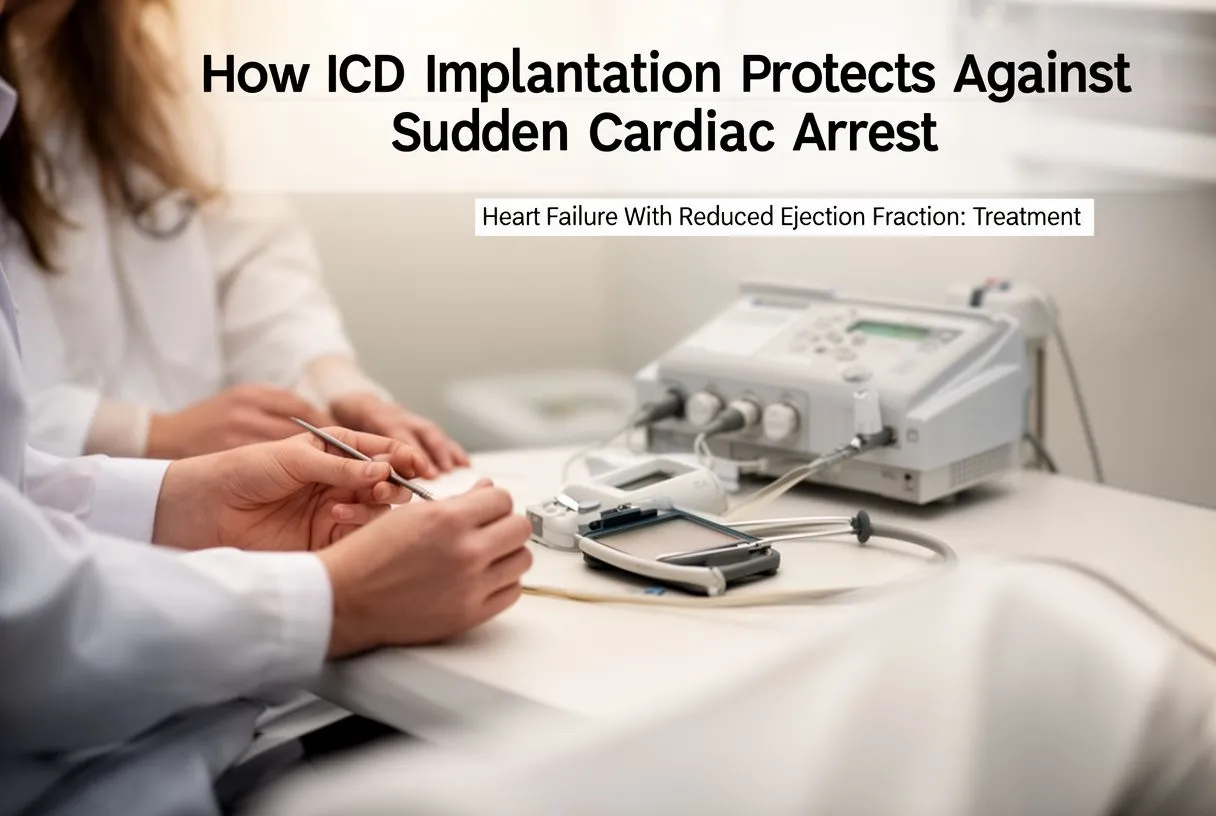
Patients with reduced ejection fraction face a higher risk of sudden cardiac arrest, usually caused by ventricular tachycardia or ventricular fibrillation, chaotic rhythms that stop effective blood flow within seconds. An implantable cardioverter-defibrillator, or ICD, is a device that monitors the heartbeat continuously and delivers an internal shock to restore normal rhythm if a dangerous arrhythmia appears. For many patients, an ICD is combined with CRT in a single device called a CRT-D.
Who should consider an ICD for primary prevention? Guidelines generally recommend prophylactic ICD implantation for patients with an ejection fraction at or below 35 percent despite at least three months of optimal medication, provided the expected survival with good quality of life is longer than one year. This is called primary prevention because the patient has not yet had a life-threatening arrhythmia. For patients who have already survived cardiac arrest or documented sustained ventricular tachycardia, the indication is clearer and called secondary prevention. The decision always balances benefit against risks, which include inappropriate shocks, infection, and lead complications over the device lifetime.
It is important to distinguish an ICD from a standard pacemaker. A pacemaker prevents the heart from beating too slowly; an ICD prevents it from stopping. An automated external defibrillator, or AED, is the external version found in public spaces and is used by bystanders during cardiac arrest emergencies. The internal device works automatically and silently until the moment it is needed. Patients with an ICD can usually lead full, active lives, though they receive specific instructions about avoiding strong magnetic fields and monitoring for signs of device infection. Knowing what the device can and cannot do removes much of the fear that surrounds it.
When Is Surgery Part of Reduced Ejection Fraction Treatment?
Reduced ejection fraction treatment is primarily medical, but surgery plays a critical role when a correctable structural problem is driving the weakness. The clearest example is severe coronary artery disease: if major vessels are blocked and the muscle is still viable, restoring blood flow through coronary artery bypass grafting can improve both symptoms and survival. The decision depends on angiography, viability imaging, and the overall surgical risk calculated with tools such as EuroSCORE II.
What valve problems can worsen heart failure? A leaky mitral valve, called mitral regurgitation, is common when the left ventricle dilates. As the chamber stretches, the valve leaflets no longer meet in the center, and blood flows backward with each beat, adding volume load to an already struggling pump. Repairing the mitral valve, sometimes with a minimally invasive clip device, can reduce this backward flow. Similarly, severe aortic stenosis, a tight aortic valve, forces the ventricle to push against high resistance and can eventually cause heart failure. Transcatheter aortic valve replacement, called TAVR, is an option for many patients, especially those at higher surgical risk, where a balloon-expandable valve is delivered through a catheter rather than open chest. When the aortic valve is the problem in the ascending aorta, surgery may be combined with graft replacement of the enlarged segment.
Another surgical option in selected centers is left ventricular reconstruction, which surgically reshapes the ventricle after a large heart attack has left it scarred and aneurysmal. This is a complex operation reserved for specific anatomy and is not routine. The key principle is that surgery in this setting is never isolated from medication. The four pillars continue before and after the operation, and the surgical plan is made jointly by a heart failure cardiologist and a cardiac surgeon. If you are told you need surgery as part of reduced ejection fraction treatment, ask your team whether the procedure is intended to improve survival, relieve symptoms, or both, and what the expected recovery timeline looks like for your specific case.
Exercise and Lifestyle in Low Ejection Fraction Management
Patients sometimes fear that activity will further harm a weakened heart, but the opposite is true. Cardiac rehabilitation, a structured and supervised exercise program, has been shown to improve exercise capacity, reduce hospitalizations, and enhance quality of life in stable heart failure patients. The program typically combines aerobic training on a treadmill or bicycle, light resistance work, and education on self-care, all monitored by trained staff.
What daily habits actually move the needle? Sodium restriction to roughly two grams per day is one of the most effective self-management steps, because excess salt drives fluid retention regardless of diuretic dose. Daily weight monitoring catches fluid accumulation before symptoms develop; a gain of more than two kilograms in three days warrants contacting the care team. Fluid intake may also be limited in advanced cases, usually to one and a half liters daily. Alcohol should be minimized or avoided, as it can depress heart function and trigger arrhythmias. Smoking cessation is essential and non-negotiable. Vaccination against influenza and pneumococcal disease reduces the risk of respiratory infections that can precipitate decompensation.
Mental health deserves attention too. Depression is common in heart failure and worsens adherence to medication and lifestyle recommendations. Addressing it, whether through counseling, medication, or social support, is part of comprehensive care. The overall message is that reduced ejection fraction treatment is not passive. The patient who understands the plan, follows the salt and fluid rules, weighs daily, and exercises within prescribed limits does measurably better than one who relies on pills alone. Lifestyle adjustment is not an add-on; it is a genuine therapeutic pillar.

Advanced Heart Failure Options When Medications Are Not Enough
When maximal medication and device therapy no longer control symptoms, the condition is classified as advanced heart failure. At this stage, options narrow but do not disappear. The conversation shifts from optimization to consideration of more aggressive interventions, each with its own trade-offs and candidacy requirements. This is the point at which referral to a specialized advanced heart failure center becomes essential.
What therapies exist for end-stage reduced ejection fraction? Mechanical circulatory support, most commonly a left ventricular assist device or LVAD, is a pump implanted inside the chest that takes over the work of the left ventricle. It is used as a bridge to heart transplantation or, in patients who are not transplant candidates, as destination therapy to improve symptoms and prolong life. The device requires permanent external power through a driveline exiting the skin, and patients take anticoagulation to prevent clots. Heart transplantation remains the gold standard for suitable candidates, offering the best long-term survival and quality of life in truly end-stage disease, though it is limited by donor availability and the need for lifelong immunosuppression. In acute, rapidly deteriorating scenarios, extracorporeal membrane oxygenation, sometimes as awake ECMO, can temporarily support heart and lung function while a decision is made.
Not every patient is a candidate for these therapies. Age, other medical conditions, frailty, and social support all factor in. Some patients choose palliative care focused on symptom relief and quality of life rather than prolongation, and this is a legitimate, respected path. An article discussing anticoagulation choices, Rivaroxaban in stroke prevention in patients with non-valvular atrial fibrillation: what facts are important for the clinician?, reminds us that even in advanced disease, managing associated conditions like atrial fibrillation remains critical to reducing stroke risk. The right choice depends on goals, values, and an honest appraisal of expected benefits and burdens, made together with the care team and family.
Recovery and Prognosis With Reduced Ejection Fraction Treatment
Recovery in heart failure does not mean a return to the heart you had before. It means stabilization, symptom relief, and the ability to live meaningfully within the new limits. Prognosis has improved dramatically over the past two decades because of the four-pillar medication approach and device therapy, but outcomes still vary widely based on cause, adherence, and how early treatment began. Patients diagnosed promptly and treated aggressively can live many years with good quality of life.
What does the recovery timeline actually look like? In the first weeks after starting or adjusting medication, symptom improvement is often noticeable as diuretics clear excess fluid and the breathing eases. Full medication titration to target doses takes several months, with regular clinic visits and blood tests to monitor kidney function and potassium. After CRT or ICD implantation, local healing takes two to four weeks, with restrictions on heavy lifting and driving during that period. Cardiac rehabilitation usually begins once the team confirms clinical stability and continues for three months, after which patients transition to a self-directed maintenance program. Some recovery milestones to expect:
- First month: diuretic adjustment, symptom relief, baseline labs
- Months two to six: medication titration to target doses, cardiac rehab
- Long-term: regular echocardiograms, device checks, lifestyle maintenance
Knowing when to seek help is part of living with this condition. Sudden weight gain, worsening breathlessness, fainting, or a single ICD shock warrant a prompt call or emergency visit. Multiple shocks in a short period are an emergency. Regular follow-up, typically every three to six months once stable, allows the team to catch subtle changes before they become crises. The prognosis with reduced ejection fraction treatment is far better than it was a generation ago, but it requires partnership. If you or a family member are facing this diagnosis, the most important step is to connect with a cardiac team that listens, explains, and builds a plan around your specific situation. We encourage you to bring your questions to a consultation and discuss your options in person rather than relying on self-assessment.






