Inferior Vena Cava Filter: When It Is Placed and Removed
Inferior vena cava filter removal explained: when placement is temporary, how removal works, recovery time, and warning signs.
Many patients leave the hospital after a blood clot diagnosis with a small metal device in a major vein and very few plain answers about it. If you are facing inferior vena cava filter removal — or you are simply trying to understand why a filter was placed — this guide walks through the whole story: what the device is, where it sits, how it is inserted, how long it can stay, how it is taken out, and what recovery really looks like. Our aim is that by the end you can ask your own surgical team sharper, calmer questions.
What Is an Inferior Vena Cava Filter?
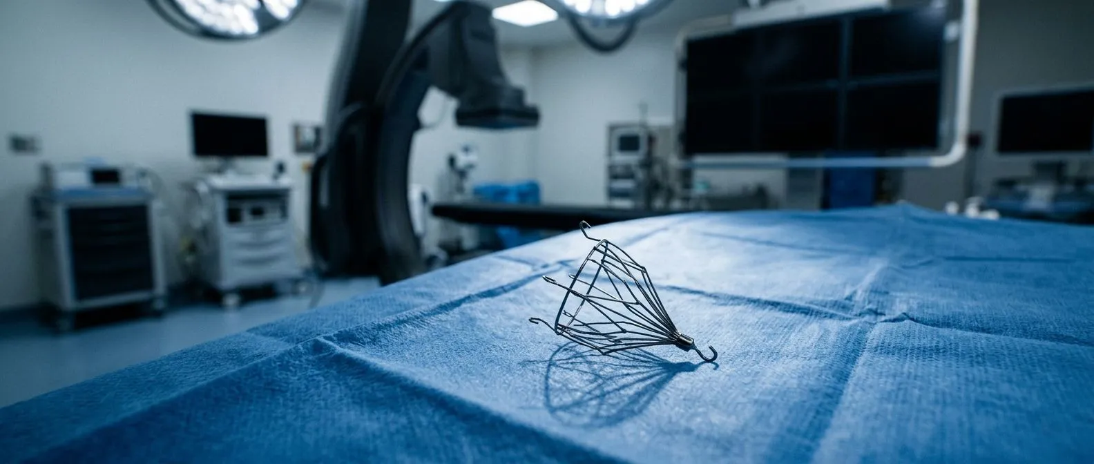
An inferior vena cava filter is a small, umbrella-like device made of thin metal wires, usually nitinol or stainless steel. It is placed inside the body's largest vein to catch blood clots that are travelling upward from the legs toward the lungs. The wires work like the safety net in a climbing wall: blood flows freely through the gaps, but a clot large enough to be dangerous gets trapped before it can reach the heart.
Does an IVC filter have metal? Yes — every commercially available filter is metallic, which is why it shows up clearly on X-rays and CT scans. The metal is biocompatible, meaning the body tolerates it without rejecting it, but it is not designed to dissolve or disappear. Some modern designs use resorbable materials that fade away over months, but these are still being studied and are not yet standard care.
Filters come in two broad families: permanent and retrievable. A permanent filter is intended to stay for life. A retrievable filter — sometimes called an optional filter — is built so it can be removed once the danger of clots has passed. Today most filters placed around the world are retrievable, because medicine has learned that leaving a filter in for years creates its own problems.
Where Is the Inferior Vena Cava Located?
The anatomy in plain words. The inferior vena cava is the largest vein in the human body. It runs up the right side of your spine, roughly behind the liver, and collects all the blood returning from your legs, pelvis, and abdomen. Near the top of the chest it empties into the right atrium, the lower-right chamber of the heart. Its partner, the superior vena cava, drains blood from the head, neck, and arms.
Knowing this location matters for two reasons. First, it explains why a filter placed in the inferior vena cava below the kidneys can intercept clots rising from the legs — the blood has no other route to the lungs. Second, it explains the routes doctors use: the vein is close to the surface at the groin (the femoral vein) and at the neck (the jugular vein), which is where catheters are inserted.
For a filter to sit safely, it must be positioned at the right level of the cava — typically below the renal veins, the veins that carry blood from the kidneys. If it drifts up or down, it can interfere with kidney drainage or fail to catch clots, which is one more reason proper imaging and experienced hands matter at every stage.
Why Do Doctors Place an IVC Filter?
Almost every filter is placed for one core reason: a patient has or is at high risk of a pulmonary embolism — a clot that travels to the arteries of the lungs and blocks blood flow — and cannot be protected with blood thinners. The usual first treatment for deep vein thrombosis, a clot forming in the deep veins of the legs, is anticoagulant medication. Filters are the backup plan when medication is not an option or has failed.
Common indications you will hear about:
- A large pulmonary embolism or deep vein thrombosis while the patient is bleeding or has a recent major surgery, making blood thinners unsafe.
- A new or recurring clot despite several weeks of properly taken anticoagulant medication.
- Certain high-risk situations, such as a massive clot in the legs before emergency surgery, or a free-floating clot in the iliac veins or the inferior vena cava itself.
It is worth understanding what a filter does not do. It does not treat the clot itself, it does not stop new clots from forming in the legs, and it is never a substitute for blood thinners when those are safe to use. Guidelines from vascular and radiology societies are clear: anticoagulation remains the foundation of treatment, and a filter is an add-on for specific situations.
How IVC Filter Placement Works — Is It Major Surgery?
No — placement is a minor image-guided procedure. An IVC filter is placed through a thin catheter, usually inserted in the femoral vein at the groin or the jugular vein at the neck, under local anesthetic and mild sedation. The doctor watches the catheter on ultrasound or X-ray guidance, positions the filter at the correct spot in the cava, and releases it. The whole procedure typically takes under an hour, and most patients go home the same day.
Because there is no open incision into the abdomen or chest, this is not surgery in the traditional sense. The puncture site is closed with pressure or a small stitch. You will be asked to keep the leg (or neck) relatively still for a few hours, then you can usually walk normally.
What we check before recommending any heart or vascular intervention: Before recommending any procedure like this, our multidisciplinary team — led by Dr Alejandro Molina — reviews the patient's imaging and history together against international practice guidelines, and the patient receives a clear proposal with the alternatives before deciding anything. That review matters because a filter is only the right answer in a specific set of circumstances; when anticoagulants are viable, they usually are the better plan.
How Long Does an IVC Filter Stay In?
The short answer: a retrievable filter should stay in only as long as the risk of pulmonary embolism is unmanageable by other means — often weeks to a few months. Many filters are approved for retrieval windows of roughly 30 to 60 days after placement, though some devices can be retrieved safely later. As soon as the patient can tolerate blood thinners again, or the temporary danger has resolved, removal should be considered.
The problem is that many filters are placed and then simply forgotten. Studies and national registries have shown that a large share of retrievable filters are never retrieved, which is why professional guidelines — including those summarized by the Cleveland Clinic — emphasize planning removal at the time of placement. Set a follow-up date in the calendar on the day the filter goes in; do not wait for someone to call you.
When the filter should stay longer or become permanent:
- A lifelong, ongoing reason why blood thinners remain unsafe — for example, a bleeding disorder or repeated major bleeding events.
- Retrieval attempts that are technically impossible, such as a filter that has tilted so far against the vein wall that removal carries more risk than leaving it.
- A patient whose clot risk is expected to return with every interruption of anticoagulation, where the team judges the filter's protection still outweighs its long-term drawbacks.
What our team watches for in the window after placement: In our consultations we track how long a patient's filter has been in place from the first video call, because the earlier the conversation about removal begins, the smoother the retrieval tends to be. Time is the quiet variable here — the longer the filter dwells, the harder it can grip the vein wall.
IVC Filter Removal Procedure: What Happens Step by Step
Removal is similar in spirit to placement: a small, image-guided procedure, not open surgery. Before anything is scheduled, the team confirms with imaging — usually an ultrasound or CT — that the filter has no clots trapped inside it and has not tilted or migrated into a position that makes retrieval unsafe.
Here is what a typical IVC filter removal procedure looks like in practice:
- 1 Pre-procedure imaging: ultrasound or CT scan of the abdomen to check the filter's position and confirm no trapped clot.
- 2 Access: a needle puncture into the jugular vein at the neck (for most retrievals) or the femoral vein at the groin, under local anesthetic and light sedation.
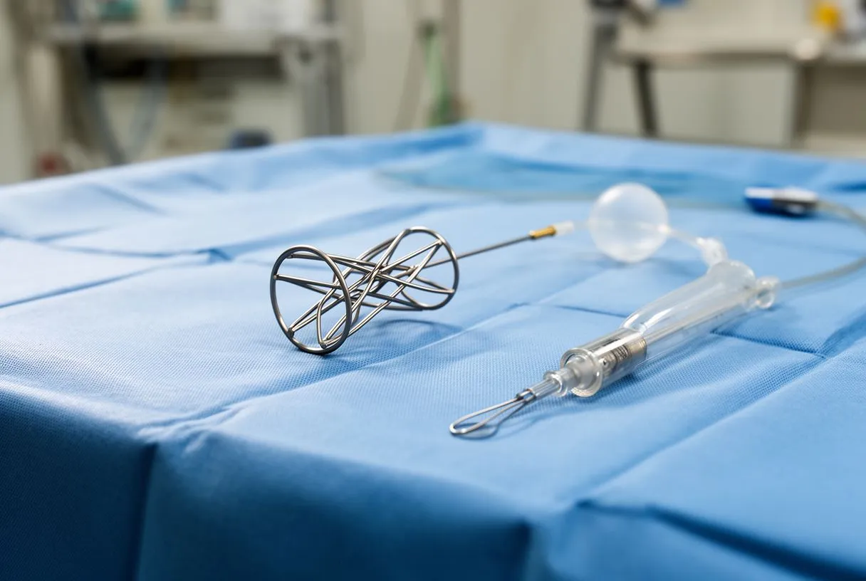
- 3 Catheter placement: a thin catheter with a snare or hook is guided to the filter's hook under live X-ray (fluoroscopy) guidance.
- 4 Retrieval: the snare grips the filter's hook, and the filter is gently pulled into a sheath and out of the body.
- 5 Closure and recovery: pressure is applied to the puncture site; you rest flat briefly, then usually go home the same day.
Some retrievals use advanced techniques when the standard snare fails — for example, repositioning a tilted filter with a balloon, or using a bronchoscopy forceps through a jugular sheath to free an embedded filter. These endovascular rescue techniques exist precisely because filter dwell time and positioning vary so much between patients. Planning the retrieval route carefully before the procedure day, rather than improvising it, matters.
Is IVC Filter Removal Painful?
Most patients report very little pain. The procedure is done under local anesthetic with sedation, so the needle puncture is numbed and many people describe the experience as pressure rather than pain. The filter itself has no nerves — the sensation comes from the catheter track and the puncture site, not from the vein where the filter sat.
Afterward, you may have some soreness or bruising where the catheter entered, especially if it was the groin. That typically fades over a few days and is managed with simple measures: acetaminophen rather than aspirin, pressure on the site for the first hours, and avoiding heavy lifting for about a day. If you are on blood thinners, the team will tell you exactly when to pause and resume them around the procedure.
When to call afterward: Expanding bruising at the puncture site, swelling of the leg, chest pain, or shortness of breath should never wait for the next appointment. Serious complications are uncommon, but a good team would rather hear about a false alarm than a missed one.
Do IVC Filters Get Clogged? Warning Signs
Yes, filters can become clogged — and this is one of the most common reasons patients start worrying about a device they have had for years. A filter's job is to trap clots, and sometimes it traps one and the clot stays there, blocking blood flow through the vein below it.
Signs that an IVC filter may be clogged:
- New or worsening swelling of both legs, sometimes with heaviness or skin darkening.
- Visible varicose veins or pelvic pressure that did not exist before.
- Abdominal or back discomfort, or in severe cases reduced urine output if the kidneys are affected.
If trapped clots block the cava, blood from the legs must find detours through smaller collateral veins, which is what causes the swelling. Diagnosis is usually made with an ultrasound first, followed by a CT scan if needed. Treatment depends on the situation: anticoagulant medication alone, catheter-directed clot-dissolving therapy, or — in some cases — removal of the filter together with clearing the clot.
There is also a deeper truth worth knowing: leaving a filter in long term is associated with complications that include not only clogging but also filter fracture, tilting, and migration of parts of the device to other locations. This is the core argument behind elective IVC filter retrieval when the original reason for placement is gone. An analysis of filter design and management published in a Ukrainian cardiovascular surgery journal — Creation of the permanent inferior vena cava filter for prevention of pulmonary artery embolism — walks through exactly how filter geometry determines both clot capture and long-term risk, and it is a useful read for anyone who wants the engineering side of the story.
IVC Filter Complications and When Removal Becomes Urgent
Most filters sit quietly and do their job. But because the device lives in a high-flow vein, several complications are recognized well enough in the literature that every patient with a filter should know the names of them.
Migration means the filter has moved from where it was placed — sometimes only a few centimeters, sometimes as far as the heart or the pulmonary arteries. Tilting means it has leaned against the vein wall, which makes the retrieval hook hard to reach. Fracture means a wire or the hook has broken off and can travel. Perforation means a strut has pushed through the vein wall toward nearby organs. Each of these changes the calculus: what was an elective, planned retrieval becomes one with a real reason to act sooner.
Warning signs that need same-day attention: A sudden change in heart rhythm, new chest pain, or imaging that shows the device has moved should be reviewed promptly, not at the next routine visit. A filter found in the heart or a pulmonary artery usually still can be retrieved by experienced interventional teams, using cardiac catheterization techniques and, in rare cases, surgical backup.
The takeaway is not to be frightened but to be informed: every complication on this list becomes more likely the longer a retrievable filter stays in place. If your filter was placed more than a year ago and no one has discussed retrieval with you, that conversation is overdue. We regularly see patients who have had a filter in place for years in another city or country, and one of the first things our team does is measure the filter's exact position and dwell time on imaging before recommending any plan.
IVC Filter Removal Recovery Time and Choosing a Specialist
Recovery is usually measured in hours, not weeks. After a routine retrieval you will rest flat for a few hours so the puncture site seals, then walk, then go home. Most people are back to normal activity — including work — within a day or two. There are no stitches to care for beyond a small bandage, and no restrictions on diet. The main "recovery" task is follow-up imaging if the team wants to confirm the vein's appearance after removal.
When you are choosing who performs the IVC filter retrieval, ask about volume and preparedness rather than promises. Useful questions include: How many retrievals does the team perform, including difficult ones with tilted or embedded filters? Is surgical support available in the building if something unexpected happens? Will imaging be reviewed by more than one specialist before the plan is finalized?
What our team offers before you commit to anything: The first contact with us is a free video consultation: we get to know the patient, explain in plain words what the imaging actually shows, and tell you exactly which studies are still needed so you are not ordered unnecessary tests — a face-to-face visit at our consulting rooms in Guadalajara, Querétaro or Hermosillo follows only when you decide to go ahead. We also treat asking for a second opinion as a normal, sensible step in deciding about any procedure, not as an insult to your current doctor.
If you have read this far because you or a family member is living with an inferior vena cava filter, the single most useful step is a clear conversation with a qualified cardiac team: how long has the filter been in, is it still needed, and what is the safest plan for removal or long-term monitoring. Inferior vena cava filter removal, when done in the right window by the right hands, is a short, image-guided procedure with a recovery measured in days — but the decision of when and whether belongs to you and your doctors together, not to a calendar alone. Bring your imaging, bring your questions, and do not self-diagnose; book a consultation and let a team that reviews every case against international guidelines help you decide.