

Inferior Vena Cava: Its Role in Heart Surgery
Inferior vena cava heart surgery explained: how surgeons access this vital vein, what procedures involve it, and recovery expectations.
When people picture heart surgery, they usually think of the heart itself — the valves, the arteries, the beating muscle. But understanding the role of the inferior vena cava in heart surgery matters just as much, because this large vein is the highway that returns blood from your legs and abdomen back to your heart. Surgeons connect to it, protect it, and in some cases operate on it directly. If you or a family member has been told a procedure involves this vessel, this guide explains what it does, what can go wrong with it, and what the treatment and recovery actually look like.
What Is the Inferior Vena Cava and Why Does It Matter?
The inferior vena cava is the largest vein in the human body. It runs upward through the abdomen, collects oxygen-poor blood from the legs, pelvis, kidneys and liver, and delivers it into the lower right chamber of the heart. From there, the blood is pumped to the lungs to pick up fresh oxygen.
Why surgeons care about this vein: nearly every open heart operation depends on it. To stop the heart safely, the surgical team must temporarily redirect the circulation to a heart-lung machine. That redirection happens through large tubes placed in the vena cavae — the two great veins that feed the heart. Without reliable access to them, the operation simply cannot proceed.
Where the two great veins differ: the superior vena cava drains the head, neck and arms, while the inferior vena cava carries everything from below the diaphragm. Both are joined into the heart-lung circuit during surgery, but the inferior one carries the greater share of the body's returning blood, which makes its handling especially important.
Is the Inferior Vena Cava on the Right Side of the Body?
Yes — in most people, the inferior vena cava sits on the right side of the spine, running up through the abdomen beside the aorta, which lies slightly to the left. This is why, during open heart surgery, the surgeon usually works on the right side of the heart's receiving chambers to reach it.
Right-sided placement has practical consequences: when surgeons open the right atrium — the heart chamber that receives blood from both cavae — they can see the openings of both veins directly. This view is essential for procedures on the tricuspid valve or for placing the cannulation tubes safely.
Natural variation exists: a small number of people are born with a duplicated vein, a left-sided vein, or a vein that takes an unusual route through the chest. These congenital variations are usually harmless on their own, but they change the surgical plan, because the team must know exactly where the vein runs before connecting to it. Good imaging before surgery — usually a CT scan with contrast dye — maps this anatomy in detail. In our practice, every case is reviewed by the multidisciplinary team against international practice guidelines before a recommendation is given, and imaging like this is exactly what that review depends on.
What Happens If the Inferior Vena Cava Is Blocked?
A blockage in the inferior vena cava prevents blood from draining freely out of the legs, pelvis and abdomen. Because the vein is so large, the body often finds detours through smaller collateral veins, and some blockages cause few symptoms at first. But when blood cannot escape quickly enough, pressure builds up behind the obstruction.
Common warning signs include:
- Swelling of both legs: fluid accumulates because blood cannot drain upward through the blocked vein.
- Abdominal swelling or pain: pressure can push fluid into the belly, and enlarged collateral veins may be visible through the skin.
- Blood clots: sluggish flow encourages clot formation, and clots can travel to the lungs, which is a serious emergency.
What causes the blockage: the usual culprits are deep vein thrombosis that scars the vein, compression from tumors or an enlarged aorta, scarring after prior surgery, or rare narrowing of the vein from birth. In our team's experience, patients often arrive having lived with leg swelling for months, assuming it was a circulation problem in the legs themselves — the vein above the legs is frequently the true cause.
Inferior vena cava obstruction treatment depends on the cause and severity. Options range from blood-thinning medication and compression stockings to balloon widening, a mesh stent that holds the vein open, or — when the blockage comes from a tumor — surgery that reconstructs the vein itself.
How Vena Cava Cannulation Works in Open Heart Surgery
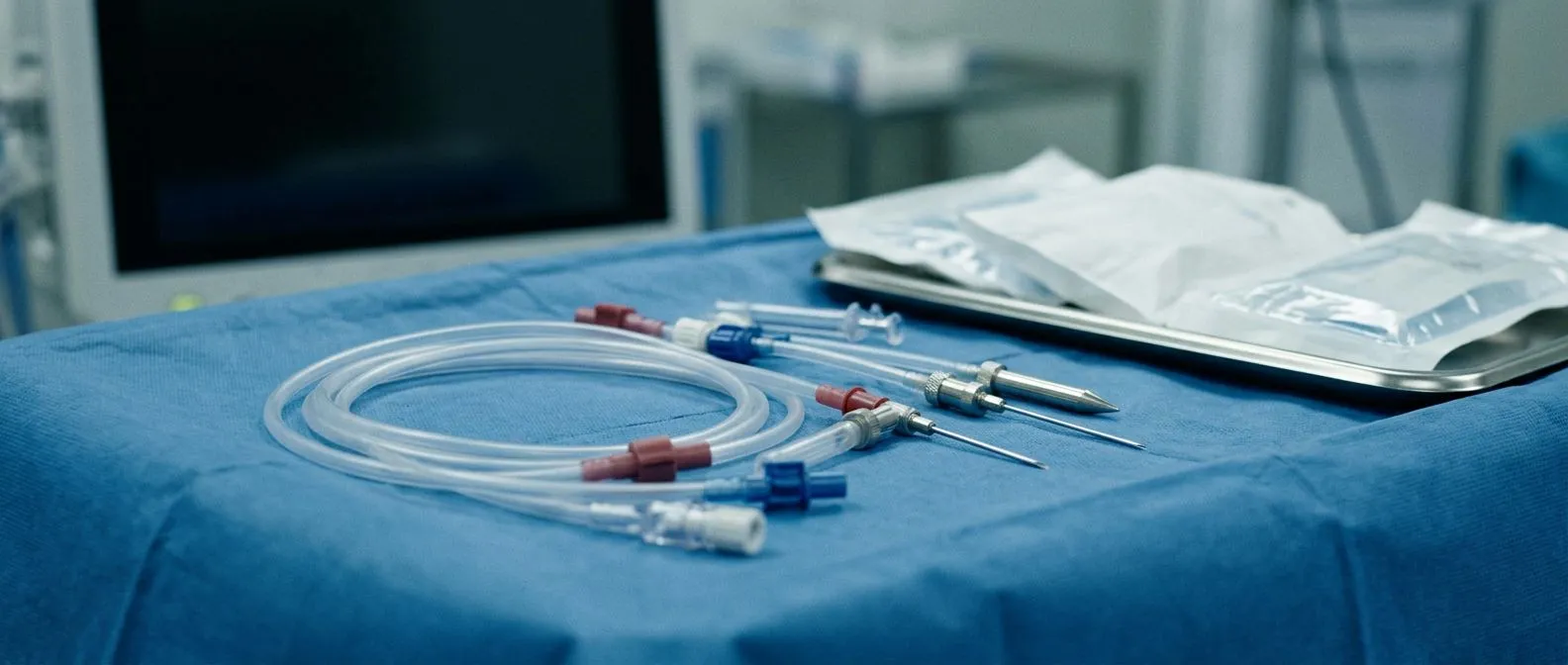
Vena cava cannulation is the step in open heart surgery where the team places large plastic tubes into the great veins so the heart-lung machine can take over circulation. Once the tubes are in, blood flows out of the body, through the machine — where it picks up oxygen and removes carbon dioxide — and back into the patient's aorta, bypassing the heart entirely.
Two cannulation techniques exist:
- Bicaval cannulation: a tube in each cava, one above and one below the heart. This lets the surgeon open the right atrium with a completely dry, blood-free field — necessary for valve work inside that chamber.
- Single venous cannulation: one long tube threaded through the right atrium that drains both veins at once, used when the right atrium does not need to be opened.
Where the tubes are placed: during conventional open surgery, the tubes are sewn directly into the right atrium through the sternum incision. In minimally invasive and robotic procedures, the cannulation can often be done through small incisions in the groin or neck, avoiding the need for a large central approach. This is one of the quiet reasons minimally invasive heart surgery has become safer over the years — the technique of peripheral cannulation has been refined enormously. Dr Molina trained in cardiac, minimally invasive and robotic surgery at the University of Montreal.
Why it matters to you as a patient: cannulation is routine, but it is also the step where the heart-lung circuit is established. A well-planned cannulation strategy — decided before the incision is ever made — is one of the marks of a careful surgical team.
What Is Inferior Vena Cava Surgery?
When people search for "inferior vena cava surgery," they are usually asking about one of several very different procedures. The vein can be repaired, widened, rebuilt, or cut open to remove something inside it. The common thread is that all of these operations involve the great vein itself rather than the heart's valves or arteries.
The main categories are:
Inferior vena cava repair: reconstructing the vein after injury or removing a section that is diseased. Surgeons may patch the vein with material to keep it wide, or replace a segment with a graft. Because the vein carries the whole lower body's return flow, repair must preserve its full width — even modest narrowing causes problems downstream.
Vena cava stenosis repair surgery: when the vein has narrowed — from scarring, prior clot, or external compression — the simplest fix is often a balloon catheter followed by a metal stent, done through a puncture in the groin rather than open surgery. Open reconstruction is reserved for blockages a stent cannot fix.
Vena cava tumor resection: the most demanding category. Kidney cancers in particular can grow into the vein and extend up toward the heart, and removing them means opening the vein under controlled conditions, sometimes with the heart-lung machine and deep cooling. This is where the vein meets cardiac surgery most directly. These cases are usually planned together with urologic surgeons, and a multidisciplinary review determines whether the operation proceeds with or without the heart-lung circuit.
Heart surgery vena cava connection: in some congenital operations — discussed below — the vein is deliberately rerouted and connected to the lung arteries, bypassing the heart's right side altogether. Here the "surgery on the vena cava" is actually a redesign of the circulation.
What Is an Inferior Vena Cava Filter and When Is It Removed?
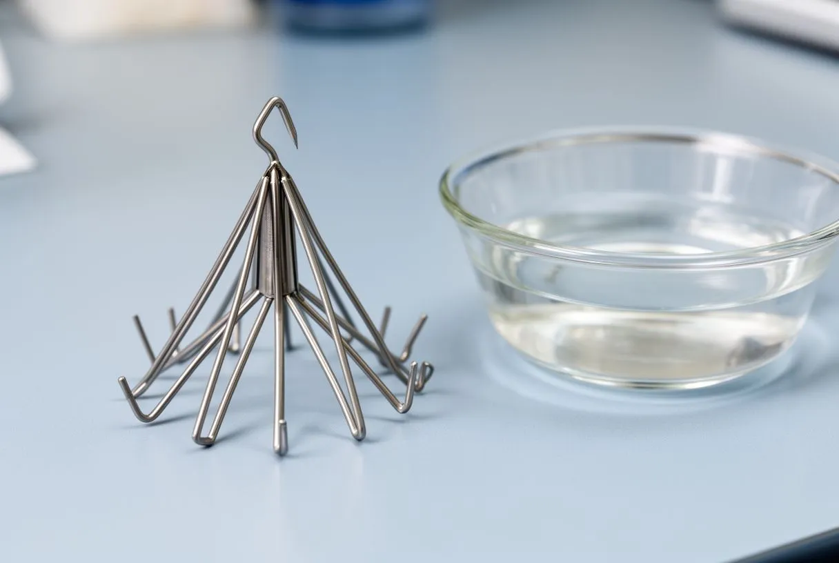
An inferior vena cava filter is a small wire basket placed inside the vein to catch clots traveling from the legs toward the lungs. It is inserted through a needle puncture in the groin and opened inside the vein — no cutting, no stitches. The concept dates back decades; the permanent filter design was created specifically to prevent pulmonary embolism in patients who could not take blood thinners (see this Creation of the permanent inferior vena cava filter for prevention of pulmonary artery embolism).
Who needs a filter: the classic patient has a dangerous clot in the leg veins plus a reason blood thinners are unsafe — active bleeding, recent major surgery, or severe trauma. The filter is a bridge for a risky window, not usually a lifetime solution.
Vena cava filter removal surgery: most modern filters are retrievable and should come out once the danger period passes — often within weeks to a few months. Removal uses the same groin puncture: a catheter grips the filter's hook and pulls it into the retrieval device. If a filter is left too long, scar tissue can grow into its struts and make retrieval difficult, which is why timely follow-up matters so much.
When a filter stays in: if the patient remains permanently unable to take anticoagulants, a permanent filter may be the plan. Long-term filters are generally harmless, but they are monitored because very occasionally a filter tilts, migrates, or pierces the vein wall, and those situations can require endovascular — or rarely open — retrieval. When a patient with a filter develops a new cardiac problem, the filter's status is usually reviewed as part of the surgical plan, because the vein needs to be free and unobstructed before it is used.
How Are Congenital Vena Cava Heart Defects Corrected?
Congenital vena cava heart defect surgery covers operations where the great veins connect to the heart abnormally from birth. The most common issues include a vein draining to the wrong chamber, a duplicated or left-sided inferior vena cava, and more complex conditions where the whole right side of the heart is underdeveloped.
The Fontan connection: in children born with a single working heart ventricle, surgeons eventually reroute the venous return directly to the lungs. The lower-body blood — from the inferior vena cava — is channeled through a tube or tunnel into the pulmonary arteries, so it flows to the lungs without ever being pumped by a heart chamber. This staged operation transforms survival for these children, though it also creates a circulation with unusual pressures that requires lifelong follow-up.
Simpler variations: a left-sided inferior vena cava that drains through an enlarged vein into the right atrium needs no treatment at all. It only becomes a surgical issue if the child needs heart surgery for another reason, when the surgeon must account for the unusual anatomy during cannulation.
What families should expect: pediatric vena cava heart surgery is performed in specialized centers with pediatric imaging, pediatric perfusion teams and intensive care. The planning is highly individual. In our team's experience, parents facing these decisions benefit most from hearing the full set of alternatives laid out plainly — the patient receives a clear proposal with the alternatives, and the final decision is always theirs.
What Is Vena Cava Syndrome and How Is It Treated?
Vena cava syndrome cardiac treatment usually refers to obstruction of one of the great veins — most often by external pressure — causing the congestion described earlier. The syndrome is named for whichever vein is blocked: superior vena cava syndrome affects the vein draining the head and arms, producing facial swelling and neck fullness, while inferior vena cava obstruction produces the leg and abdominal swelling discussed above.
What typically causes it: cancer pressing on or invading the vein is the most common cause worldwide. Benign causes include fibrous scarring in the chest, an enlarged aorta, or blood clots that have scarred the vein shut.
How treatment unfolds: the first question is always the cause. Cancer-related obstruction is usually treated by treating the cancer, with a stent to reopen the vein quickly for symptom relief. Benign narrowing responds well to balloon widening and stenting. Open surgical bypass — creating a new channel around the blockage — is reserved for patients in whom catheter-based options have failed or are not possible. The vein is a forgiving structure in one sense: because collaterals enlarge over time, some patients can be managed with medication, elevation of the legs and compression alone while the body builds its own detours.
When it becomes urgent: sudden swelling of the face, throat tightness, or rapidly worsening leg swelling with shortness of breath should be evaluated immediately. Slow, chronic congestion can be worked up calmly; sudden obstruction cannot.
Recovery After Inferior Vena Cava Surgery: What to Expect
Recovery after inferior vena cava surgery depends entirely on which procedure was done. It is worth separating the three levels of invasiveness, because they recover very differently.
Catheter-based procedures: stent placement and filter retrieval are done through a needle puncture. Most patients go home the same day or the next morning, walk normally within days, and return to desk work within a week. The main restrictions relate to the groin puncture — no heavy lifting for a short period.
Open vein reconstruction: operations that open the abdomen and rebuild the vein follow the rhythm of major abdominal surgery — several days in hospital, weeks of gradual recovery, and months before full strength returns. Blood thinners are often prescribed afterward to keep the repaired vein open, and regular imaging confirms the reconstruction stays patent.
Combined heart-and-vein surgery: when the vein is opened as part of a heart operation, recovery follows the cardiac surgery timetable — typically hospital discharge within about a week, driving after a few weeks, and a staged return to full activity over six to twelve weeks. I always tell patients the first six weeks are about healing and walking, and the strength comes back after that.
What is monitored afterward: leg swelling, signs of clot recurrence, and imaging of the vein at set intervals. Swelling that appears suddenly on one side, or pain with redness in a calf, is reported right away. Close follow-up through the first months matters, because the vein's behavior over time determines whether the repair holds.
How to Choose a Heart Surgeon for Vena Cava Surgery
Because the inferior vena cava sits at the intersection of heart surgery, vascular surgery and cancer surgery, choosing the right team matters more here than in many straightforward cardiac cases. The best heart hospital for vena cava surgery in Guadalajara — or wherever you live — is the one whose team has reviewed your specific anatomy and can explain the options honestly.
Questions worth asking any team:
- Experience with your specific problem: how many of this exact procedure has the team performed, and what does their published experience look like?
- Multidisciplinary planning: will vascular surgeons, cardiologists and — if a tumor is involved — oncology specialists all weigh in before the plan is fixed?
- Plain-language explanation: can the team explain what the studies actually show, in words you understand, and answer every question before you decide?
What our team offers: we are a multidisciplinary team of cardiac surgeons led by Dr Alejandro Molina, with consulting rooms in Guadalajara, Querétaro and Hermosillo. The first contact is a free video consultation: we get to know the patient, talk through the condition, and say which studies are still needed so no unnecessary ones are ordered. A face-to-face visit at the consulting rooms follows when the patient decides to go ahead. We perform conventional, minimally invasive, robotic and transcatheter procedures, and we recommend the less invasive route only where the evidence supports it for that patient.
A note on second opinions: we encourage second opinions and treat seeking one as a normal part of deciding about heart surgery. Any team confident in its plan will welcome another set of eyes on it. The Johns Hopkins Medicine patient guides on vascular conditions are a good place to learn the standard questions to ask before any vein-related procedure.
Bringing it all together: the inferior vena cava is far more than an anatomical footnote. It is the return highway for most of your body's blood, the gateway your surgical team connects to in nearly every open heart operation, and — in some patients — the organ that needs treatment in its own right. Whether your question involves a filter, a blockage, a tumor, or a congenital variation, the path forward starts with accurate imaging and a team that explains the options clearly. If you or a family member has been told a procedure involves this vein, discuss your specific situation with a qualified cardiac team rather than deciding alone — and consider booking a consultation to have your questions answered before any decision is made.






