

Leaflet Resection in Mitral Valve Repair
Leaflet resection mitral repair restores valve function without replacement.
If your cardiologist has told you that your mitral valve is leaking because a leaflet is prolapsing — ballooning backward with every heartbeat — you may have heard the term leaflet resection in mitral repair and wondered what it actually means. In simple words, it is a surgical technique in which a surgeon removes a small, redundant segment of the valve leaflet tissue and then rebuilds the valve so it closes completely again. It is one of the most established and durable ways to fix a leaking mitral valve without replacing it, and understanding how it works will help you ask better questions and make a calmer, better-informed decision about your own care.
What Is Leaflet Resection in Mitral Repair?
The core idea. The mitral valve sits between the left atrium and the left ventricle, the two chambers on the left side of your heart. It has two delicate, sail-like flaps — leaflets — that must meet perfectly with every beat so blood flows forward only. When a segment of leaflet becomes too long or its supporting cords stretch, part of the leaflet flops backward into the left atrium and blood leaks backward, a condition called mitral regurgitation. Leaflet resection in mitral repair means cutting out precisely that excess, diseased segment and closing the gap so the valve seals properly again.
Why repair instead of replace? Keeping your own valve tissue is almost always better than replacing it. A repaired valve keeps its natural anatomy, avoids the lifelong blood-thinning medication that mechanical replacements require, and tends to last for decades when performed by experienced teams. This is why repair is the preferred option for most degenerative valve disease, and resection of the prolapsing segment is one of the classical techniques that make repair possible.
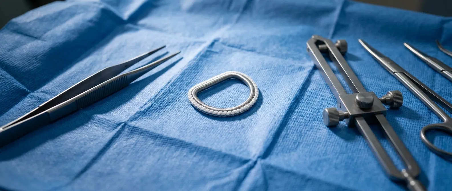
Where this technique fits. Leaflet resection is one tool in a larger toolbox. Surgeons may also use artificial chords, ring annuloplasty (a supporting band around the valve), folding techniques, or edge-to-edge approaches depending on the anatomy. The choice is individual, and a good surgical team explains which technique fits your specific valve and why.
What Is a Mitral Leaflet and Why Does It Matter?
Anatomy in plain words. Each valve leaflet is a thin, flexible flap of tissue anchored by chordae tendineae — fine, string-like cords that connect the leaflets to small muscles inside the ventricle, a bit like the guy-ropes of a tent. When the ventricle contracts, these cords hold the leaflets steady so they meet in the middle and block backward flow. When a cord stretches or ruptures, or when the leaflet tissue itself is redundant, the leaflet prolapses and the valve leaks. A clear overview of how these chordae anchor and move the leaflets is available in the literature on the chordae tendineae and their role in mitral valve repair (Journal of Tehran Heart Center).
Two leaflets, two very different jobs. The anterior leaflet is broader and generally commands a larger portion of the valve opening; the posterior leaflet is longer relative to its supporting ring and is the one most often affected by excess tissue and prolapse. That anatomical fact explains why most resection techniques were developed for the posterior leaflet: it usually has redundant scallops — named P1, P2 and P3 — and removing a small segment of the middle scallop, P2, is a well-established, highly reproducible repair.
Why precision matters. The leaflets are only fractions of a millimetre thick. Resecting too much can narrow the valve; resecting too little leaves a residual leak. This is why the technique demands intraoperative echocardiography — an ultrasound performed during surgery — so the team can confirm the valve seals perfectly before closing the chest.
Why Do Surgeons Use Leaflet Resection for Posterior Leaflet Prolapse?
Barlow's disease and fibroelastic deficiency. Most patients who need this surgery have one of two underlying conditions. In fibroelastic deficiency, one segment of the posterior leaflet has stretched and lost its strength. In Barlow's disease, the whole valve is thickened and redundant, with multiple prolapsing segments. Both can cause significant mitral regurgitation, and both are commonly treated with repair that includes resection of the affected tissue.
The goal: a competent valve with good mobility. Resection removes the excess segment that prolapses. The surgeon then re-approximates the leaflet edges, reinforces the repair with a supporting ring, and tests the valve with pressurised saline to confirm it closes evenly. The result should be a valve that leaks no more and opens freely — a balance between competence and mobility.
Evolution of the technique. Early resection methods removed larger wedges of tissue. Modern practice, influenced by techniques that "respect" rather than resect the leaflet, tends toward smaller, more targeted resections — including the so-called butterfly resection — combined with folding or artificial chords when needed. The guiding principle today is to remove only what is necessary and preserve as much native tissue as possible.
How Does Leaflet Resection Mitral Repair Work, Step by Step?
Before the operation. You will have a transthoracic and usually a transoesophageal echocardiogram (ultrasound probes on the chest and in the oesophagus) to map exactly which segment prolapses and how severe the leak is. A coronary angiography or CT scan may be added to check the heart's arteries. In our practice, every case is reviewed by the multidisciplinary team against international practice guidelines before a recommendation is given, and the patient receives a clear proposal with the alternatives — the final decision is always theirs.
During the operation. Under general anaesthesia, the surgeon accesses the heart — through a full sternotomy, a small right-side minithoracotomy, or robotically — places you on the heart-lung bypass machine, and opens the left atrium to expose the valve. The prolapsing segment is identified, the excess tissue is resected, the leaflet edges are stitched together, the repair is reinforced with an annuloplasty ring, and the valve is tested. Finally, saline testing and intraoperative echo confirm the result before the heart is closed.
After the operation. You wake in intensive care with monitoring lines in place. Most patients are sat up in a chair within a day, transferred to a regular ward within two to three days, and discharged between five and seven days after surgery, depending on how the recovery progresses.
Resect or Respect: Which Technique Is Right for You?
A genuine surgical debate. For years, surgeons have debated whether to resect the prolapsing posterior leaflet or to "respect" it — keep the tissue in place and stabilise it instead, for example with artificial chordae or folding. Randomised trials comparing resection with preservation have found that both approaches can achieve excellent, durable results, and modern practice often blends the two. A helpful overview of this "resect or respect" discussion for prolapsing posterior leaflets comes from experienced mitral surgeons (Annals of Cardiothoracic Surgery).
How the choice is made. The decision depends on the anatomy: how much redundant tissue there is, which scallop prolapses, the quality of the leaflet, and the surgeon's experience with each technique. For a single prolapsing P2 segment with excess tissue, a limited resection remains a straightforward, highly reliable option. For more complex or multi-segment prolapse, a combination of respect-and-resect strategies may give the best result.
- Resection favours: thickened, redundant posterior leaflet tissue where removing a small segment produces a predictable, durable seal.
- Respect favours: thinner or delicate tissue where preserving the leaflet and adding artificial chords or folding avoids removing native structure.
- Either way: what matters most is not the label but the result — a competent, mobile valve confirmed on echo before you leave the operating room.
In our consultation we take the time to explain what the studies actually show, in plain words, and to answer every question before any decision.
Is Mitral Valve Repair a Big Surgery?
Honest answer: it is real heart surgery, but not as heavy as it once was. Any operation on the heart is serious, and it is fair for patients to take it seriously. At the same time, mitral valve repair is one of the most refined procedures in cardiac surgery, performed routinely in experienced centres with a low rate of complications. The traditional approach uses a full breastbone incision; many patients today qualify for a minimally invasive approach through a small incision on the right side of the chest, or a robotic approach through even smaller ports.
Minimally invasive and robotic options. Small-incision and robotic repair use the same surgical principles — including leaflet resection — but avoid splitting the breastbone, which means less pain, less blood loss and a faster return to normal activity. Not every anatomy or patient is suitable, however, so candidacy has to be assessed individually. Our work covers valve disease, coronary disease, the aorta and structural heart conditions; we perform conventional, minimally invasive, robotic and transcatheter procedures, and we recommend the less invasive route only where the evidence supports it for that patient.
What to ask your team. Ask which approach they propose for your valve and why, what their repair rate is (how often they repair rather than replace), and how the result will be verified before you leave the operating room. A team that can answer these clearly is a team you can trust with the operation.

How Long Does It Take to Fully Recover from Mitral Valve Repair Surgery?
The first six weeks. Most patients leave hospital within about a week. Over the following weeks, energy returns gradually: walking daily, then climbing stairs, then light household activity. Sternum precautions apply if the breastbone was divided — no lifting more than roughly 5 kilograms and no pushing or pulling for six weeks. With minimally invasive or robotic approaches, these restrictions are lighter and shorter.
Three to six months: the full picture. Most patients feel substantially back to normal within six to eight weeks, but complete recovery — full stamina, return to demanding work and unrestricted exercise — generally takes about three months. Cardiac rehabilitation, a supervised programme of graded exercise and education, speeds and steadies this process and is strongly encouraged.
Follow-up. You will have an echocardiogram before discharge, another within the first months, and periodic checks thereafter. A successfully repaired mitral valve should remain stable for many years, and follow-up echo is how your team confirms it.
What Is the Average Life Expectancy After Mitral Valve Repair Surgery?
What the evidence shows. When mitral regurgitation is properly repaired at an experienced centre, long-term survival in many patients approaches that of the general population of the same age — the operation largely removes the burden the leaking valve placed on the heart. The exact outlook always depends on age, overall health, heart function before surgery and how promptly the repair was done, which is why earlier repair of severe regurgitation tends to protect the heart muscle.
Why timing matters. Severe mitral regurgitation makes the left ventricle work against a "escape route" — with each beat, some blood flows backward instead of forward. Over time this enlarges and weakens the ventricle. Current guidelines recommend repair before that damage becomes irreversible, which is one reason cardiologists monitor leak severity with regular echocardiograms and set thresholds for operating.
Durability of the repair. A well-performed repair, particularly for posterior leaflet prolapse, is durable: the great majority of repaired valves remain competent for decades, and fewer than about one in a hundred patients per year need a reoperation. Your own follow-up echo will track this over time.
Are There Risks with Leaflet Resection Mitral Valve Repair?
Standard surgical risks. As with any open-heart operation, the main risks include bleeding, infection, stroke, kidney strain, and rhythm disturbances such as atrial fibrillation, which is common in the first weeks and usually settles. The overall risk of serious complications at an experienced centre is low, and it is influenced strongly by age, other conditions, and how advanced the valve disease is.
Risks specific to the technique. The particular risks of leaflet resection are a residual leak if too little tissue is removed, or a narrowed valve if too much is taken — a condition called systolic anterior motion of the anterior leaflet, in which the valve obstructs outflow. Both are largely avoidable with careful planning, precise technique, and confirmation on intraoperative echo; when the surgeon sees a problem on the intraoperative test, it is corrected before you leave the operating room.
The operative plan should be matched to the patient's own anatomy — which segment prolapses, how thick the tissue is, how the chords are holding — because the safest repair is the one matched to the valve, not to a template.

How to Choose a Mitral Valve Repair Surgeon?
Volume and specialisation matter. The single strongest predictor of a durable repair is the experience of the surgeon and team. Experienced, high-volume mitral centres tend to achieve higher repair rates for degenerative disease. It is entirely reasonable — and wise — to ask a surgeon directly how many mitral repairs they perform per year and what their repair rate is for your type of valve problem.
Training and technology. Look for a team trained in the full range of approaches — conventional, minimally invasive, robotic and transcatheter — so the recommendation reflects what is genuinely best for you rather than the only technique available. Dr Molina trained in cardiothoracic surgery at the Universidad de Guadalajara, in cardiac, minimally invasive, robotic and structural surgery at the University of Montreal, and in structural cardiology and TAVR at CHU Bordeaux; the team also trained at Jena, Bad Berka and Buffalo.
Communication and second opinions. You should leave the consultation understanding your valve, your options, and the reasoning behind the recommendation. We encourage second opinions and treat seeking one as a normal part of deciding about heart surgery. A team confident in its plan welcomes scrutiny; that confidence is itself a good sign.
Practical matters for international patients. Patients travelling to Guadalajara, Querétaro or Hermosillo for surgery should plan for arrival a few days before the operation for studies and evaluation, and a stay of roughly two to three weeks after surgery before flying home. Ask in advance which tests are truly needed. The first contact is a free video consultation: we get to know the patient, talk through the condition, and say which studies are still needed so no unnecessary ones are ordered; a face-to-face visit at the consulting rooms follows when the patient decides to go ahead.
Bringing it together. Leaflet resection in mitral repair is a precise, well-established technique: the surgeon removes a small redundant segment of the prolapsing leaflet, rebuilds the valve, and confirms on ultrasound during the operation that it closes perfectly. Combined with a supporting ring and, where needed, artificial chords, it offers most patients with a leaking valve a durable repair that keeps their own tissue and avoids the commitments of replacement. The technique you read about matters less than the fit between your anatomy and your surgical team's experience — so take the results of your echocardiogram to a qualified cardiac team, ask every question you have, and book a consultation to discuss your own situation rather than deciding alone.






