

Left Atrial Appendage Closure: Device and Surgical Options
Explore left atrial appendage closure options: occlusion device vs surgical techniques.
When atrial fibrillation changes the way your heart beats, the conversation about stroke prevention often turns to blood thinners. But for many patients, long-term anticoagulation is not safe, not tolerated, or simply not the path they want to follow for the rest of their lives. That is where the discussion of left atrial appendage closure enters the room. LAA closure is a mechanical strategy that removes the source of most AFib-related strokes without forcing a lifetime of medication. Whether you are weighing a catheter-delivered left atrial appendage occlusion (LAAO) device or considering a surgical approach during another cardiac operation, understanding the full range of left atrial appendage closure options helps you sit across from your cardiac team and ask the questions that matter. This guide walks through each choice in plain language, from anatomy to recovery, so you can approach your consultation with clarity rather than anxiety.
What Is LAA Closure and Why Is It Done?
The left atrial appendage is a small, finger-like pouch that hangs off the upper-left chamber of the heart, the left atrium. In a normal rhythm, this pouch squeezes along with the rest of the heart and empties completely with every heartbeat. When atrial fibrillation takes over, that coordinated squeezing fails, and blood can pool and stagnate inside the appendage. Stagnant blood clots, and a clot that escapes this pouch can travel upward through the arterial circulation to the brain, causing a stroke. Roughly nine out of ten strokes linked to non-valvular atrial fibrillation are believed to originate from clots that form in exactly this spot. LAA closure seals off that pouch so clots cannot escape, addressing the stroke source directly rather than thinning the blood system-wide.
The decision to pursue closure rather than, or in addition to, anticoagulation usually grows out of a practical problem. Some patients bleed dangerously on blood thinners, others fall frequently, and some simply cannot adhere to the dietary monitoring that warfarin demands. Guidelines encourage clinicians to weigh stroke risk against bleeding risk using validated scores. The threshold for action is not one-size-fits-all: a high CHA₂DS₂-VASc score combined with a high HAS-BLED score is the classic scenario where mechanical closure shines. The goal is the lowest-risk approach for your situation, not a universal recommendation.
Why the appendage specifically? Because the left atrium remodels structurally in AFib, the appendage becomes the anatomical dead-end where flow is slowest. During a careful physical examination that may include auscultation, a physician can detect an irregular rhythm, but imaging studies are what reveal the appendage's size and shape. Closure can be achieved with a device placed through a catheter or with surgical techniques that stitch, staple, or clip the pouch shut. Both routes aim for the same biological outcome: no clot, no escape route, no stroke from this source.
How Does an LAA Occlusion Device Work for Atrial Fibrillation?
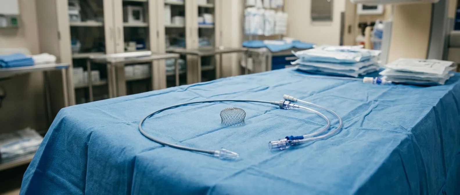
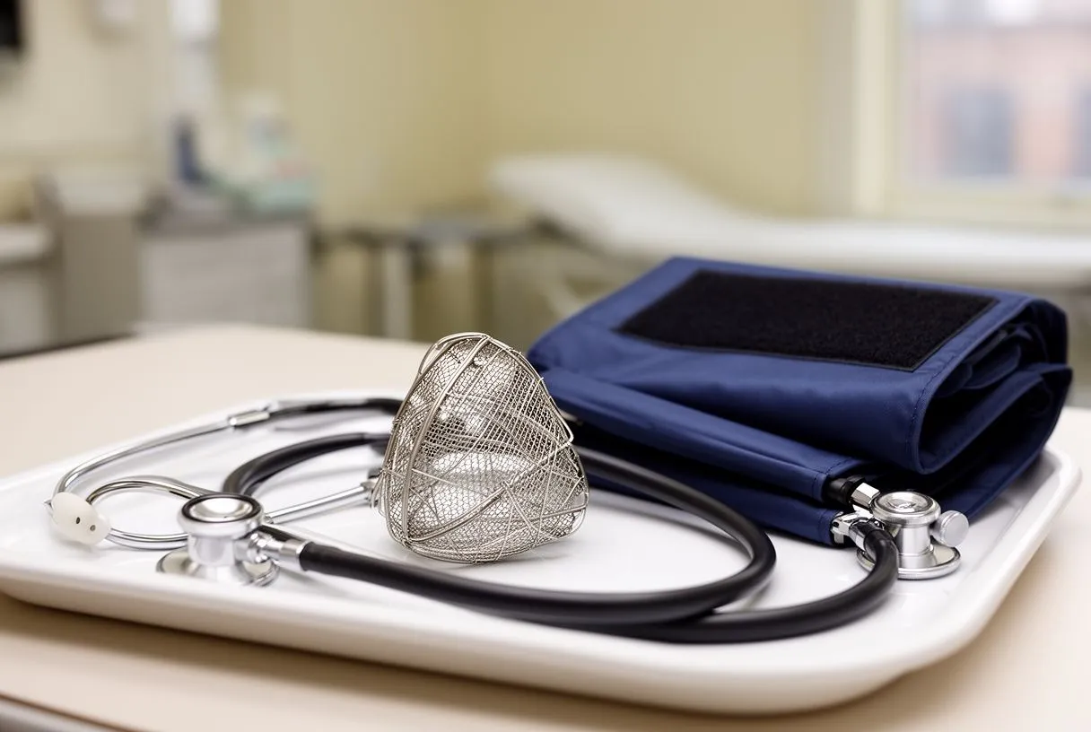
A percutaneous LAA occlusion device is a self-expanding implant, usually built on a nickel-titanium alloy frame covered with a membrane; its exact shape, for example umbrella-like, varies between devices. It is delivered through a catheter threaded from a vein in the leg, across the wall between the two upper chambers, and into the mouth of the left atrial appendage. Once deployed, the device springs open and wedges itself against the inner walls of the appendage. Over the following weeks, heart tissue grows over the membrane, forming a smooth seal that permanently blocks the appendage from the circulation. This tissue ingrowth is what makes the closure durable: the device does not simply sit there, it becomes part of the heart's inner lining.
The procedure is performed under general anaesthesia or deep sedation, typically with transoesophageal echocardiography guiding every step. Most patients spend one night in hospital and go home the next day. According to cardiology experts at Harvard Health, patient selection and imaging precision are the factors that most influence outcomes, far more than the speed of the procedure itself. The implantation takes about an hour, but the planning that precedes it, measuring appendage width and depth, ruling out thrombus, confirming anatomy, is where the real work happens.
What about the post-implant medications? The medication plan after implantation depends on the device and on your bleeding risk. A common plan is a blood thinner for roughly 45 days to prevent clots from forming on the device surface while tissue grows over it, followed by imaging to confirm whether the seal is complete. If closure is confirmed, the blood thinner is typically stopped and replaced with aspirin, which continues for several months. After six months, many patients transition to aspirin alone or even no antithrombotic therapy at all, depending on their individual risk profile. A staged medication plan of this kind is typical of the percutaneous device pathway and a major reason patients seek it out.
Who Are the Best Candidates for Left Atrial Appendage Closure?
Ideal candidates share a specific clinical fingerprint: non-valvular atrial fibrillation, an elevated stroke risk that justifies intervention, and a compelling reason to avoid long-term oral anticoagulation. That reason might be a history of serious bleeding, a high fall risk, a occupation or lifestyle incompatible with anticoagulation, or an inability to maintain the consistent monitoring that warfarin requires. The candidate evaluation is thorough and individualised. It includes a transthoracic and transoesophageal echocardiogram to measure the appendage, a CT scan in some centres for three-dimensional planning, and a bleeding-risk assessment that balances the HAS-BLED score against the CHA₂DS₂-VASc stroke score.
Not every patient with AFib is a candidate. Patients who tolerate blood thinners well and have no bleeding history are generally better served by continuing anticoagulation, because the medication also protects against clots that might form elsewhere in the heart. Patients with rheumatic mitral valve disease or a mechanical prosthetic valve are usually excluded, because their stroke risk sources differ and anticoagulation remains essential. The decision is nuanced, and a cardiac electrophysiologist or surgeon should review your full history before recommending any closure strategy.
Patients already scheduled for cardiac surgery. If you are facing a coronary bypass, a valve repair, or surgery on the ascending aorta and you also have atrial fibrillation, your surgeon can close the appendage from the outside during the same operation. This is the most efficient scenario: one anaesthetic, one recovery, and the appendage is addressed with a suture or clip while the chest is already open. For these patients, surgical left atrial appendage closure adds minimal additional risk and eliminates the need for a separate catheter procedure later.
Device vs Surgery: Which LAA Closure Option Is Right for You?
The choice between a catheter-delivered device and surgical closure hinges on three variables: whether you need another heart operation, your anatomy, and your bleeding history. A percutaneous occlusion device suits patients who are not undergoing open surgery and who want a minimally invasive, single-night-stay procedure. Surgical closure suits patients who are already in the operating room for another reason, or whose appendage anatomy is unsuitable for a device. The two approaches are not competitors in every patient; they serve different clinical situations, and a thoughtful cardiac team will recommend the route that fits your whole picture, not just the appendage.
Device closure and surgical closure achieve the same goal through different mechanics, but the evidence base differs in maturity. Device closure has been studied in large randomised trials following thousands of patients over several years, with published results comparing it directly to warfarin. Surgical closure has a longer historical track record but fewer large-scale randomised comparisons, partly because it is often performed as an adjunct to another operation. A study indexed at PubMed illustrates how researchers continue to refine understanding of closure efficacy, residual leaks, and long-term stroke reduction across both approaches.
Key trade-offs at a glance: the device route offers rapid recovery and no surgical incision, but requires temporary blood-thinning or antiplatelet medication afterward and carries a small risk of pericardial effusion. The surgical route adds no extra incision when already operating, achieves robust physical closure, but carries the risks inherent to the primary surgery itself. Neither option is universally superior; the right answer is the one that aligns with your anatomy, your other cardiac needs, and your tolerance for follow-up imaging.
What Are the Types of Surgical Left Atrial Appendage Closure?
Surgical closure encompasses several techniques, each with its own evidence and learning curve. The classic approach is direct suture ligation, in which the surgeon sews the base of the appendage closed from the outside of the beating or arrested heart. This requires precision: an incomplete suture line can leave a residual channel, and a too-aggressive purse-string can tear the fragile atrial tissue. Surgeons with high volumes of LAA work tend to favour methods that produce the most consistent seal, and the technique chosen often reflects both the patient's anatomy and the surgeon's experience.
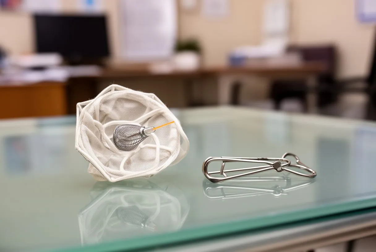
Staple and clip devices offer an alternative to hand-sewn closure. A surgical stapler can fire across the base of the appendage, creating a row of titanium clips that fold and seal the tissue in one motion. Dedicated surgical appendage clips apply a pre-formed clamp that excludes the appendage from the atrium and encourages tissue scarring. These devices standardise closure, potentially reducing the variability seen with hand suturing. The appendage is not removed in most cases; it is simply excluded from the circulation so clots cannot escape.
Open versus thoracoscopic approaches. Closure can be performed through a full sternotomy during open-heart surgery, or through minimally invasive thoracoscopic ports for a stand-alone procedure. The thoracoscopic route appeals to patients who need closure but not other cardiac surgery, though it requires specialised surgical expertise and careful patient selection. Regardless of technique, postoperative imaging is recommended to confirm complete exclusion, because even a small residual leak can allow clots to pass. Common techniques include:
- Direct suture ligation: hand-sewn closure of the appendage base, effective in experienced hands but technique-dependent.
- Surgical stapler exclusion: titanium staples fold and seal the tissue, offering reproducibility and speed.
- Clip device application: a pre-formed clamp excludes the appendage and promotes organised scarring.
- Excision and oversew: the appendage is amputated and the edge is sutured closed, the most definitive but most invasive option.
LAA Closure Recovery: What to Expect After the Procedure
Recovery after percutaneous device implantation is brief for most patients. You will spend one night in hospital for observation, with monitoring for any signs of pericardial effusion or bleeding at the groin access site. The morning after the procedure, an echocardiogram may be performed to confirm stable heart function before discharge. Most people return to light daily activities within a week, though strenuous exercise and heavy lifting should be avoided for several days to allow the femoral access site to heal fully. The recovery is comparable in length to other catheter-based cardiac procedures.

Surgical LAA closure recovery follows the timeline of the primary operation. If closure was performed during a coronary bypass or valve surgery, your hospital stay and rehabilitation reflect that larger procedure, typically five to seven days in hospital followed by several weeks of gradual recovery at home. If closure was performed as a stand-alone thoracoscopic procedure, the hospital stay is shorter, often two to three days, and full recovery occurs within a few weeks. In every case, the surgical team will outline milestones specific to your operation and your overall cardiac health.
What follow-up is required? After device implantation, a transoesophageal echocardiogram, commonly at around 45 days depending on the device, confirms whether the seal is complete enough to stop blood thinners. Further checks, often at six months and sometimes at one year, provide ongoing assurance. After surgical closure, a transthoracic echocardiogram in the early postoperative period and again at routine intervals confirms exclusion. Any patient who experiences new chest pain, shortness of breath, arm pain, or signs of stroke at any point after closure should seek emergency evaluation immediately; these symptoms are not expected and warrant urgent assessment regardless of the closure method used.
Are There LAA Occlusion Device Complications Patients Should Know About?
No procedure is risk-free, and honest conversations about complications are essential before any intervention. The most significant early risk after implantation of the occluder is pericardial effusion, fluid accumulation around the heart that occurs in a small percentage of procedures. Most effusions are minor and resolve without intervention, but a large effusion can require drainage or even surgical repair. This risk is highest in the first 24 hours, which is why overnight observation is standard. Device embolisation, where the implant dislodges and travels within the heart or vasculature, is rare but serious and typically requires retrieval. Careful sizing and imaging reduce this risk substantially.
Later complications include residual leaks, where the tissue seal over the device is incomplete and a small channel remains between the atrium and the appendage. Most residual leaks are small and clinically insignificant, but larger leaks may require continued anticoagulation. Device-related thrombus, a clot forming on the surface of the device before tissue ingrowth is complete, is another concern; this is why short-term blood-thinning or antiplatelet medication after implantation is standard protocol. The staged medication plan exists specifically to manage these transitional risks while the device heals into place.
Putting risk in perspective. The complication rates for device closure have declined with each new generation of devices and with operator experience. Large registries now report procedural complications in the low single digits. The risk of doing nothing, an ongoing stroke risk from untreated atrial fibrillation, must be weighed against the procedural risk. A shared decision-making conversation with your electrophysiologist, anchored in your individual stroke and bleeding scores, is the right way to contextualise these numbers rather than comparing them in isolation.
Cost is a practical reality for every patient considering LAA closure, and for international patients it carries additional layers. The device itself, the catheterisation laboratory time, the anaesthesia, the imaging, and the follow-up echocardiograms all contribute to the total expense. In many countries, national health systems or private insurers cover percutaneous LAA occlusion when clinical criteria are met, typically requiring documented atrial fibrillation, an elevated stroke score, and a contraindication to long-term anticoagulation. Patients travelling to Mexico for evaluation and treatment should request a transparent, itemised quote that includes the procedure, hospital stay, imaging, and the scheduled follow-up studies.

Surgical LAA closure performed as an adjunct to another operation does not always carry a separate charge; the closure is folded into the global surgical fee for the primary procedure. Stand-alone thoracoscopic closure is billed as its own operation. For patients without insurance coverage, or whose insurers do not cover closure under their plan, the cost comparison between a catheter device and a surgical approach can differ considerably.
Practical questions to ask before travelling. International patients should confirm whether their follow-up imaging can be performed locally and the results shared with the implanting centre remotely, whether the scheduled follow-up checks can be coordinated with a local cardiologist, and what the protocol is if a complication arises after return home. A well-coordinated plan that bridges the procedure abroad with follow-up at home reduces both cost and anxiety.
Left atrial appendage closure cost and insurance considerations for international patients
Cost is a practical reality for every patient considering left atrial appendage closure, and for international patients it involves additional factors. The device itself, time in the catheterisation laboratory, anaesthesia, imaging and follow-up echocardiograms all contribute to the total expense. In many countries, national health systems or private insurers cover percutaneous appendage occlusion when clinical criteria are met, which usually means documented atrial fibrillation, a high stroke-risk score and a contraindication to long-term anticoagulation. If you travel to Mexico for assessment and treatment, ask for a detailed, transparent estimate that covers the procedure, the hospital stay, imaging and the scheduled follow-up studies.
Surgical closure of the left atrial appendage performed alongside another operation does not always carry a separate charge; the closure is included in the overall surgical cost of the main procedure. Standalone thoracoscopic closure is billed as an operation in its own right. If you have no insurance coverage, or your insurer does not cover closure under your plan, the cost comparison between a catheter device and a surgical approach can vary considerably.
Practical questions to ask before you travel. If you are an international patient, confirm whether you can have the follow-up imaging done locally and share the results remotely with the centre that implanted the device, whether you can coordinate the scheduled follow-up checks with a local cardiologist, and what the protocol is if a complication arises after you return home. A well-coordinated plan that links the procedure abroad with follow-up at home reduces both cost and anxiety.
Non-Pharmacologic Stroke Prevention: How LAA Occlusion Compares to Blood Thinners
Blood thinners remain the first-line stroke prevention strategy for most patients with atrial fibrillation, and trials such as ARISTOTLE established the safety and efficacy of direct oral anticoagulants like apixaban. These medications reduce stroke risk substantially, but they require lifelong adherence and carry a bleeding risk that accumulates over years. For patients who tolerate them well, anticoagulation is effective and straightforward. The appeal of mechanical LAA occlusion is not that it outperforms anticoagulation in every patient, but that it offers a viable alternative for those who cannot or will not take blood thinners long-term.
Comparative studies between device closure and anticoagulation have generally shown non-inferiority, meaning the device provides comparable stroke protection to warfarin for appropriately selected patients. The advantage becomes more pronounced over time: as years pass, the bleeding risk on anticoagulation continues, while the device's protective effect persists without ongoing medication. However, device patients still need an initial period of blood-thinning or antiplatelet medication and sometimes continued aspirin, so the medication-free promise is not absolute on day one. Ongoing research, including studies registered on ClinicalTrials.gov examining novel ablation and rhythm-control strategies for persistent atrial fibrillation, continues to shape how clinicians integrate closure with broader AFib management.
What the comparison really means for you: the decision is rarely a simple head-to-head choice. Your cardiac team considers your stroke score, your bleeding risk, your valve status, your other conditions such as atheromatous plaque burden, and your personal preference. Closure is not a replacement for anticoagulation in every scenario; it is a tailored solution for a specific group of patients. The right comparison is not device versus drug in the abstract, but device versus drug in your body, with your history, and on your timeline.
Left Atrial Appendage Closure Alternatives and When to Seek Help
Before committing to any closure strategy, it is worth understanding the full menu of stroke prevention options. Anticoagulation with a direct oral anticoagulant is the default for most patients. Rate or rhythm control with medication addresses the underlying atrial fibrillation and may reduce stroke risk independently, particularly if normal sinus rhythm is maintained. Catheter ablation, including pulmonary vein isolation and emerging pulsed-field ablation techniques, can eliminate AFib in selected patients and may reduce the rationale for appendage closure if sinus rhythm is durable. Each of these approaches has its own evidence base, recovery profile, and role in a comprehensive care plan.
For some patients, closure is not the next step but a later one. If rhythm control succeeds and AFib does not recur, the appendage may no longer be the threat it was during active fibrillation. Conversely, if AFib persists despite ablation, closure becomes more compelling. The sequence matters, and a cardiac electrophysiologist can map out a phased strategy: rhythm control first, reassessment, and closure if needed. Understanding atrial flutter as a related but distinct arrhythmia is also important, because flutter carries its own stroke risk and may require different management alongside any LAA strategy.
When to seek help and how to decide. If you have atrial fibrillation and have never had a formal stroke risk assessment, that is the first conversation to have. If you are bleeding on blood thinners or struggling to take them consistently, ask whether mechanical closure is appropriate. If you are already facing heart surgery and also have AFib, ask your surgeon whether appendage closure will be included. The decision is yours to make with guidance, not yours to make alone. We encourage every patient to bring their questions, their imaging, and their concerns to a consultation with a qualified cardiac team. Our practice offers evaluation in Guadalajara, Querétaro, and Hermosillo, and we welcome international patients seeking a second opinion or a fresh assessment. Book a consultation rather than self-diagnosing; your anatomy and your history deserve individual attention before any plan is set in motion.






