Mediastinal Germ Cell Tumours: Diagnosis and Surgery
Mediastinal germ cell tumour diagnosis and surgery explained by specialists.
What is a mediastinal germ cell tumour?
A mediastinal germ cell tumour is a rare, distinct growth that develops in the mediastinum, the central compartment of the chest cavity situated between the lungs. This area contains critical cardiovascular structures, and the development of an abnormal mass here can be alarming for patients and their families. These tumours originate from germ cells—reproductive cells that typically develop into sperm or eggs—but during embryonic development, a small number of these cells can migrate abnormally and settle in the chest. While the exact reason these cells grow into tumours later in life is not entirely understood, recognising the anatomical reality helps clarify why a reproductive cell tumour appears in the chest.
Anatomically, the mediastinum is divided into different compartments, with the anterior (front) section being the most common site for these specific growths. This region sits directly behind the sternum, or breastbone, and closely surrounds the ascending aorta—the major artery carrying oxygen-rich blood from the heart to the body—as well as the upper chambers of the heart known as the atrium. Because of the tight space and the vital structures packed into this area, even a relatively small mass can begin to compress surrounding tissues, leading to noticeable clinical signs. Understanding the location is the first step in understanding the surgical and medical strategies required to treat it safely.
For patients navigating this diagnosis, the terminology can feel overwhelming. A germ cell tumour in the chest does not mean a lung cancer or a primary heart cancer; it is a distinct entity with its own biological behaviour, treatment protocols, and outcomes. Managing a mediastinal germ cell tumour requires a multidisciplinary team capable of both medical oncology and advanced cardiothoracic surgery. By breaking down the diagnosis into its anatomical and cellular components, families can better grasp the treatment roadmap and approach surgical interventions with clear expectations rather than fear.
Recognising mediastinal tumour symptoms
Many patients with a mediastinal mass initially experience no symptoms at all, with the growth often discovered incidentally during a routine chest X-ray or imaging performed for an unrelated issue. However, as the tumour gradually expands, the limited space within the chest cavity forces it to press against nearby structures. This compression eventually leads to mediastinal tumour symptoms that prompt patients to seek medical evaluation. The presentation largely depends on the size of the mass, its exact location within the mediastinum, and whether it is pressing on the airways, blood vessels, or nerves.
Chest pain and respiratory pressure: One of the most common complaints is a persistent, dull ache in the centre of the chest. If the mass compresses the trachea (windpipe) or main bronchi, patients may develop a chronic cough, shortness of breath, or a feeling of tightness when taking a deep breath. Occasionally, if the tumour presses against specific nerve pathways, it can cause referred discomfort such as arm pain, though this is less common than direct chest symptoms. During a physical examination, a physician may note subtle changes in breath sounds using auscultation—the act of listening to the internal sounds of the body—which can hint at airway compression before imaging even takes place.
Systemic signs and vascular compression: In more advanced cases, the tumour might press on the superior vena cava, the large vein that returns blood from the upper body to the heart. This leads to superior vena cava syndrome, characterised by facial swelling, visible distension of the neck veins, and a sensation of head fullness. Some malignant germ cell tumours also secrete hormones or proteins that cause systemic symptoms like unexplained weight loss, fatigue, or night sweats. Recognising these signs early is critical, as prompt medical imaging can prevent severe complications and expedite the diagnostic timeline.
How mediastinal mass diagnosis works
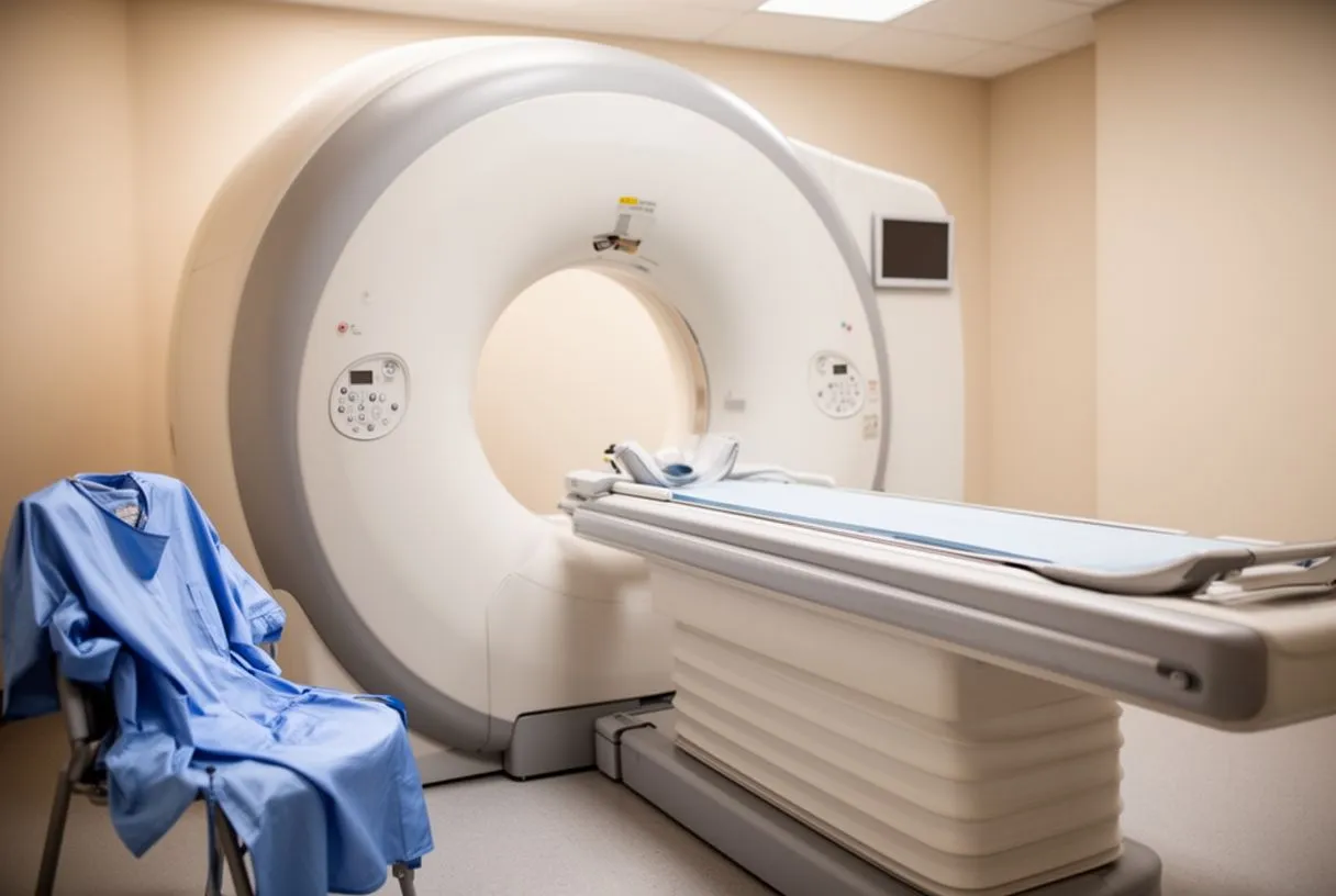
When mediastinal tumour symptoms are present, or an abnormality is spotted on a basic X-ray, the diagnostic process shifts to high-resolution imaging. A computed tomography (CT) scan of the chest is the gold standard for mediastinal mass diagnosis. This scan provides detailed cross-sectional images of the chest, allowing the surgical team to see the exact size of the tumour, its borders, and its relationship to the heart, great vessels, and airways. In some cases, magnetic resonance imaging (MRI) is utilised to provide even better soft tissue contrast, particularly if the tumour is suspected of invading local nerves or the spinal canal.
Beyond imaging, blood tests play a crucial role in characterising the mass. Certain germ cell tumours produce specific protein markers, such as alpha-fetoprotein (AFP) and human chorionic gonadotropin (beta-hCG). Elevated levels of these markers in the blood strongly suggest a malignant germ cell origin, guiding the medical team toward chemotherapy before any surgical intervention is attempted. Comprehensive research indexed in databases like PubMed consistently highlights the importance of these tumour markers in dictating the sequence of treatment. A tissue biopsy is often required to confirm the cellular type, usually performed via a minimally invasive technique like mediastinoscopy or a needle biopsy guided by CT imaging.
The diagnostic phase is also when the surgical team assesses the patient’s overall cardiovascular health. If a patient has a history of smoking or advanced age, the surgeon must check for concurrent issues that could complicate an operation. This comprehensive evaluation ensures that the team is not only treating the tumour but is also prepared to manage any underlying cardiac or respiratory vulnerabilities during the eventual surgical resection. Accurate staging and typing of the mass ultimately dictate whether the patient proceeds directly to surgery or requires a medical oncology consultation first.
Types of mediastinal germ cell tumours
Once the diagnostic workup is complete, the medical team will categorise the growth into one of several distinct types of mediastinal germ cell tumours. The classification is vital because benign and malignant tumours behave entirely differently and require divergent treatment pathways. Broadly, these tumours are divided into teratomas and malignant germ cell tumours, with the latter further split into seminomatous and non-seminomatous subtypes. Understanding which type is present allows the surgical and oncology teams to predict the tumour's behaviour and plan the most effective, lowest-risk intervention for the patient's specific situation.
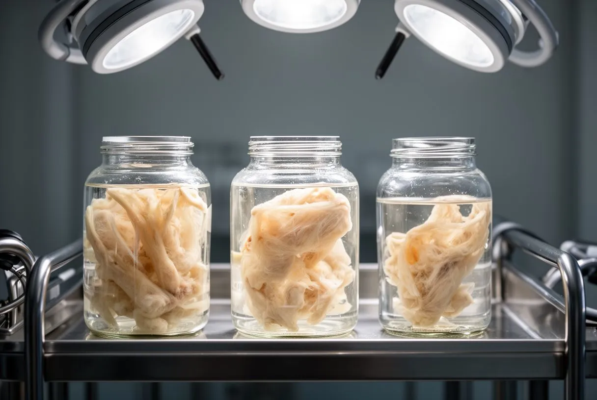
Mature teratomas are the most common type and are almost always benign. They are unique because they contain a mixture of mature tissues, such as hair, muscle, or bone, that arise from the displaced germ cells. While they do not spread to other parts of the body, they can continue to grow locally, causing structural compression. Before planning the removal of a teratoma, surgeons often evaluate the patient’s vascular health to ensure there is no significant atheromatous plaque in the coronary arteries that could complicate the surgery. In cases of large tumours requiring significant resection, the surgical team may arrange for autologous blood to be donated or salvaged during the procedure to minimise the need for donor transfusions.
- Mature Teratomas: These are typically benign growths that contain various tissue types, such as hair, muscle, or bone, and are highly curable with surgical removal alone.
- Seminomatous Tumours: Malignant growths that are highly sensitive to radiation and chemotherapy, often presenting in young men and usually having an excellent response to non-surgical treatments.
- Non-seminomatous Tumours: More aggressive malignant forms, including yolk sac tumours and choriocarcinomas, which require intensive multi-drug chemotherapy followed by surgical resection of the remaining mass.
Malignant mediastinal germ cell tumours, particularly the non-seminomatous variants, are aggressive and require systemic chemotherapy as a first-line treatment. The goal of chemotherapy is to shrink the tumour and eradicate microscopic disease before surgery. The surgical resection is then performed to remove the residual mass, which may contain a mix of dead tumour cells, scar tissue, and occasionally viable cancer cells. This multimodal approach is crucial for preventing recurrence and ensuring long-term survival.
Germ cell tumour treatment options
The landscape of germ cell tumour treatment options relies heavily on whether the mass is benign or malignant. For a benign mature teratoma, surgical removal is the definitive cure. Because these tumours do not respond to chemotherapy or radiation, the surgeon must meticulously excise the entire mass along with its capsule to prevent future regrowth. The decision to operate immediately is typically straightforward, provided the patient is healthy enough to undergo a major thoracic procedure. The goal is to relieve the mechanical compression on the heart and lungs before respiratory or vascular complications arise.
Chemotherapy for malignant masses: If the mediastinal mass diagnosis reveals a malignant germ cell tumour, treatment begins with systemic chemotherapy rather than a scalpel. Platinum-based chemotherapy regimens are highly effective at shrinking these aggressive tumours. Just as clinicians distinguish complex valvular pathologies—such as those detailed in studies like Re‐do edge-to-edge repair in patients with functional mitral valve regurgitation and advanced heart failure—from primary tumour masses, oncologists must precisely characterise the tumour markers to select the correct chemotherapeutic agents. This pre-surgical treatment is vital for reducing the tumour's vascular supply and making the eventual surgical resection safer and more complete.
The role of post-chemotherapy surgery: After chemotherapy, a repeat CT scan is performed to assess the tumour's response. Even if the mass has significantly shrunk, surgery is almost always recommended for non-seminomatous tumours. The residual tissue must be removed because it may contain teratoma components that are resistant to chemotherapy and could continue to grow, or it may harbor hidden malignant cells. Accurately distinguishing these residual masses from other distinct cardiac or systemic issues—just as researchers differentiate valve diseases in publications like Mitral valve disease with rheumatic appearance in the presence of left ventricular endomyocardial fibrosis—is an essential step in finalising the surgical plan.
Surgery for mediastinal mass removal
Surgery for mediastinal mass removal is a major cardiothoracic procedure that demands precise planning and a highly skilled surgical team. The most common approach is a median sternotomy, which involves making an incision down the center of the chest and dividing the sternum to provide direct, unobstructed access to the anterior mediastinum. This approach allows the surgeon to clearly visualise the tumour, the heart, and the great vessels, ensuring that the mass can be dissected away from these critical structures without causing inadvertent damage. The extent of the surgery depends on how much the tumour has encroached upon surrounding anatomy.
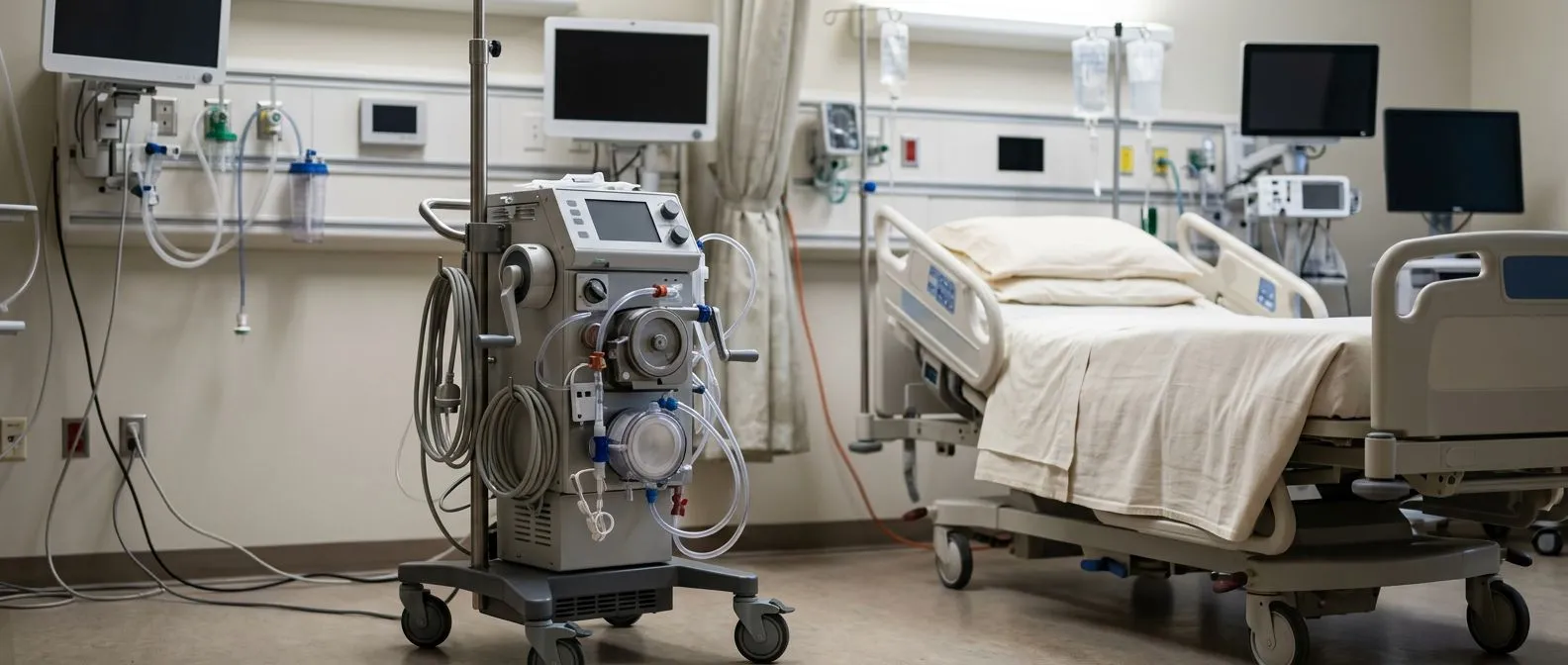
During the operation, continuous hemodynamic monitoring is essential. An arterial line is placed in the patient's wrist to provide real-time, beat-to-beat blood pressure readings, allowing the anesthesiologist to manage the cardiovascular system precisely. In cases where the tumour is densely adhered to the heart or major blood vessels, the surgeon may need to establish cardiopulmonary bypass, a machine that temporarily takes over the function of the heart and lungs. This involves inserting an arterial cannula to redirect blood flow. In extremely complex scenarios where respiratory or cardiac function is severely compromised preoperatively, advanced support systems like Awake ECMO (Extracorporeal Membrane Oxygenation) might be discussed pre-operatively as a bridge to surgery, though this is reserved for the most critical patients.
The actual dissection of the tumour requires patience and meticulous technique. The surgeon carefully peels the mass away from the pericardium (the sac surrounding the heart) and the phrenic nerves, which control breathing. If a portion of the pericardium or lung is heavily invaded by the tumour, it may be resected en bloc (together with the tumour) to ensure clear margins. The complexity of this surgery underscores the need for an experienced cardiac surgery team capable of navigating both the oncological and cardiovascular challenges presented by these rare masses.
Mediastinal germ cell tumour prognosis
Understanding the mediastinal germ cell tumour prognosis requires looking at both the tumour type and the success of the treatments applied. For patients with a benign mature teratoma, the prognosis is excellent. Once the mass is completely removed through surgery, the patient is considered cured. Recurrence is exceedingly rare, and life expectancy returns to normal. The primary focus post-surgery for these patients is simply recovering from the sternotomy and gradually returning to their normal daily activities without the burden of the compressive mass.
For malignant tumours, the prognosis is highly dependent on how the tumour responds to chemotherapy and whether the surgeon can achieve a complete resection. Seminomatous tumours generally have an excellent response to chemotherapy, and many patients do not even require surgery if the mass disappears entirely on imaging. Non-seminomatous tumours are more aggressive, but with modern platinum-based chemotherapy protocols followed by aggressive surgical removal of residual disease, long-term survival rates are highly favourable. The prognosis can be more guarded if the tumour has spread beyond the chest or if it transforms into a more aggressive sarcoma, highlighting the necessity of early and accurate intervention.
Long-term follow-up is a critical component of managing the prognosis. Patients will require regular CT scans and blood tests to monitor tumour markers for several years after treatment. This surveillance ensures that any recurrence is caught at an early, treatable stage. By adhering to the follow-up schedule, patients can move forward with confidence, knowing that their medical team is actively monitoring their recovery and maintaining a proactive stance against any potential return of the disease.
Recovery after thoracic germ cell tumour surgery
Recovery after thoracic germ cell tumour surgery is a gradual process that requires both physical healing and careful medical monitoring. Immediately following the operation, the patient is transferred to the intensive care unit (ICU) for close observation. Here, the medical team monitors heart rate, blood pressure, and respiratory function to ensure the body is stabilising after the stress of major surgery. Pain management is a priority during these first few days, utilising a combination of epidural anesthesia and intravenous medications to keep the patient comfortable enough to breathe deeply and cough effectively.
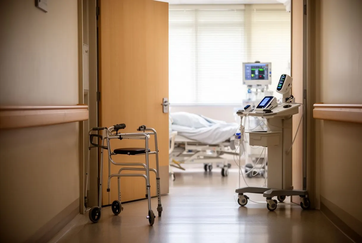
Once discharged from the ICU, the focus shifts to mobilisation and pulmonary hygiene. Deep breathing exercises and incentive spirometry are crucial to prevent lung collapse or pneumonia. Medications are carefully managed during this phase; for instance, while Aspirin is typically associated with cardiovascular prevention, it or other anticoagulants may be prescribed based on the individual’s risk of deep vein thrombosis during reduced mobility. If the patient received prolonged chemotherapy prior to surgery, their nutritional status and metabolic panels—including checking Apolipoprotein B levels if lipid metabolism is a concern—are closely monitored to support wound healing and overall recovery.
- First 24-48 hours: Monitored in the intensive care unit to ensure respiratory stability, manage postoperative pain, and watch for any signs of bleeding around the heart.
- Days 3 to 5: Early mobilisation begins, focusing on deep breathing exercises to prevent lung complications, and transitioning from intravenous to oral pain medications.
- Weeks 2 to 4: Gradual return to light daily activities at home, with strict restrictions against heavy lifting to allow the sternum to heal properly before cardiac rehabilitation.
Full recovery from a sternotomy typically takes six to eight weeks, though fatigue may persist for several months. Patients are advised to avoid driving and heavy lifting during the initial healing phase. A structured cardiac rehabilitation program may be recommended to help patients safely rebuild their cardiovascular endurance. Throughout this journey, the surgical team provides ongoing support, ensuring that the patient not only heals from the physical operation but also regains their confidence and quality of life.
When to seek help for chest masses
Knowing when to seek help for chest masses is critical for preventing severe complications. If an individual experiences persistent chest pain, unexplained shortness of breath, or a chronic cough that does not resolve with standard treatments, they should seek medical evaluation promptly. While these symptoms can be caused by many benign conditions, the presence of a mediastinal mass can only be ruled out through proper imaging. Delaying evaluation can allow a tumour to grow larger, making eventual surgical resection more complicated and increasing the risk of vascular or airway compression.
Patients who have already undergone treatment must be vigilant for signs of complications or recurrence. Sudden onset of chest palpitations or a racing heart could indicate a rhythm disturbance like atrial flutter, which can sometimes occur after surgery involving the pericardium. Similarly, severe chest pain accompanied by sweating and nausea should never be ignored, as it could signify an atypical infarction (a heart attack presenting with unusual symptoms) or a coronary issue unrelated to the tumour. If coronary disease is discovered, interventions like balloon angioplasty might be necessary before or alongside tumour treatment to ensure the heart can withstand surgery.
It is also important to recognise the systemic signs that warrant immediate attention, such as unexplained weight loss, night sweats, or swelling in the face and neck. These can be indicators of superior vena cava syndrome or an aggressive malignant process. By maintaining open communication with their medical team and attending all scheduled follow-up appointments, patients can ensure that any post-treatment anomalies are investigated swiftly and accurately, maintaining the best possible outlook for their long-term health.
Working with a mediastinal tumour specialist
Navigating the diagnosis and treatment of a mediastinal mass requires the expertise of a dedicated mediastinal tumour specialist. Because these tumours sit in the complex anatomical crossroads of the chest, the surgical team must possess deep experience in both oncological resection and cardiovascular surgery. At Cirugía de Corazón, the practice of Dr Alejandro Molina, our teams in Guadalajara, Querétaro, and Hermosillo understand the anxiety that accompanies a chest mass diagnosis. We bridge complex cardiology with clear, calm explanations, ensuring that patients and their families understand every step of the diagnostic and surgical journey.
For international patients and families travelling for care, logistics, insurance, and travel arrangements are important considerations. We prioritise anatomical precision and patient safety, using established guideline thresholds—such as cardiovascular risk scores—to drive our surgical decisions. We never claim a procedure is 'best' or 'painless'; instead, we focus on providing the lowest-risk surgical option for your specific situation, managed with evidence-based medical protocols.
Ultimately, facing a mediastinal germ cell tumour diagnosis is a significant challenge, but you do not have to face it alone. We encourage you to discuss your specific symptoms, imaging results, and concerns with a qualified cardiac and thoracic surgical team rather than self-diagnosing. If you or a loved one are dealing with a suspected mediastinal mass, please reach out to book a consultation with our specialists to develop a tailored, precise treatment plan designed for your unique anatomical needs.