

Mitral Stenosis Treatment: Balloon or Valve Surgery
Mitral stenosis treatment options compared—balloon valvuloplasty vs valve replacement.
If you have been told you have Mitral Stenosis, you already know the news lands heavily. The mitral valve is the doorway between the two chambers on the left side of your heart, and when that doorway narrows, blood struggles to pass through. The good news is that mitral stenosis treatment has more options today than ever before, from a balloon catheter to full valve surgery, and the right choice depends on your valve, your symptoms and your life. This guide walks through every option in plain language, so you can walk into any consultation already knowing the questions to ask.
What Is Mitral Stenosis?
The valve as a doorway. Your mitral valve has two thin flaps, or Valve Cusp tissue, that swing open to let blood flow from the left atrium into the left ventricle, the main pumping chamber. In stenosis, those flaps stiffen, thicken or fuse together at their edges. The opening that used to be the width of a large coin narrows to a slit.
When the opening narrows, the heart has to push blood through a smaller hole at higher pressure. Pressure builds backward into the lungs, which is why breathlessness is usually the first clue. Over years, this backlog can stretch the upper chamber and lead to irregular rhythms such as atrial fibrillation.
Knowing your heart: the mitral valve makes this easier to picture. It is the only valve with two cusps instead of three, and it handles the full pressure of every heartbeat. That workload is exactly why a stiffened mitral valve eventually demands attention rather than simple observation.
Mitral Stenosis Symptoms and When Treatment Is Needed
Early warning signs. Most people first notice breathlessness during activity that used to be easy: climbing stairs, carrying shopping, walking uphill. Some feel a fluttering or racing heartbeat, fatigue that no rest seems to fix, or a whistling cough at night. Swelling in the ankles can appear as pressure backs up through the lungs.
Moderate symptoms to take seriously. When doctors describe moderate stenosis, the symptoms usually move from "occasional" to "weekly": breathlessness on modest effort, chest discomfort, and sometimes palpitations that come and go. If you ever cough blood or feel faint on exertion, that is a same-week call to your cardiologist, not something to watch.
Timing matters because symptoms, not just measurements, drive the decision to treat. Guidelines for mitral stenosis symptoms and treatment agree on one point: a valve that is narrowing but causing no symptoms may only need yearly monitoring, while the same valve with symptoms usually needs action. In our consulting rooms we take the time to explain what the studies actually show, in plain words, before any recommendation is made.
Why Rheumatic Fever Causes Most Cases
An infection with a long memory. Most mitral stenosis worldwide traces back to rheumatic fever, an inflammatory illness that can follow an untreated strep throat, often in childhood. Weeks or years later, the immune system's attack on the bacteria also scars the valve tissue. The cusps thicken, their edges fuse, and calcium slowly settles in.
This is why rheumatic mitral stenosis treatment planning looks different from other valve disease. The scarring is diffuse and often involves the supporting cords below the valve, so the anatomy has to be studied carefully before anyone promises a particular procedure. Echocardiography, the ultrasound of the heart, grades the narrowing as mild, moderate or severe and shows whether the valve is suitable for a balloon.
Rheumatic valve disease remains common in Latin America. Recognising the rheumatic pattern early, often decades after the original fever, is an important part of any valve review.

What Are the Mitral Stenosis Treatment Options?
Four main paths. Mitral stenosis treatment options fall into four broad groups: watchful monitoring, medication, a catheter-based balloon procedure, and surgery. The choice depends on how narrow the valve is, whether the cusps are fused or calcified, whether other valves are involved, and how fit you are for an operation. There is rarely a single "correct" answer that applies to everyone.
- Monitoring: regular echocardiograms for mild stenosis without symptoms, typically once a year or sooner.
- Medication: drugs that ease the workload, control heart rhythm, or prevent clot formation while the valve is watched.
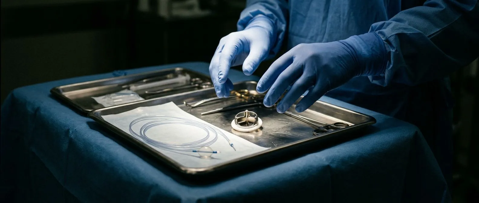
- Balloon procedure: a catheter threaded from the groin widens the valve from the inside, with no chest incision.
- Surgery: either repairing the patient's own valve or replacing it with an artificial one, through a full or small incision.
Every case is reviewed by our multidisciplinary team against international practice guidelines before a recommendation is given, and the patient receives a clear proposal with the alternatives. The final decision is always yours, and we actively encourage second opinions as a normal part of deciding about heart surgery.
How Does Balloon Valvuloplasty for Mitral Stenosis Work?
The procedure itself. Percutaneous mitral balloon valvulotomy, usually called balloon valvuloplasty, is done under local anaesthesia with sedation. A thin catheter is guided from a vein in the groin across the wall between the atria, up to the narrowed valve. A deflated balloon sits across the valve and is inflated briefly, splitting the fused cusps apart.
Balloon valvuloplasty mitral stenosis candidates are chosen carefully. The ideal valve has fused edges but little calcium, no significant leak, and no clot inside the atrium. When those conditions are met, the procedure can open the valve dramatically with a one- or two-night hospital stay and no surgical scar.
It is not for every valve, however. Heavily calcified valves, valves that also leak, or valves with clots need surgery instead, because a balloon can tear a fragile valve or dislodge a clot. In our practice we recommend the less invasive route only where the evidence supports it for that specific patient, and we say plainly when a valve is not a balloon candidate.
Mitral Valve Repair vs Replacement: Which Is Better?
Why repair is preferred when possible. Mitral Valve Repair keeps your own tissue, which avoids the lifelong blood-thinning medication that many artificial valves require and preserves the natural pumping dynamics of the left ventricle. In stenosis caused by rheumatic disease, repair is possible when damage is limited, though it is more often offered for leaking valves than stiffened ones.
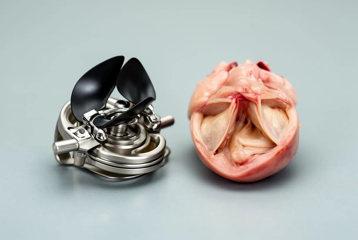
When replacement wins. A Mitral Valve Replacement becomes the better option when the valve is heavily calcified, badly damaged below the cusps, or when a previous repair has failed. Surgeons choose between mechanical valves, which last decades but require lifelong anticoagulation, and tissue valves, which avoid blood thinners but wear sooner. A Balloon-expandable Valve can also be placed through a catheter in selected cases, and a Valve-in-valve procedure can re-open a failing surgical valve without repeat open surgery.
The honest answer to mitral valve repair vs replacement is that the valve decides, not the preference of patient or surgeon. In the consultation we explain what the imaging shows about your own valve, why one option suits it better, and what each choice means for follow-up appointments and medication in the years ahead.
What Is Minimally Invasive Mitral Valve Surgery?
Smaller openings, same repair. Minimally invasive mitral valve surgery reaches the valve through a small incision on the right chest, often under the nipple line, rather than splitting the full breastbone. Robotic techniques go further, using instrument arms controlled by the surgeon through even smaller ports. The valve work inside is identical; only the route in differs.
The benefits are real but should be stated plainly: less pain, lower infection risk, faster return to work, and smaller scars. Not every anatomy is suitable, and emergency situations or heavily calcified vessels can make a conventional approach safer. Dr Molina performed the first robotic mitral valve repair in the Ángeles Health System and the first robotic heart surgery in Guadalajara, and trained in minimally invasive and robotic techniques in Montreal.
If you have heard about edge-to-edge repair (TEER) for mitral regurgitation, note that it treats leaking valves, not narrowed ones, and Transcatheter Mitral Valve Replacement for the mitral position is still reserved for very select patients who cannot undergo surgery. For most people with stenosis, the realistic minimally invasive choices remain balloon valvuloplasty or a small-incision operation.

What Is the Recovery Time After Mitral Valve Surgery?
The first weeks. Typical mitral valve surgery recovery time follows a predictable arc. Hospital stay runs five to seven days for a conventional operation, and less after a minimally invasive or balloon procedure. Most people walk short distances within a day or two, and driving is usually possible again around four to six weeks after open surgery.
Return to normal life. Full recovery, meaning unrestricted activity and return to work, generally takes six to twelve weeks depending on your age, fitness and the approach used. Cardiac rehabilitation, a supervised programme of gradually increasing exercise, shortens that journey safely and is worth asking about before you leave hospital.
Recovery also has an emotional side that families often underestimate: tiredness, low mood and vivid dreams are common in the first month and fade with time. We tell every patient before surgery what the realistic milestones are, week by week, because knowing the map makes the journey far less frightening.
How Is Severe Mitral Stenosis Managed Without Surgery?
Medication buys time, not cure. Severe mitral stenosis management without surgery is possible in specific situations: when symptoms are mild, when other conditions make surgery risky, or while a patient waits for a procedure. Medication does not widen the valve. It manages the consequences of the narrowed valve instead.
The usual medicines include beta blockers or rate-control drugs that slow the heartbeat, giving the valve more filling time between beats; diuretics that clear fluid from the lungs and ankles; and anticoagulants to prevent clots if atrial fibrillation develops. Infections matter too, so dental hygiene and antibiotic prophylaxis before certain dental procedures are part of standard advice for valve patients.
What medication cannot do is reverse the scarring. If symptoms progress despite good medical therapy, delaying a definitive procedure usually costs more than it gains. Our team reviews each case against international guidelines precisely to catch that tipping point early, so intervention happens while it is still the lowest-risk choice for your situation.
Choosing the Right Team
What actually defines the best hospital for mitral valve surgery. It is not marble lobbies; it is volume, multidisciplinary review, and imaging quality. A valve programme should include cardiac surgeons, interventional cardiologists, anaesthesiologists experienced in cardiac cases, and echocardiographers who assess the valve before and during the procedure. Ask any team directly: how many of these procedures do you perform each year, and what is your strategy if the balloon or the repair fails on the table?
Cost, honestly framed. Mitral stenosis treatment cost in Mexico varies widely with the procedure chosen, the hospital, and whether a valve prosthesis is implanted. A balloon procedure generally costs less than open surgery, and mechanical valves carry ongoing anticoagulation costs that tissue valves do not. Rather than quoting figures that change, our first contact is a free video consultation where we get to know the patient, talk through the condition, and say which studies are still needed so no unnecessary ones are ordered.
Research on repeat edge-to-edge repair in patients with functional mitral valve regurgitation and advanced heart failure illustrates how far structural heart treatments have evolved, and reviews such as those published by Springer document how transcatheter approaches now sit alongside surgery in modern guidelines. Rare causes also exist; a report of mitral valve disease with rheumatic appearance in the presence of left ventricular endomyocardial fibrosis reminds clinicians to look beyond the common diagnosis when imaging does not fit the story.
Living with Mitral Stenosis today means you have genuine choices, not a single forced path. The right one depends on your valve's anatomy, your symptoms and your goals, and that judgement deserves a team that explains everything before recommending anything. If you are weighing mitral stenosis treatment for yourself or someone you love, start with a proper conversation rather than a search engine: book a consultation with a qualified cardiac team, ask your questions, and decide from understanding rather than fear.






