Myocardial Viability Study: Is Bypass Worth It
A myocardial viability study reveals if heart bypass surgery will help you.
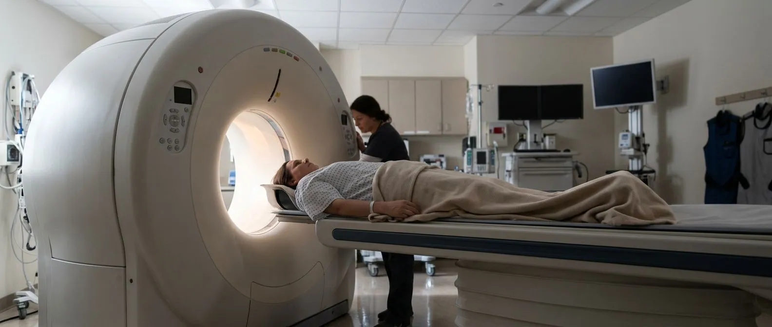
If your cardiologist has recommended a myocardial viability study, you are probably facing a big question: is bypass surgery actually going to help me? When the heart's pumping power is weak and the coronary arteries are blocked, surgery only makes sense if part of that weakened muscle is still alive and could recover. A myocardial viability study is the test that answers this. It maps which areas of heart muscle are stunned but salvageable, and which are permanently scarred. In this guide we walk you through what the test involves, what the results mean, and how they shape the decision about Coronary Artery Bypass Grafting — surgery that creates new routes for blood around blocked arteries.
What Is a Myocardial Viability Study?
Definition in plain language. A myocardial viability study is an imaging test that tells doctors whether damaged heart muscle is dead or merely sleeping. When a coronary artery narrows, the muscle it supplies may pump poorly — not because the cells are gone, but because they are receiving too little blood to work at full strength. Viability imaging separates that reversible weakness from permanent scar tissue.
Why this distinction changes everything. If a large part of the poorly pumping wall is alive, restoring blood flow through Myocardial Revascularisation — the medical term for re-establishing blood supply, whether by bypass surgery or a stent — can improve pumping function and symptoms. If the wall is mostly scar, opening the artery offers little benefit and adds surgical risk for nothing. This is why the test exists: it protects patients from operations that cannot help them.
How the test "sees" living tissue. Living heart cells actively take up glucose and certain radioactive tracers; scar tissue does not. By injecting a tiny tracer and imaging the heart with a PET or nuclear camera, the study shows a metabolic map — bright areas are alive, dark areas are scar. The principle is simple: function follows fuel.
Why Do Doctors Order a Viability Study Before Bypass?
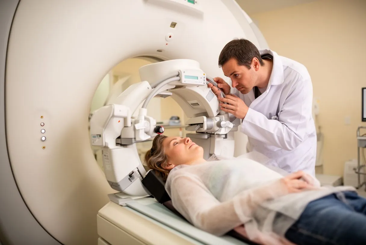
The core question: heart bypass candidacy. Before any team proposes a heart bypass candidacy test pathway, they want evidence that surgery will genuinely improve your life. An angiogram shows where arteries are blocked, but not whether the muscle behind those blocks can still be saved. A myocardial viability study before bypass fills that gap, turning a guess into an evidence-based recommendation.
When it matters most. The study is most valuable when the heart's ejection fraction — the percentage of blood the left ventricle pumps out with each beat — is reduced, or when a large territory of muscle looks poorly functioning on an echocardiogram. In strong hearts with small blockages, the answer is often obvious without it.
Our practice's approach. Every case at our team is reviewed by the multidisciplinary group against international practice guidelines, and a myocardial viability study is one of the tools we use to decide — together with the patient — whether revascularisation is worth the risk.
Hibernating Myocardium Treatment: Why Sleeping Heart Muscle Matters
What hibernating myocardium is. Hibernating myocardium is heart muscle that has downshifted to a low-energy survival mode. The cells are alive but chronically underfed by a narrowed artery, so they contract weakly. They are not dead — they are on standby. Hibernating myocardium treatment means restoring blood flow so these cells wake up and work again.
Stunned versus hibernating versus scarred. Three states of "sick but salvageable or not" muscle exist:
- Stunned muscle: temporarily weak after an Acute Myocardial Infarction — a heart attack with sudden full blockage — usually recovers on its own within weeks.
- Hibernating muscle: chronically underfed, alive, and recoverable if blood flow is restored.
- Scarred muscle: dead fibrous tissue that can never contract again, whatever you do.
Why you should ask about it. If your doctor proposes surgery because your heart is weak, ask which parts of the muscle are hibernating and how much of the wall is scar. A myocardial viability study gives exact answers, and the proportions drive the recommendation more than any single number on the angiogram.
Viability Study vs Angiogram: What's the Difference?
Two different cameras, two different questions. A viability study vs angiogram comparison confuses many patients, and understandably so. The angiogram (coronary catheterisation) uses a thin tube threaded from the wrist or groin into the coronary arteries, with dye and X-rays showing the plumbing: where arteries are narrowed and how severely. It answers where the blockages are.
The viability study answers a different question. It answers so what — does the muscle supplied by that blocked artery still live? You can have a 90% blockage over dead scar (surgery will not help that territory) or a 70% blockage over thriving, hibernating muscle (surgery may transform heart function). Both tests are complementary, not competing; almost every surgical decision uses both.
Why we never rely on one alone. In our consultation we take the time to explain what each study actually shows, in plain words, so the patient sees exactly how the angiogram's blockages and the viability map fit together before any decision.
Types of Cardiac Viability Scan: PET, MRI and Nuclear
PET scan myocardial viability — the modern standard. The most accurate option is a PET scan myocardial viability protocol, which combines two tracers: one measuring blood flow and one measuring glucose metabolism (usually FDG). Living muscle switches its fuel preference under low-flow conditions, so a segment that is dark on the flow scan but bright on the metabolism scan is hibernating — the classic mismatch that predicts recovery after revascularisation. PET offers the best image quality and the lowest radiation of the nuclear options.
MRI viability study heart imaging. An MRI viability study heart protocol uses a different trick: gadolinium contrast "lights up" scar tissue, so the scanner shows exactly how much of each wall is dead. Cardiac MRI also measures pumping function with great precision and needs no radiation. It is excellent when the main question is scar burden.
Nuclear (SPECT) perfusion imaging. The older, more widely available option uses single-photon cameras and tracers such as thallium or technetium. Myocardial perfusion viability testing with SPECT is less precise than PET or MRI but still clinically useful and more accessible in many hospitals. The right choice depends on availability, your kidney function, radiation considerations and the exact question being asked.
How Long Does a Viability Test Take?
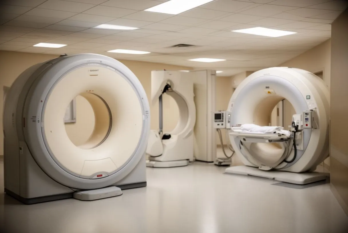
Appointment length versus total day. Patients often ask how long does a viability test take, and the honest answer depends on the type. A SPECT study typically involves one or two scans, each 15–30 minutes, with a rest phase and sometimes a delay of hours or a second day between injections. Total time in the department: a few hours, or a half-day.
PET and MRI timings. A PET viability study is usually done in a single session of two to three hours, including tracer injection, a waiting period while the heart takes up the FDG, and the scan itself — often 20–30 minutes lying in the scanner. Cardiac MRI takes 45–60 minutes on the table and requires holding your breath briefly at intervals. Add registration and recovery time, and plan the better part of a morning.
Practical tips. For FDG PET you will usually be asked to fast beforehand and may receive a glucose drink or small insulin adjustment to encourage the heart to take up the tracer — people with diabetes get individual instructions. Wear comfortable clothes, take your regular medicines unless told otherwise, and bring someone to drive you home if a stress component is included.
What drives the price. It varies widely by country, facility and technique. PET is generally the most expensive because of tracer production and scanner time; SPECT is usually the least; MRI sits in between. Costs also rise when a stress component — exercise or medication that simulates exercise — is added, and when same-day interpretation by a specialist cardiologist is included.
Insurance and international patients. For patients in Mexico and international visitors, coverage differs between insurers and between public and private systems. Ask the imaging centre for a written quote that names the exact protocol (PET with FDG plus flow tracer, SPECT rest-stress, or MRI with viability sequences), because quotes for vaguely described "cardiac scans" often omit key components.
Cost in context. The study is a fraction of the cost of bypass surgery itself, and its real value is preventing an operation that would not help. At our first contact — a free video consultation — we get to know the patient, talk through the condition, and say which studies are still needed so no unnecessary ones are ordered and no money is spent on duplicate tests.
Are Viability Studies Safe?
Radiation: lower than most people fear. A common question is are viability studies safe. PET and SPECT involve a small dose of ionising radiation from the tracer — comparable in scale to other diagnostic nuclear scans and far below levels associated with measurable harm in adult diagnostic imaging. Cardiac MRI uses no radiation at all, which makes it attractive for younger patients and those needing repeat imaging. General safety context for cardiac testing is well summarised by Medical News Today.
Contrast and tracers. MRI uses gadolinium contrast, which is avoided or adjusted in severe kidney disease — tell the team your full medical history and any prior contrast reactions. The radioactive tracers in PET and SPECT are cleared from the body over hours; drinking water helps. Allergic reactions are uncommon.
Stress components. If your study includes a stress test, you will be monitored continuously, and the medications used (or a supervised treadmill) carry the same small, well-understood risks as any stress test performed in a hospital setting. Staff are present and equipped to manage any reaction immediately.
Do I Need Heart Bypass? How Results Guide the Decision
Reading the report: what "viable" means in practice. A typical report states how many heart-wall segments are viable, the scar burden, and the left ventricle's pumping function. A common clinical threshold is that meaningful benefit from revascularisation is expected when a substantial portion of the poorly contracting muscle is viable. Below that, medication often offers equal or better outcomes than surgery.
Bypass versus stent — the team weighs both. If hibernating muscle is confirmed, the next question is how to restore flow: Off-pump Bypass (surgery performed without the heart-lung machine), On-pump Bypass (the conventional approach using Cardiopulmonary Bypass — a machine that takes over circulation so the heart can be operated on still), or angioplasty with stents. The choice depends on how many arteries are diseased, valve problems, Bypass Time considerations, and your overall health. Myocardial Protection — techniques that keep the heart muscle safe during surgery — and Weaning From Bypass are handled by the surgical team, and poor viability is exactly the situation where these details matter most.
Red flags and second opinions. A viability study does not force one answer; it informs one. You should always understand why surgery is being proposed and what happens if you decline. Warning signs that need urgent attention regardless of test results include chest pain at rest, breathlessness that wakes you at night, or swelling with rapid weight gain — seek emergency care for those.
- Viable muscle present: revascularisation (bypass or stent) is likely to improve pumping function and symptoms.
- Predominant scar: medical therapy and device options are usually discussed first, since opening blocked arteries over dead tissue adds risk without benefit.
- Borderline findings: a reasonable step is a second opinion — many teams consider that normal and helpful.
Heart Surgery Evaluation Guadalajara: What to Expect
What a proper evaluation looks like. A thorough heart surgery evaluation Guadalajara patients can trust starts long before an operating theatre. Expect a full review of your history, an echocardiogram, an angiogram, and — where the heart is weak — a viability study for coronary artery disease. The team should explain each result in everyday language and present treatment options with their trade-offs, not just one path.
Our team's experience and range. We are a multidisciplinary team of cardiac surgeons led by Dr Alejandro Molina, with consulting rooms in Guadalajara, Querétaro and Hermosillo; our work covers valve disease, coronary disease, the aorta and structural heart conditions, and we recommend the less invasive route only where the evidence supports it for that patient. Dr Molina trained in cardiothoracic surgery at the Universidad de Guadalajara, in cardiac, minimally invasive, robotic and structural surgery at the University of Montreal, and in structural cardiology and TAVR at CHU Bordeaux.
What happens at the first appointment. The first contact is a free video consultation: we get to know the patient, talk through the condition and the studies already done, and only then say which additional tests are genuinely needed — and every final decision rests with the patient, who receives a clear proposal with all the alternatives. If you have been told your heart is weak and bypass is on the table, the most useful next step is a conversation with a qualified cardiac team rather than more internet searching: bring your existing reports, ask whether hibernating muscle has been looked for, and let a myocardial viability study — where indicated — turn a frightening question into an answer you can act on.