
Right Coronary Artery Blockage: Symptoms and Treatment
Right coronary artery blockage symptoms and treatment explained by Guadalajara's heart surgery specialists.
What Is Right Coronary Artery Blockage?
Understanding a right coronary artery blockage begins with visualizing how the heart receives its own blood supply. The right coronary artery (RCA) is a major vessel that branches off the aorta and wraps around the bottom of the heart, delivering oxygen-rich blood to the right pumping chamber and critical electrical nodes. When this artery becomes narrowed or completely blocked, the heart muscle it supplies can suffer from oxygen deprivation.
This narrowing is most often caused by atheromatous plaque, a sticky buildup of cholesterol, calcium, and cellular waste. As plaque accumulates, it restricts blood flow. The risk of this buildup accelerating is closely monitored by measuring Apolipoprotein B, a protein that carries bad cholesterol through the blood. According to The Society of Thoracic Surgeons, identifying and addressing these blockages early is crucial for preventing severe cardiac events.
The RCA specifically supplies blood to the right ventricle and the sinoatrial node, the heart's natural pacemaker, which sits in the right Atrium. Because of this unique territory, a blockage here can cause both structural muscle damage and dangerous electrical malfunctions. Recognizing the anatomy of this blockage helps patients understand why specific symptoms and treatments differ from blockages in the left coronary arteries.
What Are RCA Blockage Symptoms?
Recognizing RCA blockage symptoms is vital for early intervention. The most common sign is chest pain or angina, which often feels like a heavy pressure or squeezing sensation in the center of the chest. Unlike blockages on the left side, right coronary artery blockage pain frequently radiates to unusual areas, including the jaw, neck, and sometimes even causing referred Arm Pain.
Sometimes, the symptoms are less typical, leading to an Atypical Infarction. Instead of crushing chest pain, a patient might experience severe shortness of breath, profound fatigue, cold sweats, or nausea. In older adults, women, and people with diabetes, these silent or atypical symptoms are more common, making it easy to dismiss the warning signs of a heart issue.

If you experience sudden dizziness, fainting, or an irregular pulse alongside chest discomfort, it could indicate that the blockage is affecting the heart's electrical system. Because the right coronary artery supplies the electrical nodes, an obstruction can slow the heart rate dangerously. Any combination of these symptoms warrants immediate medical evaluation to prevent irreversible muscle damage.
How Does Right Coronary Artery Disease Develop?
Right coronary artery disease develops gradually over years or even decades. It begins when the inner lining of the artery wall becomes damaged, often due to high blood pressure, high cholesterol, or smoking. Once the wall is compromised, low-density lipoproteins infiltrate the tissue and form fatty streaks, which eventually harden into obstructive plaques.
As these plaques grow along the vessel near the base of the Ascending Aorta, they reduce the lumen size, restricting blood flow. Physical examinations using Auscultation, where a doctor listens to the heart with a stethoscope, might reveal abnormal sounds or murmurs if the blood flow becomes turbulent. Research highlighted in Cardiovascular Disease in Advanced Chronic Kidney Disease in a Tertiary Institution, Johannesburg, South Africa demonstrates how systemic conditions rapidly accelerate this plaque progression.
The progression is usually silent until the artery is significantly narrowed. In some cases, a plaque ruptures, forming a blood clot that immediately occludes the artery. This abrupt closure transforms a stable, manageable narrowing into an acute emergency, cutting off blood supply to the heart muscle and causing immediate cellular distress.
How Do Doctors Diagnose RCA Stenosis Treatment Needs?
Diagnosing RCA stenosis treatment needs involves a combination of functional tests and direct imaging. Physicians typically begin with an electrocardiogram (ECG) to monitor the heart's electrical activity, looking for specific patterns that suggest ischemia, or reduced blood flow, in the right ventricle. Blood tests are also drawn to check for cardiac enzymes that leak into the bloodstream when heart muscle is damaged.
To visualize the blockage directly, a coronary angiogram is performed. During this procedure, a specialized Arterial Line is placed, usually through the wrist or groin, to guide a catheter to the heart. Contrast dye is injected into the coronary arteries, and X-ray images capture the exact location and severity of the blockage, determining if it is a proximal RCA occlusion or a mid RCA blockage.

Advanced imaging, like cardiac MRI or CT angiography, may be used for patients with complex anatomy or borderline stenosis. These tools provide a highly detailed, three-dimensional view of the heart, allowing Dr Alejandro Molina and his team to calculate precise intervention strategies. The goal is to determine whether the blockage can be managed with medication or requires a physical reopening of the artery.
What Are the Types of Proximal RCA Occlusion?
Proximal RCA occlusion refers to a blockage located near the origin of the right coronary artery, immediately after it branches off the aorta. This location is critical because a blockage here affects the entire downstream territory of the artery, including the right ventricle and the electrical nodes, posing a high risk for extensive muscle damage and severe rhythm disturbances.
Another specific and complex type is the right coronary artery chronic total occlusion (CTO). A CTO is a complete blockage that has formed slowly over time, allowing the heart to sometimes develop collateral, or tiny bypass, vessels. Treating a CTO requires specialized techniques and experienced operators to safely reopen the hardened, long-standing blockage.
- Acute occlusion: A sudden, total blockage causing an immediate heart attack (inferior MI RCA).
- Chronic total occlusion: A gradual blockage that may cause stable angina and requires specialized wiring techniques.
- Subtotal stenosis: A severe narrowing that allows some blood flow but causes symptoms during exertion.
It is important to distinguish these coronary artery blockages from valve issues. While procedures like Balloon Aortic Valvuloplasty are used to open narrowed heart valves, coronary blockages require entirely different catheter-based tools to clear the vessels supplying the heart muscle itself.
Right Coronary Artery Angioplasty vs Coronary Artery Bypass Graft RCA
Choosing between right coronary artery angioplasty and a coronary artery bypass graft RCA depends heavily on the blockage's anatomy and the patient's overall health. Angioplasty is a minimally invasive procedure where a catheter equipped with a tiny balloon is threaded to the blockage site. Balloon Angioplasty pushes the plaque against the artery walls, and a Balloon-expandable Valve mechanism is often used to deploy a stent that keeps the vessel open.
In cases where the right coronary artery has complex disease, diffuse plaque buildup, or chronic total occlusions, surgical intervention might be safer. A coronary artery bypass graft (CABG) creates a new route for blood to bypass the blockage entirely. Surgeons use an Arterial Graft, such as the internal mammary artery, or a harvested vein to connect the aorta to the coronary artery beyond the blockage. Insights from Stakeholders Want a Menu of Choices: Findings from a Consultation Workshop on Improving Access to Secondary Prophylaxis of Rheumatic Fever and Rheumatic Heart Disease emphasize the importance of offering multiple surgical pathways tailored to individual patient needs.
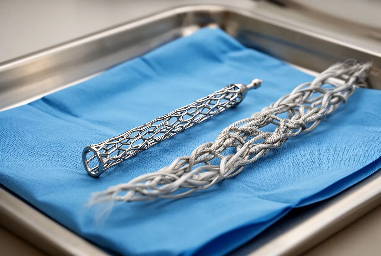
The decision requires evaluating the patient's EuroSCORE II, a system that estimates the risk of mortality after heart surgery. If the risk of a prolonged minimally invasive procedure is too high, or if multiple arteries are diseased, surgery is usually the recommended path. Dr Molina’s team carefully weighs these factors to provide the lowest risk option for your specific situation.
What to Expect During RCA Intervention Surgery
When undergoing RCA intervention surgery, patients are placed under general anesthesia, and the heart is carefully accessed. During a traditional bypass operation, an Arterial Cannula is inserted into the heart to connect the patient to a cardiopulmonary bypass machine, which takes over the job of pumping blood and oxygenating it while the heart is stopped.
Surgeons prioritize using the patient's own tissue for the best long-term outcomes. An Autograft, such as the internal thoracic artery, is preferred for its durability and resistance to future plaque buildup. Additionally, utilizing Autologous Blood—blood that is collected from the patient and reinfused during the operation—minimizes the need for donor blood transfusions and reduces the risk of complications.
After the bypass grafts are securely sewn into place, the heart is restarted, and the bypass machine is gradually withdrawn. The entire procedure requires extreme precision and real-time monitoring to ensure the new grafts have excellent blood flow. The surgical team ensures the chest is closed meticulously to promote optimal healing.
Inferior MI RCA: Recognizing a Heart Attack
An inferior MI RCA, or inferior myocardial infarction, occurs when the right coronary artery suddenly becomes completely blocked, starving the lower wall of the heart. This specific type of heart attack often presents with distinct electrical changes on an ECG and requires immediate emergency treatment to restore blood flow and minimize permanent damage.
Because the right coronary artery feeds the heart's electrical nodes, an inferior MI can trigger dangerous arrhythmias like Atrial Flutter, where the upper chambers beat rapidly and out of sync with the lower chambers. If the heart rhythm collapses into a lethal pattern, medical teams may need to use an Automated External Defibrillator to deliver a controlled electric shock and reset the heart's rhythm.
- Sudden severe chest pressure: Often radiating to the back or jaw, requiring a 911 call.
- Bradycardia: A dangerously slow heart rate caused by damage to the SA node.
- Right ventricular failure: Fluid backup causing swelling in the legs and shortness of breath.
Time is muscle during an inferior MI. The longer the right coronary artery remains occluded, the more irreversible damage occurs to the heart tissue. Immediate transport to a facility equipped for emergency angioplasty is the standard of care to extract the clot and place a stent.
RCA Blockage Recovery and Long-Term Outlook
RCA blockage recovery begins in the intensive care unit, where the heart is closely monitored for any signs of rhythm instability or graft failure. Patients are encouraged to begin gentle movement relatively quickly to prevent blood clots and promote lung function. Pain management is carefully balanced to keep the patient comfortable while avoiding excessive sedation.
In severe cases where the heart muscle has suffered profound damage leading to acute failure, temporary life support like Awake ECMO (Extracorporeal Membrane Oxygenation) might be utilized. This advanced system oxygenates the blood outside the body, giving the heart time to heal while the patient remains awake and responsive.

Long-term management relies heavily on medication. Daily Aspirin is prescribed to prevent new clots from forming in the stent or native arteries, alongside statins to stabilize any remaining plaque and lower cholesterol. Patients must also commit to heart-healthy lifestyle changes, including a balanced diet and supervised cardiac rehabilitation, to maintain the results of their RCA intervention surgery.
When to Seek Help for Right Coronary Artery Syndrome
Knowing when to seek help for right coronary artery syndrome can be the difference between a manageable procedure and a life-threatening emergency. If you have a known history of right coronary artery disease and notice that your angina occurs with less exertion, lasts longer, or does not respond to your prescribed nitroglycerin, this is an urgent warning sign that your blockage may be worsening.
Patients who have undergone interventions for atrial fibrillation might be on specific blood thinners. Decisions regarding these medications are often guided by clinical trials like ARISTOTLE, which established protocols for balancing stroke risk against bleeding risk. If you experience any unusual bleeding or bruising while on these medications, contact your cardiology team immediately.
Ultimately, this information is intended to help you understand the anatomy and treatment of right coronary artery blockage, but it is not a substitute for professional medical advice. Every patient’s anatomy is unique, and treatment decisions must be made based on personal health factors. We encourage you to discuss your specific situation with a qualified cardiac team and to book a consultation with Dr Alejandro Molina to create a personalized care plan rather than self-diagnosing.