Silent Brain Infarction After Heart Surgery
Learn about silent infarction after heart surgery risks, detection, and prevention from Guadalajara heart specialists.
Every year, hundreds of thousands of people undergo coronary bypass, valve repair, or other cardiac operations, and most walk out of the hospital feeling the surgery accomplished exactly what their heart needed. What many families never hear about, however, is a quieter phenomenon that researchers have documented repeatedly: small areas of brain injury that occur during or shortly after the procedure without causing any obvious symptoms. This is known as a silent infarction after heart surgery, and understanding it — how it happens, who is most at risk, and what can be done about it — helps patients and their loved ones make calmer, better-informed decisions. In this guide, we walk through the whole picture in plain language, from the operating room to the recovery chair.
What Is a Silent Infarction After Heart Surgery?
The basic definition. An infarction is an area of tissue that has lost its blood supply and been damaged as a result. When that happens in the heart itself, it is called a Myocardial Infarction — the medical name for a heart attack. When it happens in the brain, it is a cerebral infarction, or stroke. A silent infarction is one that produces no symptom the patient or family can identify: no weakness on one side, no slurred speech, no obvious confusion. It is usually discovered later, either on a brain scan done for another reason or through subtle changes in memory and thinking.
Why "silent" does not mean "harmless." The absence of dramatic symptoms does not mean the brain was untouched. Imaging studies of patients after cardiac procedures have found that a meaningful proportion show new small bright spots on brain MRI — findings consistent with tiny areas of infarction — even in people who felt entirely normal. Over time, accumulating small lesions can contribute to difficulties with memory, processing speed, and attention. This is why specialists take the phenomenon seriously even though most patients never notice a single event.
How common is it? The honest answer is that published estimates vary widely depending on the type of surgery, the imaging method used, and how soon after the operation the scan is performed. What is consistently reported across studies is that silent brain lesions after cardiac surgery are far more common than clinically obvious strokes. That gap between "what the scan shows" and "what the patient notices" is precisely why the topic deserves careful explanation rather than alarm.
Why Does Brain Injury Happen During Cardiac Procedures?
Tiny particles traveling upward. The main mechanism is embolization — the release of small particles or air bubbles that travel in the bloodstream toward the brain. During operations on the heart and especially on the aorta (the large artery that carries blood from the heart to the body), the surgeon manipulates tissue that can shed microscopic fragments of plaque, fat, or clotted blood. The anatomy works against us here: the arteries supplying the brain branch directly off the aortic arch, so anything loosened in that region has a short, direct route upward.
The heart-lung machine factor. In conventional coronary artery bypass grafting (CABG), the heart is often stopped and a pump-oxygenator — commonly called the heart-lung machine — takes over circulation. This allows meticulous repair, but it also means blood is handled outside the body, passed through tubing and filters, and returned under controlled pressure. Microbubbles and tiny particulate matter can form or enter in this circuit. Off-pump techniques, which avoid the machine altogether, were developed partly to reduce these exposures, though the evidence on whether they reliably reduce silent brain injury is mixed.
Blood pressure, rhythm, and oxygen delivery. The brain is exquisitely sensitive to changes in blood flow. Periods of low blood pressure during surgery, irregular heart rhythms in the immediate recovery period, and inflammation triggered by the surgery itself can all reduce the fine blood supply to small deep areas of the brain. Atrial fibrillation — a chaotic rhythm that can develop after surgery — is a well-known source of clots that can travel to the brain, and it is one reason your team monitors your heart rhythm so closely in the days after your operation.
Silent Stroke After Cardiac Surgery Versus Obvious Stroke
What an obvious stroke looks like. A clinically evident stroke produces signs anyone can see: drooping of one side of the face, weakness or numbness in an arm or leg, difficulty speaking or understanding words, sudden vision loss, or severe unbalance. These events are emergencies, and hospital teams respond immediately with imaging and treatment. Thanks to careful surgical technique and postoperative monitoring, major disabling stroke after cardiac surgery has become progressively less common over recent decades, though it remains one of the most serious possible complications.
What a silent event looks like — and does not look like. A silent stroke after cardiac surgery, by contrast, may announce itself only as mild word-finding trouble for a few days, a vague sense of mental fog, or nothing at all. Families sometimes describe a loved one as "not quite themselves" after surgery without being able to name anything specific. Because there is no dramatic event to trigger an emergency response, these lesions are typically identified only if a brain scan is performed for another reason — or sometimes never identified at all.
Why the distinction matters for your care. Knowing that these two entities are different helps families ask better questions. An obvious stroke changes the immediate treatment plan. A silent lesion, once discovered, changes the longer conversation: about blood pressure control, rhythm management, cognitive follow-up, and risk reduction before any future procedure. Both matter; they simply call for different responses.
Who Has the Highest Brain Infarction Risk Heart Procedure-Wise?
Pre-existing brain and vascular disease. Patients who already carry heavy vascular burden — a history of prior stroke, disease in the carotid arteries of the neck, longstanding high blood pressure, or diabetes — have less reserve in the small vessels of the brain. When an embolic particle lodges or blood pressure dips, a brain that already has narrowed or stiffened small arteries is less able to compensate. Age compounds this: the aortic wall becomes more fragile and more plaque-laden with each decade, releasing more debris when it is handled.
Diabetes and the nervous system. Research into patients with type 2 diabetes has examined whether damage to the autonomic nervous system — the automatic wiring that regulates heart rate and blood pressure — increases vulnerability. A study published in the Journal of the American Heart Association found that cardiac autonomic dysfunction was associated with a higher risk of silent myocardial infarction among adults with type 2 diabetes. The broader lesson for surgical patients is that diabetes affects more than blood sugar: it blunts warning signals and weakens small vessels, which is why diabetic patients deserve especially careful neurological follow-up after any cardiac procedure.
Procedure-specific factors. The risk profile also depends on what is being done. Operations that require clamping and manipulating the ascending aorta — the segment just before the vessels to the brain — carry more embolic exposure than procedures that avoid it. Longer pump times, complex multi-valve operations, and emergency surgery all add risk. None of this means you should refuse a needed operation; it means your surgical team plans technique, monitoring, and aftercare around your individual profile.
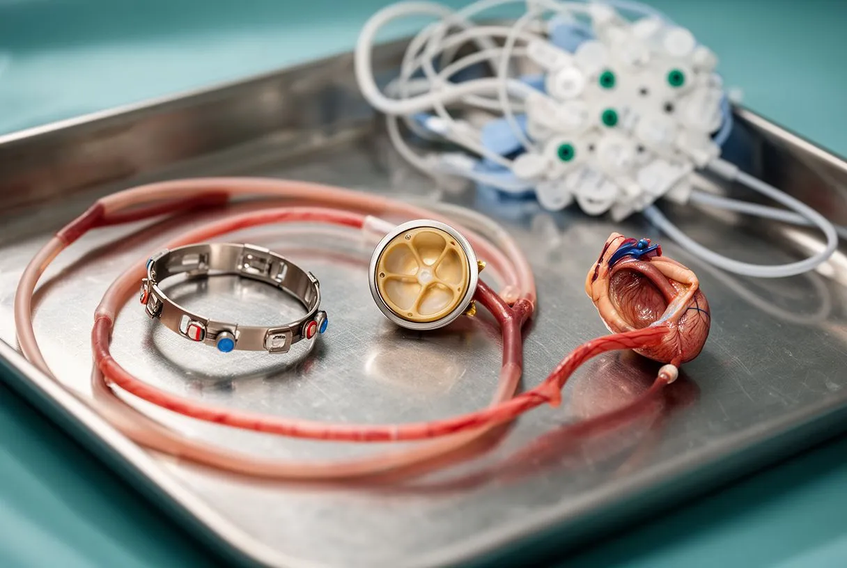
Bypass Versus Valve Surgery: Which Carries More Neurological Risk?
Coronary bypass and the aortic handling question. During coronary bypass brain injury risk is driven largely by how much the aorta must be clamped and touched. Surgeons now have options: single-clamp techniques, "no-touch" aortic approaches, epiaortic ultrasound scanning of the aorta before clamping to identify plaque-free spots, and in selected cases totally off-pump grafting using alternative inflow sites. Each choice trades off some measure of embolic risk against surgical practicality and long-term graft durability — a conversation worth having with your surgeon before the operation.
Aortic valve replacement and its particular exposure. In aortic valve replacement the surgeon works at the root of the aorta, in immediate proximity to the arteries feeding the brain. Calcified valves can shed calcium particles when they are removed, and calcified aortic walls add further debris. For patients whose aortic valve needs replacing but whose surgical risk is elevated, transcatheter aortic valve replacement (TAVR) — a catheter-based approach threaded through the arteries rather than open surgery — avoids much of that manipulation, though it introduces its own embolic exposures during catheter passage and valve deployment. Newer devices with embolic protection filters are designed to catch debris before it reaches the brain, and this remains an active area of device development.
Minimally invasive and catheter-based approaches. Many patients assume that a smaller incision automatically means a smaller brain risk. That is not reliably true. Minimally invasive heart surgery brain risk depends more on what happens inside the vessels than on the size of the skin incision. Catheter procedures can release debris too. The honest framing for patients: less invasive approaches may offer faster external recovery, but neurological risk is determined by the specific technique, the aortic disease present, and the patient's own vascular health — not by incision size alone.
Cognitive Decline Post Cardiac Surgery: What Families Notice
The first days: delirium. In the immediate postoperative period, a significant number of patients experience delirium — a temporary state of confusion, agitation, or unusual drowsiness that fluctuates through the day. It is common, usually reversible, and frequently distressing for families who do not expect it. Delirium is more frequent in older patients, those with pre-existing cognitive impairment, and those with longer operations. While most cases resolve, an episode of delirium is worth reporting and documenting, because it can be a signal of underlying brain stress.
The weeks that follow: subtle deficits. Once the fog of anesthesia and hospital disruption clears, some patients and families notice slower thinking, difficulty juggling several tasks at once, or trouble recalling names and appointments. In research this is often measured with formal neuropsychological testing, and studies show that early cognitive decline post cardiac surgery is common but that most patients improve substantially over the following months. The small silent lesions identified on imaging are one plausible contributor to these changes, alongside anesthesia effects, sleep disruption, pain medication, and the stress of major surgery itself.
The longer view. For most patients, cognition stabilizes and recovers. For a minority — particularly those with accumulating silent lesions or pre-existing vascular disease — subtle deficits can persist or progress. This is not a reason for fear; it is a reason for follow-up. Tracking memory and thinking over the first year, and treating vascular risk factors aggressively in that window, gives the brain its best chance to recover and protects against future events.

How a Brain Scan After Heart Surgery Detects Hidden Injury
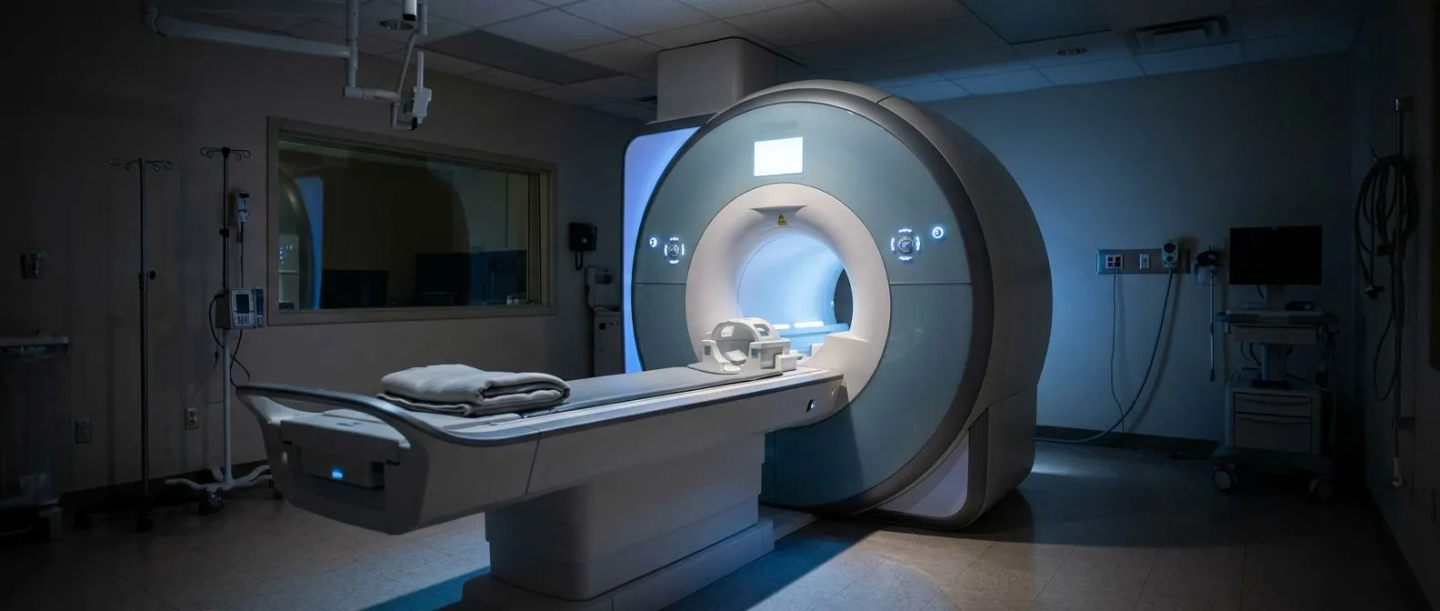
MRI and its small bright markers. The most sensitive tool for detecting silent cerebral lesions is diffusion-weighted magnetic resonance imaging (DWI-MRI), which can show new infarcts as small bright spots within hours to days of their occurrence. Cardiac surgery brain MRI findings typically appear as a handful of tiny punctate lesions, sometimes described in research as "new ischemic lesions," scattered in patterns that match an embolic shower — a burst of small particles released during the procedure. These spots often shrink or leave only faint scars, and many patients with them function normally.
When is scanning actually done? Routine brain imaging after every cardiac operation is not standard practice in most centers. Scanning is typically prompted by: any neurological sign however brief, prolonged or severe postoperative confusion, an abnormal rhythm such as new atrial fibrillation, or enrollment in research protocols. If you or a family member has persistent cognitive changes weeks after surgery, raising the question of imaging with your team is entirely reasonable.
What a finding does and does not mean. A small bright spot on a postoperative scan is not a prediction of decline. It is information. It tells your cardiologist that an embolic event occurred, which in turn informs decisions about anticoagulation, rhythm management, blood pressure targets, and the approach to any future intervention. Framed that way, the scan is less a verdict and more a data point in an ongoing plan.
Can You Prevent Silent Infarction During Surgery?
Technique and technology in the operating room. Surgeons and perfusionists (the specialists who run the heart-lung machine) have a growing toolkit for reducing embolic load. The list below summarizes the approaches most often discussed with patients:
- Aortic screening before clamping: epiaortic ultrasound lets the surgeon map plaque and choose a safe clamping site, or avoid clamping altogether.
- Reduced manipulation of the aorta: no-touch aortic techniques and single-clamp strategies shorten the time the diseased aorta is handled.
- Embolic protection devices: filters or deflectors positioned in the arteries to the brain during catheter-based valve procedures catch debris before it arrives.
- Careful perfusion management: filtered circuits, meticulous de-airing, and tighter blood pressure management on pump all reduce microemboli and low-flow episodes.
Blood pressure control as prevention. Evidence from randomized trial analysis suggests that more intensive blood pressure lowering in high-risk patients can reduce the occurrence of silent myocardial infarction — a benefit documented in a post hoc analysis published in the Annals of Noninvasive Electrocardiology. The same vascular logic applies to the brain: keeping blood pressure in a well-controlled range before and after surgery protects the small vessels most vulnerable to injury, and the European Society of Cardiology guidelines emphasize rigorous cardiovascular risk factor management as foundational prevention.
What patients can genuinely influence. You cannot choose your aortic anatomy, but you can arrive at surgery in the best vascular condition possible: blood pressure well controlled, diabetes managed, smoking stopped well in advance where feasible, carotid arteries evaluated when your team recommends it, and all medications reviewed with your surgeon and anesthesiologist. These are modest actions with meaningful effects on the whole cascade of perioperative brain injury.
Cardiac Rehabilitation and Brain Protection During Recovery
Rehabilitation is brain work too. Cardiac rehabilitation — the supervised program of graded exercise, education, and risk-factor coaching that follows most heart operations — is usually prescribed for the heart, but the brain benefits as well. Regular aerobic exercise improves blood flow, supports the small-vessel health of the brain, helps regulate blood pressure and blood sugar, and has consistent evidence for supporting cognition in older adults. Attending rehabilitation faithfully is one of the most practical cardiac rehabilitation brain protection steps available to any patient.
The vascular checklist after discharge. Recovery is the ideal time to lock in habits that protect both organs. A short list worth keeping visible at home:
- Take prescribed medications exactly as directed — antiplatelet or anticoagulant drugs, statins, and blood pressure medicines are the core of secondary prevention.
- Move daily within your team's limits — walking programs prescribed in rehab are calibrated to your recovery stage, not a generic target.
- Moderate salt and processed food — blood pressure control is a daily act, not just a clinic visit.
- Protect sleep — sleep disruption after surgery is common and worsens both cardiac and cognitive recovery; report persistent insomnia or loud snoring with daytime sleepiness.
- Attend every follow-up — rhythm checks, blood tests, and medication adjustments in the first months catch problems while they are still easy to manage.
Monitoring thinking as part of recovery. Families are often the first to notice subtle change. Keeping a simple informal record — is word-finding better or worse this month? Is bill-paying still manageable? — gives your follow-up visits real data. If changes persist beyond what your team considers typical early recovery, ask explicitly about cognitive assessment and whether brain imaging is appropriate.

When to Talk to Your Cardiac Team
Before surgery: the questions worth asking. Given your own aortic anatomy and imaging, what technique will be used and why? How will the aorta be handled during the operation? What monitoring will track brain-related complications afterward? What is the plan if a rhythm problem develops postoperatively? These are not challenging questions — good surgical teams welcome them, and the answers help you understand the specific plan built around your specific anatomy.
After surgery: the signs that deserve a call. Seek contact with your team promptly for any of the following, even if they resolve quickly: brief weakness or numbness on one side, a few seconds of speech difficulty, a sudden severe headache, new trouble with balance, or confusion that is worse at a follow-up visit than expected. Silent does not have to mean undiscussed. A brief event that resolves on its own — sometimes called a transient ischemic attack — is precisely the kind of thing your team wants to hear about, because it changes how the next months are managed.
The takeaway, and the next step. A silent infarction after heart surgery is a real, documented phenomenon — but one that modern surgical technique, careful blood pressure and rhythm management, and engaged rehabilitation can meaningfully reduce. The majority of cardiac surgery patients recover fully in body and mind, and the patients who fare best are those who understand the risks, participate actively in prevention, and stay in close contact with their team through the first year. If you are weighing a cardiac procedure or recovering from one and have questions about brain health during your recovery, the most useful next step is a conversation with a qualified cardiac team rather than self-diagnosis. Dr Alejandro Molina's practice in Guadalajara, Querétaro and Hermosillo offers consultation and patient education around every stage of cardiac care — book a consultation to discuss your own situation, your own scan results, and the plan that fits your own anatomy.