Stress Echocardiogram: Preparation and What It Shows
Stress echocardiogram preparation guide from heart specialists in Guadalajara.
What is a stress echocardiogram and why is it ordered
A stress echocardiogram is a specialized diagnostic test that combines ultrasound imaging of your heart with a monitored physical stress test to evaluate how well your heart muscles function under exertion. When you are at rest, your heart may appear to pump normally, but pushing it to work harder can reveal hidden blood flow issues. By taking ultrasound pictures before and immediately after you exercise on a treadmill or stationary bike, your cardiac team can capture real-time visuals of your heart’s pumping capacity, valve movements, and chamber sizes. This allows doctors to detect coronary artery disease early, so that you receive the most appropriate intervention for your specific situation.
Doctors typically order this test when a patient presents with symptoms like unexplained chest pain, shortness of breath, or irregular heartbeats, especially if a resting electrocardiogram (ECG) was inconclusive. During a standard physical exam, a doctor might use auscultation, or listening to your heart with a stethoscope, to identify murmurs or irregular rhythms, but a stress echo provides a much deeper anatomical view. It helps visualize the presence of an atheromatous plaque, which is a buildup of fatty deposits in the artery walls that can restrict blood flow. According to Stanford Health Care, combining exercise with imaging significantly increases the accuracy of diagnosing blocked arteries compared to a standard exercise test alone.
Patient scenario planning: Imagine you have been experiencing mild chest tightness when walking uphill, but it vanishes when you sit down. This is a classic scenario where a stress echo is invaluable. The test safely reproduces the physical conditions that trigger your symptoms in a controlled medical environment. By monitoring your heart during this specific exertion, your cardiologist can determine if your symptoms are indeed related to reduced blood supply to the heart muscle, or if they stem from a non-cardiac source. This precise diagnostic clarity is the foundation of building a safe, effective treatment plan tailored to your unique cardiovascular health.
How to prepare for stress echocardiogram
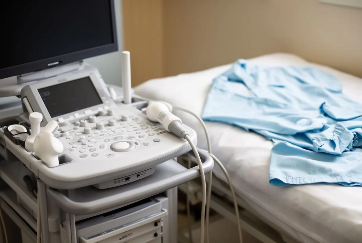
Proper preparation is essential for ensuring the accuracy and safety of your stress echocardiogram procedure. One of the most critical steps involves managing your current medications, as certain cardiac drugs can blunt the heart's response to exercise, masking vital diagnostic signs. Your cardiologist will give you specific instructions, often asking you to temporarily stop taking medications like Aspirin or beta-blockers 24 hours before the test, though you should never stop taking prescribed heart medications without explicit medical guidance. Understanding the role of medication adherence is crucial for patient safety, as highlighted by research on the Pharmacist Role in Enhancement of Medication Adherence and Clinical Outcomes in Acute Coronary Syndrome Paitents.
Fasting and dietary guidelines: To prevent nausea and ensure your heart rate responds naturally to physical exertion, you will typically be asked to fast for three to four hours before your appointment. You can usually drink small sips of water, but you should avoid caffeine, including coffee, tea, and certain sodas, as stimulants can artificially elevate your heart rate and skew the test results. Eating a heavy meal before a cardiac stress test can also divert blood flow to your digestive system rather than your heart, potentially complicating the interpretation of the ultrasound images. Wearing comfortable, loose-fitting clothing and supportive walking shoes is highly recommended, as you will be engaging in physical exercise.
When you arrive for the test, staff will usually guide you through a brief pre-test checklist. They will review your medical history, confirm your current symptoms, and take your baseline blood pressure and heart rate. It is helpful to bring a list of all your current medications and dosages, as well as any previous cardiac test results you may have. Being transparent about how you feel in the days leading up to the test allows your medical team to adjust the protocol if necessary, ensuring the procedure is both safe and effective for your specific health profile.
What happens during the stress echo procedure
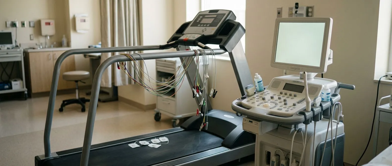
The stress echocardiogram procedure is divided into two distinct phases: the resting ultrasound imaging and the exercise phase. You will first be asked to lie down on an examination table while a sonographer applies a clear gel to your chest and places a small device called a transducer against your skin. This transducer emits high-frequency sound waves that bounce off your heart’s structures, creating real-time moving images on a monitor. The technician will capture specific views of your heart, paying close attention to the left atrium and the ventricles, documenting how effectively the heart muscle contracts while you are completely at rest.
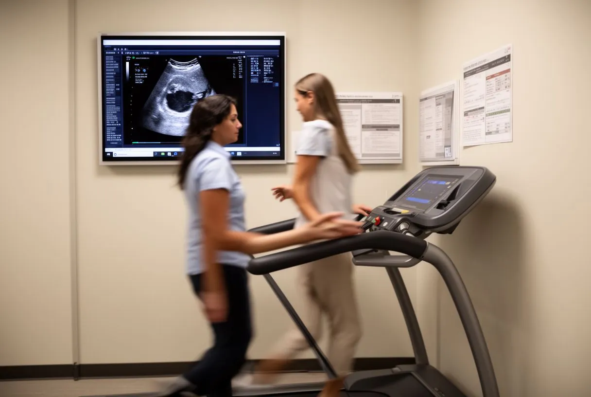
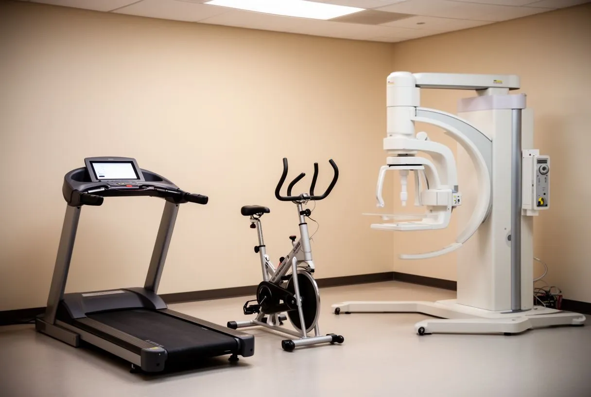
Once the resting images are recorded, the exercise phase begins. You will be hooked up to an ECG machine to continuously monitor your heart’s electrical activity, and a blood pressure cuff will be placed on your arm to track your cardiovascular response. You will then start walking on a treadmill or pedaling a stationary bike. The intensity will gradually increase by raising the speed and incline or resistance, mimicking the physical stress of climbing a steep hill. You will be encouraged to exercise until you reach a target heart rate based on your age, or until you develop symptoms like significant chest pain or fatigue that require you to stop.
Immediate post-exercise imaging: The most critical part of the stress echo occurs the moment you stop exercising. You will be quickly guided back to the examination table, and the sonographer will capture a second set of ultrasound images while your heart is still beating rapidly. This window is brief, usually lasting only one to two minutes, because the effects of physical stress on the heart muscle diminish quickly as you recover. By comparing the resting images directly with the peak-stress images, the cardiologist can identify specific areas of the heart that are not receiving adequate blood flow, pointing to the likely location of an arterial blockage.
Understanding stress echo results and what they mean
Interpreting stress echo results requires comparing the ultrasound images taken at rest with those taken immediately after exercise. A normal result means your heart muscle pumps with the same strength and efficiency during peak physical exertion as it does when you are resting. This indicates that your coronary arteries are supplying adequate blood to the heart muscle even under increased demand, effectively ruling out significant coronary artery disease. For many anxious patients, receiving a normal stress echocardiogram result provides immense relief and confirms that their symptoms may be stemming from a non-cardiac source, such as muscular strain or gastrointestinal issues.
Identifying ischemia and abnormal findings: If the results are abnormal, the ultrasound will show specific areas of the heart muscle that begin to contract weaker or more sluggishly during the post-exercise imaging. This phenomenon, known as exercise-induced ischemia, means that a portion of your heart is temporarily starved of oxygen-rich blood. Depending on the severity and the specific walls of the heart affected, your cardiologist may suspect a blockage in one or more of your coronary arteries. Recognizing these abnormal movement patterns is vital for preventing an atypical infarction, a heart attack that may present without the classic crushing chest pain but causes similar underlying damage to the heart muscle.
When abnormal results are detected, the next steps depend on the degree of ischemia observed. Mild changes might be managed with medication and lifestyle adjustments, accompanied by close monitoring over time. However, if the stress echo reveals significant regional wall motion abnormalities, indicating severe or multi-vessel coronary artery disease, your cardiac team may recommend a more invasive diagnostic procedure like a coronary angiogram. This detailed roadmap of your heart's blood vessels will help Dr Molina determine if you are a candidate for interventions such as stenting or bypass surgery to restore proper blood flow and prevent future cardiac events.
Stress echo vs other cardiac stress tests
While all cardiac stress tests aim to evaluate how your heart performs under physical stress, the specific technology used to monitor the heart varies significantly. A standard exercise stress test relies solely on an electrocardiogram (ECG) to monitor the electrical activity of your heart while you walk on a treadmill. While this is an excellent baseline screening tool, it only records the electrical signals and cannot directly visualize the physical movement of the heart muscle. A stress echocardiogram goes a step further by adding ultrasound imaging, allowing the cardiologist to actually see the heart walls contracting and relaxing, which dramatically increases the test's diagnostic accuracy.
Another common alternative is the nuclear stress test, which uses a small amount of radioactive tracer injected into your bloodstream to highlight blood flow to your heart muscle using a specialized camera. While highly accurate, nuclear imaging exposes the patient to a small dose of radiation and often requires a longer appointment time, sometimes taking several hours to complete due to the necessary waiting periods between resting and stress imaging. For patients who cannot exercise physically, a pharmacological stress test is used, where medication is given intravenously to simulate the effects of exercise on the heart, combined with either ultrasound or nuclear imaging.
Choosing the right diagnostic tool: The decision of which test is most appropriate is highly individualized based on your clinical history, baseline ECG results, and physical ability. Here is a quick comparison of why a stress echo is frequently chosen:
- Direct Visualization: Unlike standard ECG tests, the ultrasound provides real-time images of heart valves and muscle contractions, revealing structural issues alongside blood flow problems.
- No Radiation: Stress echocardiograms use harmless sound waves, making them a safer option for patients who need repeated testing or wish to avoid radioactive tracers.
- Quicker Results: Because the resting and stress imaging are done in quick succession, the total active testing time is significantly shorter than a nuclear stress test.
Stress echocardiogram risks and side effects
A stress echocardiogram is widely considered a very safe procedure, as it is performed in a controlled clinical environment under the direct supervision of trained medical professionals. The test is designed to mimic the physical exertion of daily activities, and the risk of a serious complication, such as a heart attack or a life-threatening rhythm disturbance, is exceptionally low. Your heart rate, blood pressure, and ECG are continuously monitored throughout the entire process, and the test is immediately halted if you develop concerning symptoms or if your vital signs move outside of safe parameters.
Recognizing rhythm changes: Because the test pushes your heart to beat faster and harder, it can occasionally provoke benign irregular heartbeats. Patients with underlying rhythm issues, such as atrial flutter (a rapid, organized beating in the upper chambers of the heart), need to be monitored closely, as exercise can exacerbate these conditions. In rare cases, the stress of the test may lead to more significant arrhythmias requiring medical intervention. For patients on blood thinners, maintaining a delicate balance to prevent blood clots while avoiding severe bleeding is crucial, a clinical challenge explored in the Risk factors for thromboembolic and bleeding events in anticoagulated patients with atrial fibrillation: the prospective, multicentre observational PREvention oF thromboembolic events - European Registry in Atrial Fibrillation (PREFER in AF).
Beyond the cardiac considerations, the physical act of exercising can produce minor, expected side effects. You may experience mild arm pain or leg fatigue from walking rapidly on an inclined treadmill, or slight breathlessness that resolves quickly once you stop. Some patients feel dizzy or lightheaded if their blood pressure drops slightly after the exercise ends, which is why you are monitored closely during the cool-down phase. If you have joint issues or mobility limitations that prevent you from using a treadmill, be sure to inform your cardiologist ahead of time so alternative protocols, like a supine bicycle, can be arranged to ensure your safety and comfort.
How stress echo helps evaluate chest pain
Chest pain is one of the most common and alarming symptoms patients experience, but its origin can be notoriously difficult to pinpoint without advanced testing. A stress echocardiogram is uniquely equipped to differentiate between chest pain caused by coronary artery disease and pain stemming from non-cardiac sources like acid reflux, muscle spasms, or anxiety. By visualizing the heart muscle in real time while the patient is experiencing exercise-induced symptoms, the test provides objective evidence of whether the heart's blood supply is actually compromised. If the heart walls contract normally despite the pain, the cardiologist can confidently look elsewhere for the root cause.
Visualizing the impact of blockages: When chest pain is cardiac in nature, it usually means that a portion of the heart muscle is not getting enough oxygen to meet the demands of physical exertion. The ultrasound images captured immediately after exercise will reveal this oxygen deficit as a distinct wall motion abnormality, where a specific segment of the heart muscle stops contracting vigorously. Identifying exactly which wall of the heart is affected helps the medical team deduce which specific coronary artery is likely narrowed or blocked. This precise anatomical mapping is invaluable for planning further interventions, whether that means adjusting medication or scheduling a coronary angiogram.
For patients presenting to emergency settings with ambiguous symptoms, a stress echo can be a decisive tool to prevent unnecessary hospital admissions or, conversely, to catch a severe blockage before it causes permanent damage. It allows Dr Molina and his team to make evidence-based decisions regarding the urgency of care. Having a clear, visual confirmation that a patient's chest pain is indeed linked to ischemic heart disease provides the solid justification needed to move forward with definitive treatments, ensuring that the patient is not sent home with a dangerous underlying condition left untreated. In the rare event of a sudden cardiac emergency during the test, the clinic is equipped with an Automated External Defibrillator to immediately restore a normal heart rhythm.
Recovery and what to expect after your stress echo
One of the distinct advantages of a stress echocardiogram is the complete absence of a prolonged recovery period, as the test is non-invasive and does not require any sedation or incisions. Once the post-exercise ultrasound images are captured and your heart rate and blood pressure have returned to resting levels, the test is officially over. You will be given a towel to wipe the ultrasound gel from your chest, and you will be encouraged to drink some water and sit quietly for a few minutes while the medical staff ensures you feel stable and well before standing up.
Because you were fasting and engaging in physical exertion, it is highly recommended that you bring a light snack to eat immediately after the appointment. This helps quickly replenish your blood sugar and aids in your overall recovery from the physical fatigue of the treadmill. You should be fully capable of driving yourself home and returning to your normal daily activities right after leaving the clinic. There is no downtime required, and you do not need to take time off work to recover from the procedure itself, making it highly convenient for traveling patients and their families.
Long-term monitoring after testing: While the physical recovery from the test is immediate, the diagnostic information gained from the stress echo informs your long-term cardiovascular care. If your results were normal, it provides a reassuring baseline that can be referenced in the future if new symptoms arise. If your results indicated early signs of coronary artery disease, your recovery phase transitions into a management phase. This involves implementing lifestyle modifications, adhering to newly prescribed cardiac medications, and scheduling follow-up appointments to ensure that your heart health is closely monitored over the subsequent months and years.
When to contact your cardiac team following the test
After your stress echocardiogram, a follow-up appointment is usually arranged to review the images and discuss the results in detail. This appointment is essential for understanding your cardiac health status and asking any lingering questions you might have about the findings. It is important to remember that the sonographer performing the test is not permitted to give you immediate results, as the images must be carefully analyzed and interpreted by the cardiologist. You should contact the clinic if you have not heard back regarding your scheduled follow-up within a few days of your test.
While rare, certain symptoms following a stress test warrant immediate medical attention. If you experience new or worsening chest pain, severe shortness of breath that does not resolve with rest, fainting, or a rapid and irregular heartbeat that persists for more than a few minutes after you have left the clinic, you should seek emergency medical care immediately. These could be signs that your heart condition is unstable, requiring urgent intervention rather than waiting for a routine follow-up appointment. Always err on the side of caution when it comes to cardiac symptoms.
Coordinating your ongoing care: Effective cardiac care relies on clear communication between the patient and the medical team. If your stress echo reveals the need for further testing or a change in your treatment plan, our team at Cirugía de Corazón will guide you through every step of the process. Keep the following points in mind for your post-test care:
- Medication Adjustments: If your cardiologist instructed you to stop certain medications for the test, confirm exactly when you should resume your normal dosing schedule.
- Symptom Tracking: Keep a brief diary of any new symptoms you experience in the days following the test to share with Dr Molina during your results consultation.
- Prompt Communication: Do not hesitate to call the clinic if you have concerns about post-test side effects or if you need clarification on your pre-appointment instructions for any upcoming procedures.
Planning your visit for a stress echocardiogram in Mexico
For international patients and those traveling within Mexico, planning a visit for a stress echocardiogram requires a bit of logistical preparation. Dr Alejandro Molina leads a multidisciplinary team of cardiac surgeons with consulting rooms in Guadalajara, Querétaro, and Hermosillo. When scheduling your appointment, be sure to inform the clinic staff if you are traveling from out of town. They can often assist with coordinating appointment times to minimize your stay and provide recommendations for nearby accommodations, ensuring that you and your family have a comfortable and stress-free experience while accessing top-tier cardiac care.
Understanding the cost and insurance coverage for a stress echocardiogram is a common concern for many families. Because the practice does not sell products and focuses purely on patient care, the costs associated with the test reflect the professional medical expertise and the advanced imaging technology utilized. We encourage open discussions about financial expectations before the procedure so there are no surprises.
If you or a loved one are experiencing symptoms that warrant a cardiac stress test, or if you have been referred for a stress echocardiogram and wish to receive expert care, we invite you to reach out to our team. A proper diagnosis is the first step toward protecting your heart health and improving your quality of life. Please book a consultation with Cirugía de Corazón to discuss your specific situation with Dr Molina. Do not attempt to self-diagnose based on symptoms alone; let our specialized team provide the accurate, compassionate medical evaluation you deserve.