

Vasa Vasorum: The Blood Supply of the Aortic Wall
Vasa vasorum aortic wall blood supply explained: learn how these tiny vessels nourish the aorta, why it matters for heart health, and when to see a specialist.
What Is the Vasa Vasorum Aortic Wall Blood Supply?
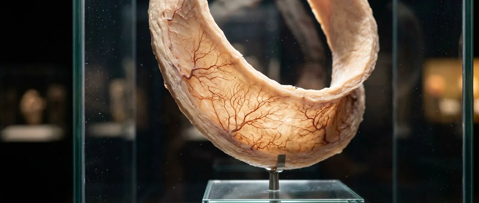
The vasa vasorum are the tiny blood vessels that supply oxygen and nutrients to the walls of larger arteries, including the aorta itself. When patients first hear that the aorta — the largest artery in the body — has its own internal blood supply feeding its walls, the concept can seem surprising. After all, the aorta carries oxygenated blood to nearly every organ, so why would its own tissue need a separate set of vessels? The answer lies in the thickness of the aortic wall: the innermost cells can absorb oxygen directly from the blood flowing through the lumen, but the outer layers are too far from that blood column to receive adequate nourishment by simple diffusion alone. The vasa vasorum aortic wall blood supply bridges that gap, keeping the structural tissue of the aorta alive and functional.
Why the aortic wall needs its own microcirculation: The aortic wall is composed of three distinct layers — the intima (inner lining), the media (thick muscular and elastic middle layer), and the adventitia (outer connective tissue wrapping). In the ascending aorta, the portion of the aorta that rises from the heart, the media is particularly thick and demands a robust adventitial blood supply to remain healthy. Research published in JAMA Network has contributed to our understanding of how disruption to this microcirculation contributes to vascular disease. Without a functioning vasa vasorum network, the outer portions of the media would become starved of oxygen, weakening the wall and setting the stage for serious complications.
For patients facing cardiac surgery, understanding this microcirculation matters because it explains why certain conditions — hypertension, atherosclerosis, and connective tissue disorders — damage the aorta in ways that go beyond simple wear and tear. When the vasa vasorum are compromised, the wall tissue they serve begins to deteriorate, and the aorta can dilate, tear, or rupture. This is why your cardiac team pays close attention not only to the diameter of the aorta but to the overall health of the wall tissue when deciding whether surgery is necessary.
How Does Vasa Vasorum Anatomy Work in the Aorta?
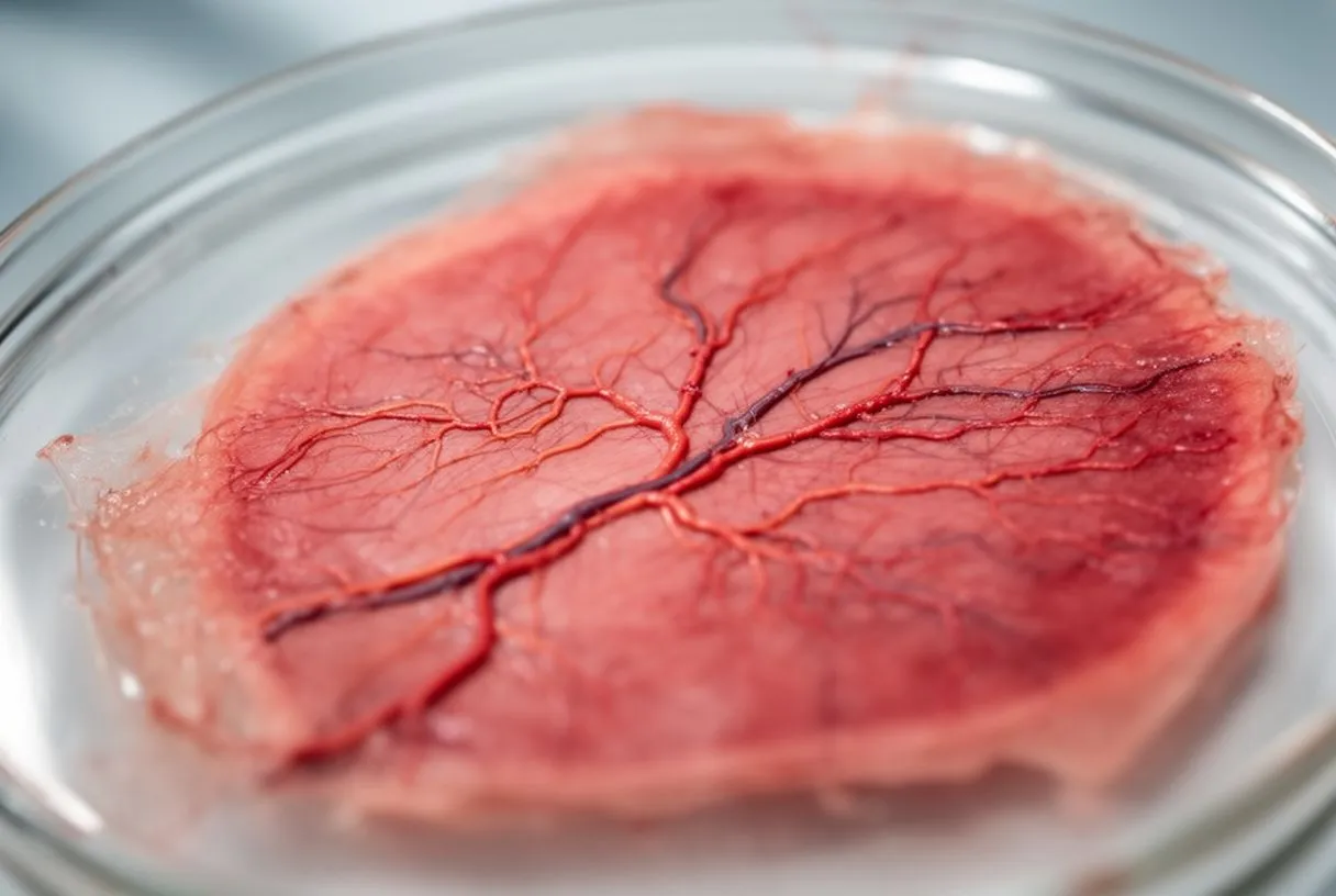
The vasa vasorum anatomy follows a branching pattern that begins in the adventitia, the outermost layer of the aortic wall. Small arterioles enter from nearby connective tissue and perivascular fat, then divide into a capillary network that penetrates inward toward the media. These capillaries run parallel to the circular smooth muscle cells of the media, delivering oxygen and removing metabolic waste. Venous return travels back outward through venules that drain into the adventitial veins. This inward-facing supply system is sometimes described as running "counter-current" to the main blood flow inside the aorta, though the precise hemodynamic relationship remains an area of active research.
Differences between thoracic and abdominal supply: The thoracic aorta, which includes the ascending aorta and the aortic arch, generally has a richer and more densely organized vasa vasorum network than the abdominal aorta. This difference in aortic wall vascularization helps explain why the two segments behave differently in disease. The abdominal aorta is more prone to atheromatous plaque formation and aneurysm formation, while the thoracic aorta is more frequently involved in dissection and connective tissue disorders. The variation in microcirculatory density is one contributing factor, alongside differences in wall composition, elastin content, and biomechanical stress.
In patients undergoing procedures that involve the ascending aorta — such as valve replacement, aortic root repair, or the Bentall procedure, which replaces both the aortic valve and the adjacent aortic segment — the surgical team must consider the impact on the vasa vasorum. When an arterial graft is sewn into place, the native adventitial blood supply is inevitably disrupted at the anastomosis, and the healing process depends partly on re-establishing microcirculatory connections. This is one of many reasons why recovery from aortic surgery is measured in weeks and months rather than days, and why your surgeon will discuss gradual return to activity rather than immediate resumption.
What Is the Function of Vasa Vasorum in the Aortic Wall?
The primary function of the vasa vasorum is to deliver oxygen and nutrients to the cells of the aortic wall that sit beyond the reach of diffusion from the lumen. Smooth muscle cells in the media require oxygen to maintain the elastic recoil of the aorta with every heartbeat, and fibroblasts in the adventitia need nutrients to produce and maintain the collagen framework that gives the wall its tensile strength. The vasa vasorum function extends beyond simple delivery, though — this capillary network also clears carbon dioxide and metabolic by-products, participates in immune surveillance by allowing inflammatory cells to access the wall, and contributes to the remodeling processes that occur during normal aging and disease.
Role in wall integrity and repair: When the aortic wall experiences mechanical stress — from high blood pressure, for example — the smooth muscle cells and extracellular matrix must adapt. The vasa vasorum supply the metabolic fuel for that adaptation. If this supply is compromised, the wall cannot repair micro-injuries as effectively, and small tears or areas of degeneration can accumulate over time. This is one mechanism by which chronic hypertension contributes to aortic dilation: not only does elevated pressure stretch the wall mechanically, but the resulting damage to the vasa vasorum also impairs the wall's ability to maintain and repair itself.
For patients taking medications such as aspirin or other blood thinners before cardiac surgery, the vasa vasorum are also relevant to how the surgical team manages bleeding risk. These tiny vessels are fragile, and the anticoagulation protocols used during cardiopulmonary bypass — where an arterial cannula diverts blood flow through a heart-lung machine — affect how the vasa vasorum respond when normal circulation is restored. Your anaesthesiologist and surgeon coordinate the timing of anticoagulation reversal carefully, balancing the need to prevent clotting against the need to protect the microcirculation of the aortic wall and other tissues.
Types of Vasa Vasorum in the Aortic Wall
Vasa vasorum histology reveals two main patterns of vessel distribution within the aortic wall. The first type consists of vessels that remain confined to the adventitia, forming a network in the outermost connective tissue layer. These adventitial vessels are present throughout most of the aorta and provide the primary supply route for the outer wall. The second type penetrates into the media, travelling inward through the muscular layer to supply the deeper portions of the wall. The degree of medial penetration varies along the length of the aorta, being more pronounced in the thoracic segments and less so in the abdominal aorta.
Vessel subtypes and their clinical relevance: Within these two broad categories, researchers have identified arterioles, capillaries, and venules, each playing a distinct role. The arterioles regulate flow into the capillary bed, the capillaries perform the actual exchange of oxygen and nutrients, and the venules drain waste-laden blood back outward. In some patients who undergo combined procedures — for example, simultaneous pectus excavatum repair using sternal plate in a patient undergoing the Bentall procedure, as described in a published case report — the surgical planning must account for how the chest wall and aortic microcirculation interact during recovery.
Understanding these vessel subtypes also helps explain why certain diseases target specific layers of the aortic wall. Conditions that impair the adventitial vasa vasorum, such as some forms of vasculitis, tend to cause inflammation concentrated in the outer wall. Conditions that affect the penetrating medial vessels may contribute to intramural hematoma, where blood collects within the wall itself without a full-thickness tear. Your cardiac team uses imaging and clinical findings to distinguish between these patterns, because the treatment approach — whether medical management, urgent surgery, or watchful waiting — depends on which part of the wall and which component of the microcirculation is involved.
How Does the Aortic Wall Get Its Oxygen Supply?
The aortic wall receives its oxygen through two complementary mechanisms: direct diffusion from the blood flowing through the lumen, and delivery by the vasa vasorum. The intima and the inner portion of the media are close enough to the luminal blood that oxygen diffuses directly across the endothelial surface. This diffusion pathway is efficient for the innermost 0.5 to 1 millimetre of wall thickness. Beyond that depth, the distance is too great for diffusion to meet metabolic demand, and the vasa vasorum become the essential supply route.
When diffusion is not enough: In a healthy adult aorta, the wall thickness ranges from roughly 2 to 4 millimetres depending on the segment and the individual. This means that a significant portion of the media and all of the adventitia depend on the vasa vasorum for survival. When the aorta dilates — as in an aneurysm — the wall often thins in some areas while the overall diameter increases, creating a mismatch between the metabolic demand of the tissue and the capacity of the existing vasa vasorum to supply it. This mismatch is thought to contribute to the progressive weakening seen in enlarging aneurysms and is one reason why surgical intervention is recommended once the aortic diameter exceeds certain thresholds, such as 5.5 centimetres for the ascending aorta in many guidelines.
Patients with elevated levels of apolipoprotein B, a protein carried on cholesterol particles, may develop atheromatous plaque in the intima that further impairs oxygen diffusion to the inner media. The plaque itself takes up space, thickens the intima, and creates a physical barrier between the luminal blood and the deeper wall cells. In this way, atherosclerosis affects the aortic wall not only by narrowing the lumen but also by starving the wall tissue of oxygen from both supply routes simultaneously — diffusion from the inside and vasa vasorum delivery from the outside are both compromised. This dual mechanism helps explain why advanced atherosclerosis is associated with weakening and aneurysm formation, not just obstruction.
What Happens When Vasa Vasorum Blood Supply Fails?
When the vasa vasorum blood supply to the aortic wall is disrupted, the consequences follow a predictable sequence. The cells of the media, deprived of oxygen, cannot maintain the elastic fibres and smooth muscle that give the aorta its compliance and strength. Over time, these cells undergo degeneration — a process sometimes called cystic medial necrosis, though the term is somewhat misleading because the changes involve gradual breakdown rather than acute cell death. The extracellular matrix weakens, elastin fragments, and collagen remodelling becomes disordered. The wall loses its ability to recoil with each heartbeat, and the aorta gradually dilates.
Links to aneurysm and dissection: This degenerative cascade is one of the mechanisms underlying aortic aneurysm formation. As the wall weakens, the pressure of each heartbeat pushes outward, increasing the diameter further. The larger the aorta becomes, the greater the wall tension according to the law of Laplace, creating a feedback loop that accelerates dilation. If the process continues unchecked, the wall may tear, producing aortic dissection — a life-threatening condition where blood enters the wall and splits the layers apart. Disruption of the vasa vasorum is therefore not merely a consequence of aortic disease but is increasingly recognized as a contributor to its initiation and progression.
For patients, this means that conditions which impair microcirculation — poorly controlled hypertension, diabetes, smoking, and certain inflammatory diseases — should be taken seriously not only because of their general cardiovascular risks but because of their specific effect on aortic wall health. Your cardiac surgeon will assess these factors when evaluating your overall risk profile, often using scoring systems such as EuroSCORE II to estimate operative risk. Managing blood pressure, quitting smoking, and controlling blood sugar are not generic pieces of advice — they directly affect whether the vasa vasorum can continue to nourish the aortic wall and keep it intact.
How Is Vasa Vasorum Disease Diagnosed?
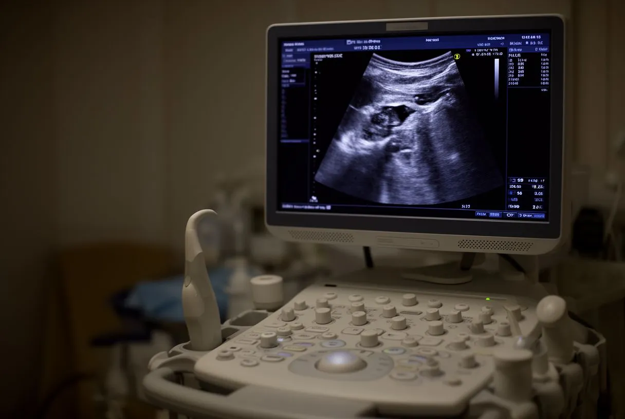
Direct imaging of the vasa vasorum in a living patient is challenging because these vessels are extremely small, often in the range of 20 to 100 micrometres in diameter. Conventional echocardiography, CT angiography, and standard MRI can visualise the aortic wall and its gross structure but cannot resolve individual vasa vasorum. However, advanced imaging techniques are beginning to provide indirect and sometimes direct evidence of microcirculatory disruption. Contrast-enhanced MRI, for instance, can demonstrate patterns of wall enhancement that suggest increased vasa vasorum density or inflammation, and research continues to refine these methods.
Imaging tools and their limitations: Magnetic resonance imaging in the evaluation of pectus excavatum and related chest wall conditions, as discussed in a published study, illustrates how cross-sectional imaging can characterise the relationship between structural abnormalities and vascular anatomy in the thorax. While this specific research focuses on chest wall deformity, the imaging principles — high-resolution tissue characterisation, contrast dynamics, and multiplanar reconstruction — are directly relevant to evaluating aortic wall health. Your cardiac team will typically combine several imaging modalities to build a complete picture of your aorta and its wall integrity.
In clinical practice, the diagnosis of vasa vasorum dysfunction is usually inferred rather than directly confirmed. Your surgeon or cardiologist will consider the overall condition of the aortic wall as seen on imaging, the presence of risk factors known to impair microcirculation, and findings such as wall thickening, intramural haematoma, or evidence of inflammation. Auscultation — listening to the heart and blood vessels with a stethoscope — may reveal murmurs or abnormal sounds that prompt further investigation, though it does not directly assess the vasa vasorum. The diagnostic process is one of assembling multiple pieces of evidence to reach a reasoned assessment of wall health and surgical risk.
Vasa Vasorum and Aortic Dissection: What Is the Link?
The relationship between the vasa vasorum and aortic dissection is an area of significant clinical interest. Traditional teaching holds that dissection begins with a tear in the intima that allows blood to surge into the media, splitting the wall layers apart. However, an alternative and complementary theory proposes that rupture of the vasa vasorum within the wall can initiate the process. According to this model, bleeding from a damaged vasa vasorum vessel creates a haematoma within the media — an intramural haematoma — which can then progress to a full dissection if the pressure builds sufficiently to rupture through the intima.
Evidence from clinical and pathological studies: This vasa vasorum rupture theory helps explain certain cases of aortic dissection that occur without a clearly identifiable intimal tear on imaging, as well as cases of intramural haematoma that evolve into classic dissection over time. The distinction matters for treatment, because intramural haematoma is sometimes managed differently from acute dissection, particularly when it is small and the patient is stable. However, both conditions require urgent evaluation by a cardiac specialist, because the risk of progression is real and unpredictable.
Patients who have undergone previous cardiac surgery, including those with an arterial graft or prosthetic valve, should be aware that the junctions between native tissue and surgical materials create areas of altered stress on the aortic wall. While the vasa vasorum supply to the native aorta is generally preserved in these regions, the healing and remodelling processes at anastomotic sites can affect microcirculatory patterns over the long term. Regular follow-up imaging is essential for these patients, typically on an annual basis, to monitor for changes in aortic diameter, wall thickness, or new findings that could signal developing problems.
Can Vasa Vasorum Rupture Cause Aortic Wall Ischemia?
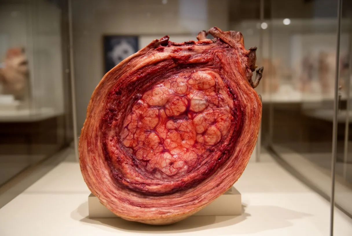
Yes, rupture of the vasa vasorum can lead to localised aortic wall ischemia — a shortage of oxygen supply to the wall tissue — though the mechanism differs from the ischemia seen in other organs. When a vasa vasorum vessel ruptures, blood escapes into the wall tissue, creating pressure and swelling within the media. This bleeding can compress adjacent capillaries, reducing flow to nearby areas of the wall and creating zones of relative oxygen deprivation. The result is a combination of direct mechanical damage from the haematoma and secondary ischemic injury to the surrounding cells.
Understanding the ischemic mechanism: Unlike a heart attack, where a coronary artery is blocked and an area of heart muscle dies, aortic wall ischemia from vasa vasorum rupture tends to be patchy and progressive rather than sudden and complete. The wall does not die all at once; instead, small areas of damage accumulate, weakening the overall structure. This gradual process is one reason why some patients experience warning symptoms — chest pain, back pain, or a tearing sensation — before a full dissection occurs, while others have no symptoms at all until a catastrophic event. Arm pain or unusual chest discomfort that does not resolve should always be evaluated promptly, particularly in patients with known aortic disease or risk factors.
For patients and families trying to understand why their cardiac team recommends surgery at a particular time, the ischemia mechanism provides important context. The decision to operate is not based solely on the current diameter of the aorta but on the assessment of wall health, the rate of change over time, and the presence of findings that suggest the vasa vasorum and wall tissue are under stress. Your surgeon balances the risk of the operation against the risk of leaving a deteriorating aorta in place, and this calculation is specific to each individual. In some cases, autologous blood — the patient's own blood, collected and re-administered — may be used during surgery to support the microcirculation and reduce the need for donor transfusion.
When to See a Cardiac Specialist About Aortic Wall Health
Several signs and situations warrant evaluation by a cardiac specialist with expertise in aortic disease. If you have been diagnosed with an aortic aneurysm, even a small one, regular monitoring is essential to track changes in diameter and wall characteristics. A family history of aortic dissection, aneurysm, or sudden cardiac death should prompt screening, as genetic factors significantly influence aortic wall health and the integrity of the vasa vasorum network. Conditions such as Marfan syndrome, Loeys-Dietz syndrome, bicuspid aortic valve, and other connective tissue disorders are associated with a higher risk of aortic complications and require specialised, lifelong follow-up.
Practical guidance for patients and families: Seek immediate medical attention if you experience sudden, severe chest or back pain that feels tearing or ripping in quality, particularly if it radiates to the back or is accompanied by differences in blood pressure between the arms. These symptoms may indicate acute aortic dissection, which is a surgical emergency. For less acute concerns — such as a newly diagnosed aneurysm, questions about whether your aortic diameter is approaching surgical thresholds, or planning for monitoring — a consultation with a cardiac surgeon or cardiologist specialising in aortic disease is the appropriate next step.
Patients travelling from outside Mexico to consult with Dr Alejandro Molina and the team at Cirugía de Corazón in Guadalajara, Querétaro, or Hermosillo should bring all prior imaging studies, echocardiogram reports, and relevant laboratory results. The practice does not sell products or recommend supplements; the focus is on evidence-based surgical and medical management tailored to your specific anatomy and risk profile. Every recommendation is grounded in established clinical understanding, and no claim is made that cannot be supported by published evidence. If you or a family member have questions about aortic wall health, the vasa vasorum, or whether surgical evaluation is appropriate for your situation, the most important step is to book a consultation and discuss your case directly with a qualified cardiac team rather than relying on self-assessment.
- Key takeaway for patients: The vasa vasorum are the lifeline of the aortic wall, and their health directly influences whether the aorta remains strong and intact.
- What to watch for: Gradual aortic enlargement on imaging, new chest or back pain, and any family history of aortic disease all warrant specialist evaluation.
- Next step: Bring your imaging and records to a consultation and discuss your individual risk profile with a cardiac surgeon who specialises in aortic disease.






