Ventricular Septal Defect in Adults: Closure Options
Ventricular septal defect adults need timely closure options.
Most people who learn they have a ventricular septal defect in adults are surprised: they may have lived decades with a small hole in the wall that separates the heart's two lower pumping chambers, and only now a new doctor has heard a heart murmur or an echocardiogram has finally explained symptoms that never had a name. A ventricular septal defect, or VSD, is a gap in the ventricular septum, the muscular wall between the left and right ventricles. Some close on their own in childhood; others persist into adult life quietly, or grow more troublesome with age. This guide walks through how an adult VSD behaves, how it is diagnosed, and the closure options available today — from open-heart repair to catheter-based devices — so you can discuss your situation with a cardiac team from a position of understanding.
What Is a Ventricular Septal Defect in Adults?
A ventricular septal defect in adults is an abnormal opening in the wall that separates the heart's two lower chambers. Normally, the left ventricle — the chamber that pushes blood to the entire body — operates at much higher pressure than the right ventricle, which sends blood only to the lungs. When there is a hole between them, oxygen-rich blood is pushed through the defect and recirculated to the lungs instead of out to the body. Surgeons and cardiologists classify VSDs by their location: muscular defects sit in the lower, muscular part of the septum; perimembranous defects lie near the valves at the top of the wall; and outlet or supracristal defects sit close to the pulmonary valve, where blood leaves toward the lungs.
The size of the defect matters more than its location when it comes to symptoms. A small restrictive VSD allows only a limited amount of blood through and often causes no symptoms at all, which is why many adults discover theirs incidentally. A large defect allows significant blood to flood the lungs year after year, straining the right side of the heart and raising pressure in the lung arteries. Adults typically fall into two groups: those who had a defect repaired in childhood that has reopened or left a residual leak, and those whose small defect was never repaired because it never caused problems.
Many adults have a VSD that was first mentioned decades ago and never followed up; reviewing the original imaging, repeating the echocardiogram with current techniques, and explaining in plain words what the defect is doing to the heart today is the right starting point. That fresh assessment is the foundation for every decision that follows.
What Causes a Ventricular Septal Defect?
Most VSDs are congenital heart defects — they form during pregnancy, when the fetal heart fails to close the wall between the ventricles completely as it develops. The ventricular septum is built from several components that must fuse together in the first eight weeks of gestation; when one edge fails to meet its neighbor, a permanent opening remains. In most cases no specific cause is identified, though certain genetic conditions such as Down syndrome raise the likelihood, and some environmental exposures during pregnancy are associated with higher risk as well.
Adults can also acquire a septal defect later in life. After a heart attack, the muscular septum can rupture — this is a rare but catastrophic complication that requires emergency surgery. After heart surgery, a septal defect can occasionally result from complications of procedures on the aortic or mitral valves. From infection, severe endocarditis — infection of the heart's inner lining — can erode tissue and create an acquired defect. These acquired VSDs behave very differently from congenital ones: they appear suddenly in a heart that has never compensated for the extra blood flow, and they almost always demand urgent attention.
The practical point for most adult readers is this: if you have a congenital VSD, nothing you did caused it, and nothing you did could have prevented it. What matters now is how the defect has behaved over the years and what it is doing to your heart at this stage of your life.
Ventricular Septal Defect Symptoms in Adults
Small defects often produce no symptoms whatsoever — the only clue is the characteristic harsh heart murmur that a doctor hears with a stethoscope. When a VSD does cause problems in adulthood, the symptoms reflect the heart working harder and the lungs receiving more blood than they should over many years. Shortness of breath during exertion is the most common complaint, and it tends to creep up slowly enough that many people attribute it to getting older or being out of shape. Fatigue and reduced exercise capacity follow the same pattern: the heart is rerouting part of its output back to the lungs, so less oxygenated blood reaches the muscles.
Palpitations — the sensation of a racing or irregular heartbeat — can signal that the heart's chambers have stretched from years of extra volume. Adults with long-standing, larger defects may develop atrial fibrillation or ventricular tachycardia, an abnormal fast rhythm arising from the pumping chambers. Signs of heart failure — swelling of the ankles, breathlessness lying flat, waking at night breathless — appear when the heart can no longer keep up, and they warrant prompt evaluation. In rare, advanced cases, chronic over-circulation of the lungs causes permanent changes in the lung arteries, a condition called Eisenmenger syndrome, in which the defect actually reverses direction and oxygen-poor blood enters the body circulation.
If you notice new breathlessness, palpitations, or swelling and you know or suspect you have a septal defect, do not wait for the next routine check. These symptoms are the heart's way of reporting that the balance has shifted, and earlier evaluation widens the treatment options considerably.

How Serious Is a Ventricular Septal Defect?
Seriousness depends almost entirely on the size of the defect and the pressures inside the heart. Small defects — the kind that produce a loud murmur but little actual shunting — often cause no measurable harm, and many people with a small VSD live a full, normal life without ever needing surgery. Moderate defects progressively overload the left side of the heart, and over years they can enlarge the left atrium and left ventricle, reduce pumping efficiency, and trigger rhythm problems. Large defects that have gone unrepaired since childhood are the most serious: chronic high flow can eventually damage the lung arteries and raise pulmonary artery pressure, at which point closing the defect may no longer be possible or beneficial.
The window for intervention is therefore a moving target. A defect that was safe to watch at 30 may need repair at 50 if the heart has begun to dilate. This is why cardiologists track specific measurements — the size of the left ventricle, the ratio of lung-to-body blood flow, and the pressure in the pulmonary arteries — and why a VSD in an adult should never be dismissed as "just a hole you were born with." One of the key risks of leaving a significant defect untreated is left ventricular dilatation, the gradual stretching of the main pumping chamber as it handles the extra volume, year after year.
We review every adult VSD case as a team against international guidelines before making any recommendation — we look at the defect's size, the chamber dimensions, and the lung pressures together, and then present the patient with the options and the reasoning behind each one. The seriousness of your particular defect is not a guess; it is something the studies can show precisely.
How Is a Ventricular Septal Defect Diagnosed in Adults?
The diagnosis usually begins with the stethoscope. A VSD produces a distinctive pansystolic murmur — a harsh sound lasting through the whole contraction of the heart — often accompanied by a vibration the examiner can feel with a hand on the chest. Many adults are first sent for testing precisely because a new physician hears this murmur during an unrelated checkup. From there, the workup is straightforward and non-invasive.
- Echocardiogram: the central test. Ultrasound images show the defect's location and size, measure the direction and volume of blood crossing it, and assess the size and pumping function of both ventricles.
- Transesophageal echocardiography: an ultrasound probe passed into the esophagus gives sharper views of the septum, and is often used when the exact anatomy is unclear or when planning device closure.
- Electrocardiogram and Holter monitoring: these record the heart's electrical activity and detect rhythm disturbances that long-standing shunting can cause.
- Cardiac catheterization: thin catheters measure pressures inside the heart and lungs and calculate the ratio of blood flow to the lungs versus the body — the number that determines whether closure is safe.
Cardiac MRI has become increasingly valuable for adults, because it measures ventricular volumes and shunt fractions with precision and can track changes over years of follow-up. For most patients, an echocardiogram plus one or two supporting studies answers every question the team needs. In the consultation we take the time to explain what each study actually shows, in plain words — what the defect looks like, what the pressures mean, and why a given measurement drives the recommendation — before any decision is made.
When to Repair a Ventricular Septal Defect in Adults?
Not every adult VSD needs closure, and knowing when to repair a VSD in adults is one of the most important judgments a cardiac team makes. The general principle: repair when the defect is causing harm or will cause harm, and repair before the damage becomes permanent. Clear indications include significant shunting that has enlarged the left ventricle, heart failure symptoms attributable to the defect, recurrent endocarditis, and progressive worsening on serial imaging. In each case the goal is to close the hole while the heart and lungs can still recover.
Watchful waiting is a legitimate strategy for small, restrictive defects with no chamber enlargement and normal pressures. These patients simply need regular follow-up echocardiograms — typically every few years — to confirm nothing is changing. The danger lies in assuming a defect is harmless without ever measuring it. A defect deemed trivial at 25 deserves re-evaluation at 45, because the heart's tolerance for extra volume declines with age, and conditions like hypertension or atrial fibrillation can change the calculus.
When closure is no longer advisable — usually when pulmonary artery pressure has risen so high that the lungs resist the redirected blood — closing the defect can actually harm the patient by straining the right ventricle against high resistance. This is why the catheterization study measuring lung pressures is so important before committing to repair. The team weighs the ratio of lung-to-body blood flow: above certain thresholds, closure is contraindicated; below them, it offers real benefit. Every case is reviewed by our multidisciplinary team against international practice guidelines before a recommendation is given — and the final decision is always the patient's, presented with the alternatives laid out clearly.
Ventricular Septal Defect Surgery in Adults
Open-heart surgical closure remains the standard treatment for large defects, complex anatomy, and defects in locations unsuitable for devices. The operation is performed under general anesthesia on a heart-lung machine, which takes over circulation while the surgeon works. Through an incision in the chest — classically through the breastbone, though many defects can be reached through a smaller right-sided thoracotomy or a partial sternotomy — the surgeon opens the right atrium or right ventricle and closes the defect with a patch of synthetic material or the patient's own tissue, sewn securely into the firm edges of the septum.
Surgical repair in adults carries a well-established track record. The operation also allows the team to address anything found along the way: a leaking valve can be repaired at the same time, and in perimembranous defects the surgeon takes care to protect the conduction pathway that runs along the edge of the defect, since injury there can require a pacemaker. Adults who had childhood repairs that have partially reopened — a residual VSD — often undergo repeat surgery, which is more technically demanding but routinely successful in experienced hands.
Which approach suits you? The choice between a full sternotomy and a minimally invasive incision depends on the defect's location, prior chest surgery, and the patient's own priorities. Our team performs conventional, minimally invasive, robotic and transcatheter procedures, and we recommend the less invasive route only where the evidence supports it for that specific patient — for Dr Molina, who trained in cardiac and minimally invasive surgery at the University of Montreal, the question is never which technique is newest, but which is safest for this heart. Transesophageal echocardiography is used during the operation to confirm the closure is complete before the patient leaves the operating room, a step shown to catch residual leaks immediately rather than months later, as documented in studies of post-repair echocardiographic assessment published in the Asian Journal of Anaesthesiology and Pain Medicine.
Catheter Closure of a Ventricular Septal Defect in Adults
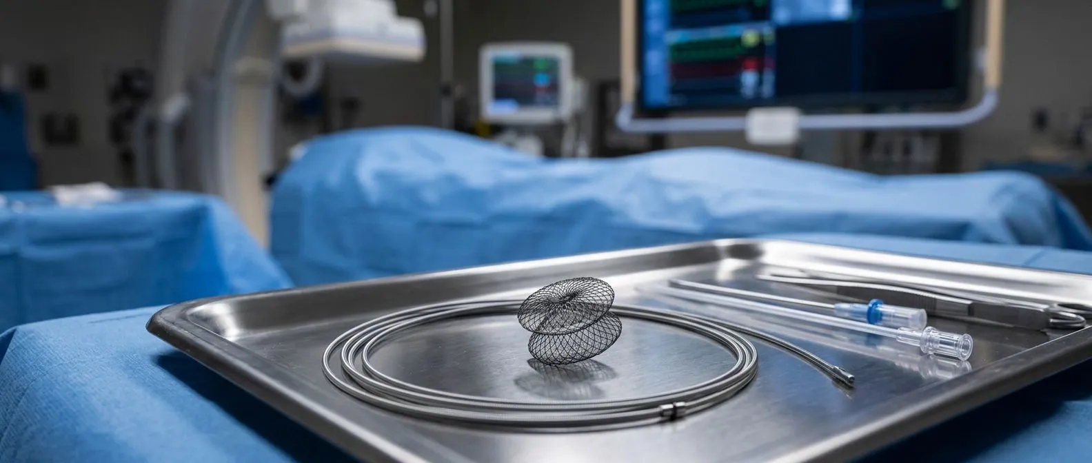
For selected defects, catheter closure of a VSD in adults offers an alternative to open surgery. The procedure is done in a cardiac catheterization laboratory: a thin catheter is threaded from a vein in the groin up to the heart, across the defect, and a mushroom-shaped occluder device — most commonly an Amplatzer-type device — is deployed to plug the hole from both sides of the septum. There is no chest incision, no heart-lung machine, and typically only one or two nights in hospital.
Who is a candidate? Catheter closure works best for muscular defects, which have thick tissue margins away from valves and conduction tissue, and for selected perimembranous defects using devices specifically designed to avoid the conduction system and the aortic valve. Defects close to the pulmonary valve or the tricuspid valve, very large defects, and defects with thin or inadequate margins usually require surgery instead. The heart team measures the defect precisely on imaging — often with the help of transesophageal ultrasound — and selects a device size that bridges the hole without impinging on neighboring structures.
The advantage is clear: a much gentler recovery and no sternum to heal. The trade-offs are equally clear: device closure is only suitable for a subset of defects, and the device leaves small metal discs within the heart that require follow-up imaging and, briefly, precautions against bacteria entering the bloodstream during dental work. Because we perform both surgical and transcatheter closure, our team can present both options honestly — including the option of doing nothing — rather than steering every patient toward whichever procedure we happen to favor. Related structural procedures, such as septal defect closure for atrial septal defects — holes in the wall between the upper chambers — and paravalvular leak closure after valve replacement, use the same catheter technology and are routinely performed by teams experienced in structural heart intervention.
Ventricular Septal Defect Closure Recovery Time
Recovery differs sharply between the two approaches. After catheter closure, most adults go home within one or two days, feel back to their normal selves within a week, and resume full activity — including exercise — within two to four weeks. The groin puncture site needs a few days of care, and there are short-term restrictions on heavy lifting while the device endothelializes, meaning while the heart's own tissue grows over the device surface to incorporate it permanently.
After open-heart surgery, recovery is longer and more structured. The breastbone, when divided, takes roughly six to eight weeks to knit back together, and patients follow lifting restrictions during that period. Hospital stay is typically five to seven days; walking begins within a day or two of surgery; driving resumes at around four to six weeks; and most adults return to work — desk work sooner, physical work later — between two and three months. Cardiac rehabilitation, a supervised program of graded exercise and education, is strongly recommended and measurably improves both recovery and long-term outcomes.
We prepare every patient for what recovery will actually look like — a week-by-week picture of energy levels, wound care, and warning signs — so the first few weeks at home hold no surprises. Fatigue is normal and lingers longest; it reflects the body's healing, not the heart failing. Any new chest pain, fever, wound drainage, or breathlessness during recovery should be reported to the surgical team immediately.
Life Expectancy and Follow-Up After Ventricular Septal Defect Closure
What is the life expectancy of someone with a ventricular septal defect? For adults with a small defect that never required treatment, life expectancy is essentially normal. For those who undergo closure before significant heart or lung damage develops, the outlook is very good — most repaired patients return to full, unrestricted lives. The prognosis deteriorates mainly when a large defect goes uncorrected until the lung arteries have been permanently damaged, which is precisely why timely evaluation matters. Long-term data on congenital heart defects in the adult population are tracked by the CDC, which notes that more adults than ever are now living with congenital heart conditions, underscoring the importance of lifelong follow-up care.
Follow-up after closure is straightforward but permanent. For surgical patients, the patch becomes incorporated into the heart and rarely causes problems; echocardiograms confirm the closure is holding and the ventricles are returning to normal size over months and years. For device patients, imaging confirms the device is seated well, and antibiotic prophylaxis before dental procedures is recommended for the first six months. Both groups need periodic echocardiography for life — not because something usually goes wrong, but because monitoring is how the rare residual leak or rhythm problem gets caught early.
The history of septal defect treatment is a story of steadily less invasive options, from early open-heart repairs to today's catheter-based devices, a progression traced in detail in a review of atrial septal defect treatment published in the Cerrahpaşa Medical Journal — and the same trajectory now benefits patients with ventricular defects.
We encourage every patient to seek a second opinion and treat it as a normal part of deciding about heart surgery — the first contact with us is a free video consultation, where we get to know the patient, talk through the condition, and say which studies are still needed so no unnecessary ones are ordered. A ventricular septal defect in adults is a condition with well-understood answers. The most valuable step you can take is to sit down with a qualified cardiac team, review your own imaging and pressures, and decide together — with the evidence in front of you — whether watching, repairing, or a middle path is right for your heart. If you suspect an undiagnosed defect or have been told years ago that you have one, book a consultation rather than self-diagnose; the studies take days, and the clarity lasts a lifetime.