Congenital Heart and Chest Wall Surgery in Adults
Congenital heart and chest wall surgery adults in Guadalajara: specialized care for adult congenital defects and chest deformities.
Many adults live for decades with a heart problem they were born with, or with a chest that never looked or felt quite like other people's, without ever knowing that surgical help exists. Congenital heart and chest wall surgery in adults is a distinct field, different in its goals and techniques from the surgery done on babies and children. This guide explains what these conditions are, how they are diagnosed, when surgery makes sense, and what recovery actually involves, so you can walk into a consultation with clear questions of your own.
What Is Congenital Heart and Chest Wall Surgery in Adults?
Two families of problems fall under this heading. The first is congenital heart disease, meaning structural faults in the heart that were present at birth, such as holes between the heart's chambers or malformed valves. The second is chest wall deformity, a malformation of the ribs, breastbone (sternum), or cartilage that shapes the front of the chest.
The two families often overlap. A chest that is sunken inward can press on the heart and shift its position, making the heart work harder and murmurs louder. A valve problem from birth may only declare itself at forty. Adult congenital cardiac surgery therefore demands a team that understands both the heart's plumbing and the chest that houses it.
The aims differ from pediatric surgery. In children, the goal is often complete correction before complications develop. In adults, surgeons weigh the repair against years of established compensation, scar tissue, calcification, and other conditions such as high blood pressure or diabetes. The question is not "can this be fixed?" but "will fixing it make this person's life better?"
What Are Congenital Heart Defects Found in Adult Life?
Some of the most common defects diagnosed in adults were silent for years or produced only vague symptoms that were never traced to the heart. Atrial septal defect, a hole in the wall between the two upper chambers, may cause fatigue and breathlessness only in middle age. A bicuspid aortic valve, where the valve has two leaflets instead of three, is among the most frequent congenital heart defects and often surfaces as a murmur or a stiffening valve in the forties or fifties. Rarer still is a unicuspid aortic valve, with a single functioning leaflet, which can mimic ordinary aortic stenosis and has been described in adult case series requiring careful preoperative imaging before congenital repair is planned.

The symptoms that bring adults to a cardiologist are familiar: shortness of breath on stairs, palpitations, a heart murmur heard during a routine exam, or unexplained fatigue. Sometimes a chest infection that keeps returning is the clue. Because the heart has adapted over decades, symptoms are often subtle rather than dramatic, and chest pain, chest pressure, or chest tightness may appear only once the heart can no longer compensate.
It is worth knowing that a murmur is a sound, not a disease. It may reflect a leaking or narrowed valve, a small leftover hole, or nothing significant at all. Surgery for a heart murmur in an adult is never decided on the murmur alone; it is decided on what the imaging shows about how the heart is coping.
How Does Pectus Excavatum Affect Adults?
Pectus excavatum, often called funnel chest, is the most common chest wall deformity. The breastbone sinks inward, sometimes deeply, creating a hollow where the ribs meet the sternum. Many adults were told as children that it was only cosmetic and would fix itself, which is generally untrue. The appearance may be the reason they finally seek help, but the physical effects matter just as much.
How it affects the heart and lungs: a deep depression can compress the heart, shift it to the left, and restrict how much the lungs can expand. Adults describe getting winded quickly during exercise, a racing or fluttering heartbeat, or a feeling of chest pressure after activity. Some have reduced exercise tolerance that they assumed was simply poor fitness.
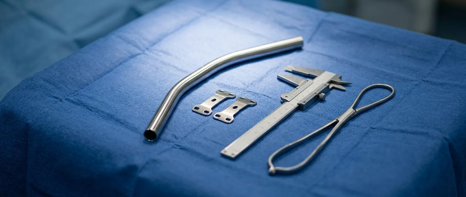
Why adults still benefit from repair: surgical correction of funnel chest in adults is more technically demanding than in teenagers because adult ribs and cartilage are stiffer. Even so, relief of symptoms and improvement in exercise capacity are well recognized, and the psychological effect of finally resolving a lifelong source of self-consciousness is substantial. Repair is usually done with a bar or bar-and-plate system placed behind the sternum to push it forward, sometimes combined with partial cartilage resection. Many adults arrive at this decision after years of doubt, and the first consultation is as much about honest counseling on what correction will and will not change as it is about the operation itself.
Pigeon Chest Correction: What Are the Options?
Pectus carinatum, the opposite deformity, pushes the breastbone outward instead of inward, producing a pigeon chest. It is less common and causes fewer internal compressions, but it brings its own challenges: visible prominence, local discomfort, and difficulty with clothing or sports that press against the chest.
In children and adolescents, external bracing can often reshape the chest, because the cartilage is flexible. In adults, cartilage has hardened, so bracing is usually ineffective and surgery becomes the realistic option. Pigeon chest correction in adults typically involves removing segments of deformed cartilage and repositioning or stabilizing the sternum, either through small incisions or, in some cases, a minimally invasive approach that avoids opening the whole chest.
The decision rests on severity, symptoms, and what the patient wants. A purely cosmetic concern is a valid reason to seek an opinion, but the surgeon will also assess whether the deformity affects breathing or posture. The trade-offs, including visible scars, healing time, and the small risk of recurrence, deserve a full conversation before any decision.
How Are Congenital Heart Defects Diagnosed in Adults?
Diagnosis usually begins with a stethoscope and a chest X-ray, which can show an abnormally shaped heart, a shifted heart position, or signs of extra blood flow. From there, echocardiography, an ultrasound of the heart, identifies holes, valve problems, and how well the heart pumps. It is painless, widely available, and often the single most informative first test.
Cardiac MRI as the definitive tool: when the anatomy is complex or surgery is being considered, cardiovascular magnetic resonance provides detailed images of the heart, the great vessels, and the chest wall in a single study. Expert consensus guidelines on cardiovascular magnetic resonance in children and adults with congenital heart disease outline exactly which measurements and protocols should guide treatment planning, and following them is part of how we prepare every case before an operation is recommended.
Sometimes a computed tomography (CT) scan is added to map calcified tissue, the coronary arteries, and the chest wall itself, especially when a combined heart and chest procedure is being considered. In our practice, the first step is a free video consultation in which we review the studies you already have and tell you precisely which ones are still missing, so no unnecessary test is ordered.
When Does a Congenital Repair Need Surgery in Adults?
Not every defect found in adulthood needs surgery. Small holes that cause no shunting of blood may be monitored for years. The decision to operate rests on a small set of well-established questions:
- Is the heart being damaged? Enlargement of a heart chamber, worsening valve function, or reduced pumping capacity all point toward repair.
- Are symptoms limiting life? Breathlessness, fatigue, or fainting that interferes with work or daily activity weighs in favor of surgery.
- What is the risk of leaving it? Some defects, such as a significant septal defect, carry a long-term risk of heart failure or abnormal heart rhythms if untreated.
When surgery is justified, the choice between open-heart surgery with a sternotomy (opening the breastbone) and a minimally invasive approach through small incisions depends on the defect, its location, and your anatomy. Minimally invasive congenital heart repair can mean smaller incisions and faster recovery, but it is not suitable for everyone. We recommend the less invasive route only where the evidence supports it for that specific patient, and every case is reviewed by the whole multidisciplinary team against international practice guidelines before a proposal is made.
What Types of Chest Wall Deformity Surgery Exist?
Chest wall deformity surgery has evolved into a family of named procedures, each suited to a different shape and severity of deformity.
- Nuss procedure: a metal bar is placed behind the sternum through small incisions on the sides of the chest and flipped to push the breastbone outward. Used for pectus excavatum, typically with the bar remaining in place for several years.
- Ravitch procedure: deformed cartilage is resected and the sternum repositioned, sometimes with a stabilizing plate. Better suited to certain deformities, stiffer chests, and older patients.
- Reverse/Nuss-type techniques for pectus carinatum: cartilage resection with struts or plates to hold the corrected position.
- Thoracic wall reconstruction: for complex or asymmetric deformities, including rib deformity, combining resection, reshaping, and customized fixation.
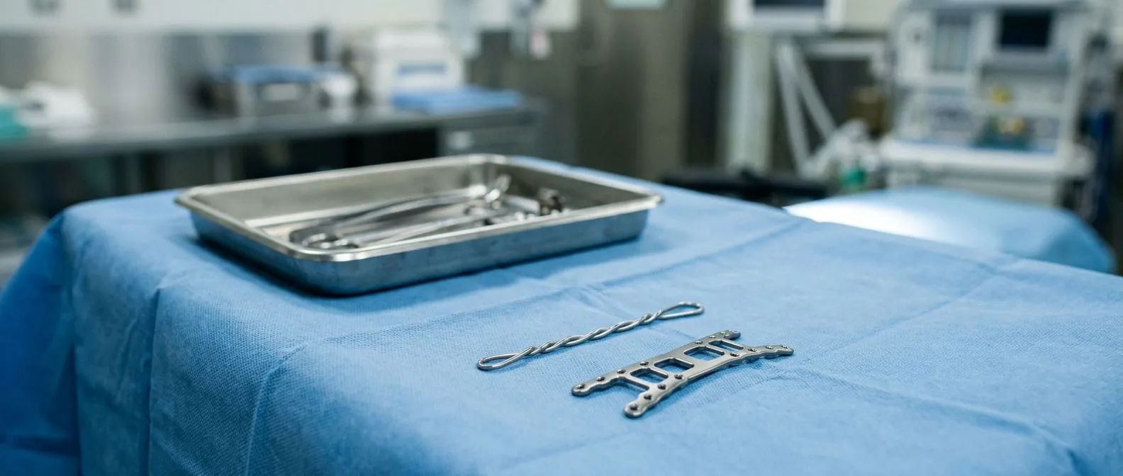
Sternal deformity correction in adults is a genuinely surgical decision about hardware and healing time. The bar or plate is temporary in most cases and is removed in a later, smaller procedure. Hardware options include titanium plates and bars that are visible on future imaging but do not prevent airport scanners from being an occasional nuisance; a letter from your surgeon solves this.
Can Heart and Chest Wall Surgery Be Combined?
Some adults face both problems at once: a congenital heart defect plus a chest wall deformity that complicates access, or a chest wall deformity that has been pressing on the heart for decades. Combined heart and chest surgery in a single operation is possible but demands careful planning. The order of operations matters, the stabilization hardware must coexist with the cardiac repair, and the anesthetic plan is more complex.
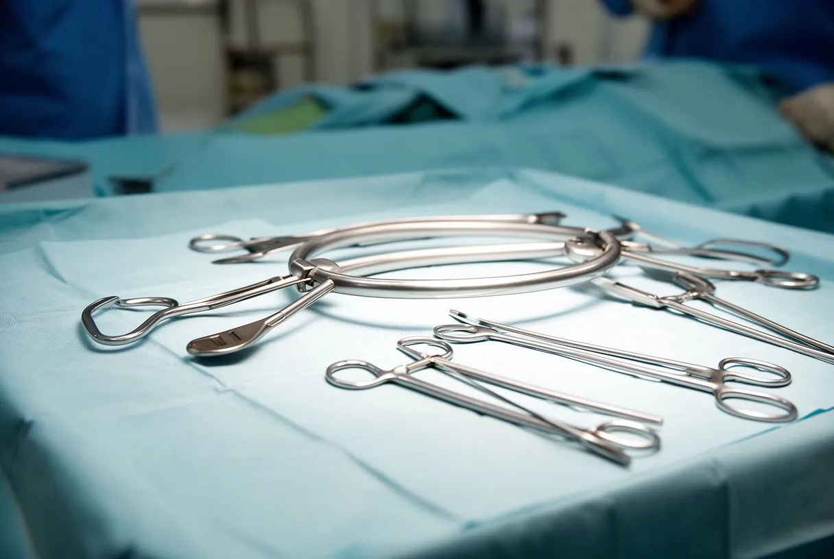
These cases illustrate why a multidisciplinary team matters. Cardiac surgeons, imaging specialists, and sometimes anesthesiologists with congenital experience plan the operation together. In our team, a case like this is reviewed against international guidelines before a single recommendation is made, and the patient receives a clear proposal with the alternatives laid out side by side, because the final decision always belongs to the patient.
It is also fair to say that sometimes the right answer is a staged approach: fixing the chest wall first and addressing the heart later, or the reverse. There is no universal answer, and a surgeon who presents only one option deserves a second opinion. We encourage second opinions and treat seeking one as a normal part of deciding about heart surgery, never as an insult.
What Does Heart and Sternum Surgery Recovery Look Like?
Recovery is where expectations meet reality, and honest expectations are worth more than optimistic ones. Most patients who undergo sternotomy, the opening of the breastbone for heart surgery, spend several days in hospital. The breastbone itself takes roughly six to eight weeks to knit, and during that time lifting, pushing, and driving are restricted. Chest wall surgery with bars or plates carries its own rules: no twisting, no contact sports, and careful attention to how you get out of bed.
The first weeks: pain is real but manageable with scheduled medication, not heroic endurance. Walking is encouraged from early on and is the main engine of recovery. Breathing exercises, often with a simple device, prevent lung complications.
Weeks to months: most people return to desk work in four to eight weeks, depending on the procedure, and to heavier activity over three to six months. A deep ache or clicking sensation around the sternum is common during healing and usually settles. Patients who have had minimally invasive chest wall surgery often recover faster, but the same principles of gradual return apply. In our consultations, we take the time to explain in plain words what the studies actually show and to answer every question about recovery before any decision, because patients who know what week one, week six, and month six will feel like cope better with all three.
Are Congenital Heart and Chest Wall Procedures Safe?
No honest answer is "completely safe," and you should distrust anyone who gives one. These are established procedures with long track records, but every operation carries risks that vary with your age, your other conditions, and the specifics of the defect. Bleeding, infection, arrhythmias (abnormal heart rhythms), and hardware-related problems such as bar displacement are the risks most often discussed. The safety of pectus surgery in adults has been examined in the medical literature, including studies indexed on PubMed, and overall results are reassuring when patients are properly selected.
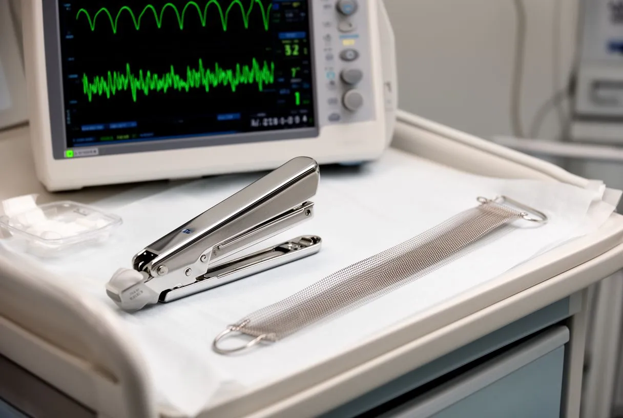
Safety also depends heavily on volume and experience, not just on the operation itself. A team that performs these procedures regularly anticipates the problems that a general team might discover during surgery. When you compare options, it is entirely fair to ask a surgeon how many of these specific operations they perform each year and what their complications have been.
Finally, safety includes the decision not to operate. Sometimes the best plan is monitoring with regular imaging, or medication to manage symptoms while the case is watched. A team that can say "we do not need to operate on you yet" is a team you can trust when it eventually says "now is the time." Dr Molina trained in cardiothoracic surgery at the Universidad de Guadalajara and in cardiac, minimally invasive, robotic, and structural surgery at the University of Montreal, and our practice exists to help you reach that kind of decision, not to push you toward an operation.
Adults with congenital heart defects or chest wall deformities are far more common than most people realize, and help is available at every stage, from a simple echocardiogram to a full combined heart and chest wall reconstruction. The most important step is not deciding on surgery yourself; it is getting an accurate picture of your own anatomy and asking a qualified cardiac team to explain it. If you suspect a congenital heart problem, have been told about a chest wall deformity, or simply want to understand what your studies show, start with a free video consultation with our team, which has consulting rooms in Guadalajara, Querétaro and Hermosillo, and decide with clear eyes.