When a Heart Valve Fails: Signs and Next Steps
When a heart valve fails, early signs like fatigue and breathlessness matter.
When a heart valve fails, the heart keeps working — but it works against itself. A valve is a one-way door between the chambers of the heart, and when that door no longer opens or closes properly, blood moves in the wrong direction or gets squeezed through a gap that is too narrow. The result is usually a slow, quiet decline that many people mistake for normal aging: getting winded on the stairs, feeling unusually tired, noticing swollen ankles by evening. This guide walks you through what valve failure actually is, the warning signs, how doctors confirm the diagnosis, and what your treatment and recovery options look like. If you or someone in your family has been told there is a "murmur" or a "leaky valve," you are in the right place.
What Happens When a Heart Valve Fails?
Your heart has four valves — the aortic, mitral, tricuspid and pulmonary — and each one opens and closes with every heartbeat, roughly a hundred thousand times a day. When a heart valve fails, it does so in one of two ways. It can become narrowed, which doctors call stenosis, so blood struggles to push through. Or it can become leaky, which doctors call regurgitation, so blood flows backward instead of forward.
Either way, the heart has to compensate. In stenosis, the pumping chamber thickens and stiffens as it pushes harder against the obstruction. In regurgitation, the chamber stretches as it holds both the returning blood and the incoming blood. For years the heart manages this extra workload, which is exactly why valve disease can hide. By the time symptoms appear, the heart may already have remodeled itself — and that remodeling is what we are trying to prevent with timely treatment.
This pattern is common: a patient believes it is "just fatigue," and the echo shows a valve that has been failing quietly for years. The important thing to understand is that valve failure is mechanical, not mysterious. Once the mechanics are understood, the options usually become much clearer.
What Are the Signs a Heart Valve Is Failing?
The heart valve failure symptoms that patients report most often follow a predictable pattern. Because the failing valve reduces the heart's efficiency, the first things to change are usually effort tolerance and breathing.
- Shortness of breath: you notice it climbing stairs, carrying groceries, or walking hills that used to feel easy.
- Fatigue: a tiredness heart valve issue produces that rest does not fully fix, because your body is working harder for the same output.
- Swollen ankles or feet: fluid backs up when the heart cannot keep up, and it shows up first at the lowest points.
- Irregular heartbeat or fluttering: stretched heart chambers can start to misfire electrically.
- Dizziness or fainting spells: more common with narrowed aortic valves, especially during activity.
Leaking heart valve symptoms and narrowing symptoms overlap considerably, which is why self-diagnosis is so unreliable. One person with severe regurgitation may feel almost nothing, while another with moderate stenosis feels exhausted. What matters is the direction of change: if last year's stair climb has become this year's rest stop, that deserves an explanation.
A classic first clue is a heart murmur — the whooshing sound a doctor hears through a stethoscope when blood moves abnormally through a valve. Murmurs are common and often harmless, but a new or worsening murmur in an adult is a reason to investigate, not to wait.
Which Heart Valve Disease Warning Signs Should You Never Ignore?
Most valve disease develops slowly. A small set of warning signs, however, signals that the situation has become urgent rather than simply worth monitoring.
Chest pain with exertion is the first. When the heart muscle strains against a narrowed valve, it can become starved of oxygen even with clear coronary arteries. Chest pressure that appears with activity and eases with rest in someone known to have valve disease is a red flag, not a nuisance.
Fainting or near-fainting during activity is the second. This suggests the heart temporarily cannot deliver enough blood to the brain. With aortic stenosis, exertional fainting is a recognized warning sign that the valve has reached a critical stage and needs prompt evaluation.
Sudden breathlessness at rest, or waking at night gasping, is the third. This pattern suggests fluid is building up in the lungs — a stage that in severe cases can progress toward congestive heart failure, the condition where the heart can no longer meet the body's demands. These symptoms do not automatically mean catastrophe, but they do mean the conversation with a cardiac team should happen this week, not this year.
How Do Doctors Diagnose a Failing Heart Valve?
The echocardiogram is the cornerstone. It is an ultrasound of the heart, entirely painless, and it shows each valve moving in real time. It measures how wide a narrowed valve opens, how much blood leaks backward through a failing one, the pressures inside the heart chambers, and how well the heart muscle is pumping. Most of the decisions in valve care are made from this one study.
Listening with a stethoscope still matters. The timing, character and location of a murmur give an experienced cardiologist a strong first impression of which valve is involved and how severely. Johns Hopkins notes that abnormal heart sounds are frequently the earliest sign of valve disease, detected long before symptoms begin — which is one reason routine checkups are worthwhile even when you feel fine.
Additional studies fill in the details. An electrocardiogram checks for rhythm disturbances. A chest X-ray shows heart size and lung congestion. In selected cases, a CT scan maps the valve's calcium burden and the access routes for catheter-based treatment, and cardiac catheterization directly measures pressures and checks the coronary arteries. When a patient comes to us, we take the time to explain what the studies actually show, in plain words, and to answer every question before any decision is made.

What Is the Difference Between Heart Valve Stenosis and Regurgitation Symptoms?
Stenosis and regurgitation both mean a heart valve fails to do its job, but they stress the heart in different ways, and their symptom timing differs too.
Aortic stenosis — narrowing of the main outflow valve — tends to announce itself late. The heart compensates for years, thickening its walls to force blood through the tight opening. The classic triad is breathlessness, chest pain, and fainting with exertion. It is most common in people over seventy, as calcium accumulates on the valve over a lifetime. Once symptoms appear, progression accelerates, which is why this valve in particular is watched closely.
Mitral regurgitation — a leaky valve between the left chambers — behaves differently. The left atrium absorbs the backward flow first, causing pressure backup into the lungs, palpitations from atrial fibrillation, and gradual fatigue. Mitral Valve Prolapse is one of the common underlying causes, where the valve's leaflets billow backward and eventually stop sealing cleanly.
Understanding which valve and which mechanism is involved changes which treatment makes sense — repair versus replacement, catheter versus surgery.
When Should You See a Heart Surgeon for Valve Problems?
Timing is the central question in valve disease. Operating too early on a valve that is doing well can expose you to risks without benefit; operating too late means the heart has already been damaged by years of strain, and recovery of function is less complete. Guidelines define thresholds — for example, an aorta above a certain diameter, or specific pressure measurements across a stenotic valve — at which intervention becomes recommended even without severe symptoms.
The honest answer is: see a specialist when the echo says so, not just when the symptoms say so. A valve can reach the surgical threshold while you still feel reasonably well. That window is precisely when surgery offers its greatest benefit, because the heart muscle has not yet been permanently stretched or scarred. If you already have a known murmur or mild valve disease, regular surveillance echocardiograms let your team operate at the right moment rather than in an emergency.
Every case at our clinics is reviewed by the full multidisciplinary team against international practice guidelines before a recommendation is given, so the timing question is answered for your specific heart, not for a textbook average. And if you have been told "we'll just watch it," it is fair to ask: watch for what, and until when?
What Are the Main Heart Valve Repair and Replacement Options?
Repair is the first choice wherever possible. A repaired mitral valve keeps your own tissue, avoids the need for blood thinners, and generally lasts better than a replacement. Robotic and minimally invasive repair techniques have made this possible through small incisions between the ribs, avoiding opening the breastbone.
Replacement is needed when repair is not feasible. The options then divide into two families, and each has honest trade-offs:
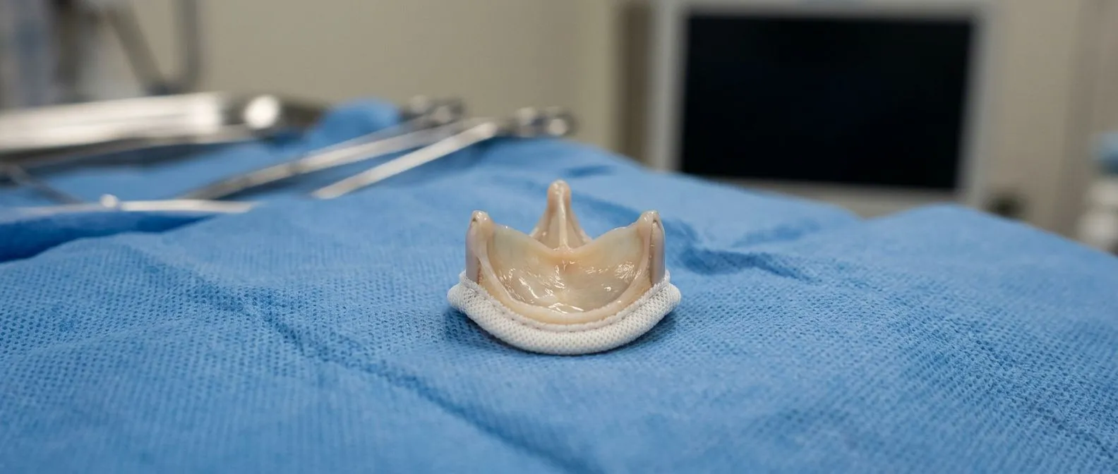
- Biological tissue valves: made from animal tissue such as a Porcine Valve or a Bovine Pericardial Valve. They function naturally and usually avoid lifelong blood thinners, but they gradually wear — a process called Valve Degeneration — often needing reoperation after a decade or more.
- Mechanical valves: extremely durable, often lasting a lifetime, but requiring lifelong anticoagulant medication to prevent clots from forming on the valve, a risk known as Valve Thrombosis.
Within each family there are many designs — from long-established mechanical valves to newer devices — and factors like your age, lifestyle, other planned procedures, and personal preference about medication all shape the right choice. We present every patient with a clear proposal that includes the alternatives, because the final decision about their own heart is always theirs.
When a previously placed tissue valve wears out, a new valve can increasingly be implanted inside the old one — a technique known as Valve-in-valve — sparing the patient a repeat open operation in many cases.
How Does Minimally Invasive Heart Valve Repair Work?
The biggest change in valve care over the past fifteen years has been the arrival of catheter-based treatment — treating a valve through a thin tube, usually from an artery in the groin, without opening the chest at all. Transcatheter Aortic Valve Implantation is the established name for the aortic version, though you will commonly hear the procedure called TAVR.
In a TAVR, a collapsed replacement valve mounted on a catheter is guided to the diseased aortic valve and expanded into place, immediately taking over the job of the old valve. For older patients or those at higher surgical risk, it has transformed what used to be a major operation into a procedure often done with a few days in hospital. Transcatheter options now extend to the mitral and tricuspid valves as well, through a growing set of dedicated devices.
Our team performs conventional, minimally invasive, robotic and transcatheter procedures, and we recommend the less invasive route only where the evidence supports it for that specific patient — sometimes conventional surgery truly is the better answer, and we will say so plainly. Dr Molina trained in structural cardiology and TAVR at CHU Bordeaux and performed the first robotic mitral valve repair in the Ángeles Health System, so our team evaluates both open and catheter options with equal firsthand experience.
The right question is never "which technique is newest," but "which approach gives me the lowest risk and the best lasting result for my anatomy and my health." That answer comes from a team that offers all the options, not just the one it happens to perform.

What Does Recovery After Heart Valve Surgery Really Look Like?
Recovery is a staged process, not a single event. After conventional valve surgery, most patients spend one or two days in intensive care, then several days on the ward, and go home within the first week. Driving, light housework and short walks resume within a few weeks; return to fuller activity typically unfolds over the following two to three months as the breastbone heals.
Cardiac rehabilitation makes a measurable difference. Structured, supervised exercise after valve surgery helps patients rebuild strength safely and regain confidence. For heart failure patients, research on nurse-coordinated home-based cardiac rehabilitation shows that structured follow-up support improves exercise capacity and quality of life while reducing the burden of repeated hospital visits — a model worth asking about in your own recovery planning.
Recovery milestones are worth knowing in advance. Here is the realistic sequence we describe to our patients:
- Weeks 1–2: walking daily, gradually increasing distance; no lifting, no driving yet.
- Weeks 3–6: driving usually resumes (per your surgeon), light activity, return to desk work for many patients.
- Months 2–3: breastbone fully healing; gradual return to most normal activities.
- Ongoing: regular echo surveillance of the new or repaired valve, usually yearly.
Recovery after catheter procedures like TAVR is typically faster — many patients walk the same day and go home within a few days — though the same principle of staged, supervised progress applies.
What Should You Expect From a Heart Valve Consultation?
The first step is simpler than most people fear. Our first contact is a free video consultation: we get to know the patient, talk through the condition, and say which studies are still needed so no unnecessary ones are ordered; a face-to-face visit at the consulting rooms follows only when the patient decides to go ahead. You do not need to travel anywhere or commit to anything to get a professional read on your situation.
Bring your questions and your existing studies. Any prior echocardiograms, ECGs or reports help the team see the trajectory of your valve — whether it is stable, slowly worsening, or changing quickly. Trajectory often matters more than any single snapshot.
A good consultation ends with clarity, not pressure. You should leave knowing: which valve is affected, how severely, whether treatment is needed now or can be watched, what the options are, and what the trade-offs of each are. We encourage second opinions and treat seeking one as a normal part of deciding about heart surgery — a decision of this size deserves confidence, not urgency imposed by anyone.
Valve disease is one of the most treatable conditions in all of cardiology — when it is caught in time. The heart valve disease warning signs worth acting on are familiar and unglamorous: breathlessness that is creeping upward, fatigue that rest does not fix, swollen ankles, a new murmur, chest discomfort with effort. When a heart valve fails, the heart has usually been compensating for years, and treatment works best before the compensation gives way. Whether the answer is surveillance, a minimally invasive repair, a TAVR, or conventional surgery, the decision belongs to you after a clear conversation with a team that can offer every option honestly. If any part of this article describes you or someone you love, book a consultation with a qualified cardiac team rather than self-diagnosing — starting with a simple video visit costs nothing and clarifies almost everything.