

Wound Dehiscence After Heart Surgery: Signs and Care
Wound dehiscence after heart surgery requires fast action.
What Is Wound Dehiscence After Heart Surgery?
Undergoing cardiac surgery is a profound journey, and the recovery phase is just as critical as the operation itself. When surgeons need to access the heart, they often perform a median sternotomy, which involves making a precise incision down the center of the chest and carefully dividing the sternum, or breastbone. This bone is then wired or banded back together at the end of the procedure, and the overlying skin is closed with sutures or staples. However, sometimes the layers of this surgical wound do not heal as expected and begin to separate, a complication medically recognized as wound dehiscence after heart surgery.
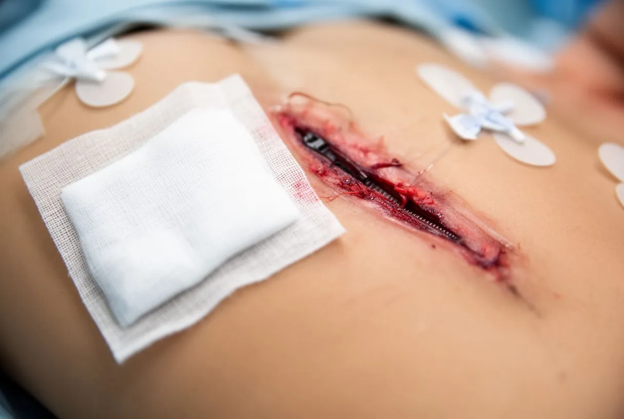
Dehiscence essentially means the unplanned reopening of a surgical incision. In the context of cardiac surgery, this can range from a superficial separation of the skin edges to a severe, deep structural failure where the sternal wires break and the bone pulls apart. The sternum is a highly dynamic bone; it moves slightly with every breath and cough. If the healing tissue cannot withstand these forces, the closure fails. Understanding this risk is essential for any patient preparing for or recovering from a major cardiac intervention.
For patients and families, the thought of an incision reopening is understandably frightening. It is important to remember that cardiac surgical teams are highly trained to manage these exact complications. While dehiscence requires immediate attention to prevent further issues like infection, it is a known risk of opening the chest. Recognizing the signs early and understanding the anatomy of the repair can empower you to seek timely medical help, ensuring your recovery stays on track.
Why Do Sternotomy Wounds Separate?
The chest wall is under constant mechanical stress during the postoperative period. Every time you breathe, cough, or move your upper body, the sternum experiences tension. Surgeons use heavy stainless-steel wires to hold the bone together, acting like internal casts, but these wires rely on the structural integrity of the bone itself. If the bone is brittle, osteoporotic, or if the patient puts too much strain on the chest before the bone has fused, the wires can cut through the bone or snap entirely, leading to a mechanical failure of the closure.
Surgical factors also play a significant role in wound stability. During complex procedures, such as those involving the ascending aorta, surgeons may need to alter their approach or use specialized grafts. If an arterial graft is harvested from the chest wall, the blood supply to the overlying skin and bone can be compromised. Without adequate blood flow, the tissues struggle to heal, making the incision vulnerable to separation. The depth of the incision and the exact techniques used to approximate the tissue layers also determine how well the wound will withstand early postoperative movement.
Biological factors, particularly infection, are another primary driver of wound separation. Bacteria can colonize the surgical site, breaking down the collagen and new tissue that form the bridge between the divided bone. When a deep infection sets in, the tissues become swollen, fragile, and unable to hold the sutures. The body’s natural inflammatory response can actually work against the mechanical closure, literally pushing the wound apart as it tries to expel the infectious material. Managing these combined mechanical and biological threats requires careful postoperative vigilance.
Early Signs of Wound Dehiscence After Heart Surgery
Recognizing the early warning signs can drastically improve outcomes for a separated sternal wound. The most common initial symptom is a noticeable change in the incision’s appearance. You might observe that the skin edges, which were previously neatly approximated, are beginning to drift apart. This gap might start small, resembling a thin red line that is widening, or you might notice a small area of the incision that simply refuses to close over with healthy tissue. Changes in the color of the skin immediately surrounding the incision are also a primary indicator that healing is not progressing normally.
Another critical sign is the presence of new or unusual drainage. While some clear or slightly bloody fluid is normal in the first few days, a sudden increase in fluid, or drainage that becomes thick, cloudy, yellow, or foul-smelling, suggests a complication. Patients should also be attuned to physical sensations. A sudden "popping" or clicking sound in the chest, particularly when changing positions or taking a deep breath, can indicate that a sternal wire has broken or the bone has shifted. Additionally, radiating shoulder or arm pain that accompanies chest instability is a significant red flag that should not be ignored.
Systemic signs of infection often accompany or precede true wound separation. If the wound is failing because of an underlying infection, you may experience a low-grade fever, chills, or a general feeling of malaise. The chest area might feel unusually warm to the touch or exhibit increased swelling and redness. Recognizing these interconnected symptoms is crucial, as early intervention with antibiotics or wound care can often prevent a superficial separation from becoming a severe structural failure.
- Visible Separation: Watch for skin edges drifting apart or a widening gap along the incision line.
- Abnormal Drainage: Notice any thick, cloudy, or foul-smelling fluid leaking from the wound.
- Popping Sensations: Listen for clicking or popping sounds when breathing or moving.
How Do Cardiac Teams Diagnose a Separated Sternum?
When a patient presents with concerns about their sternal incision, the cardiac team begins with a thorough physical examination. The surgeon will carefully inspect the wound for signs of separation, assessing the degree to which the skin and underlying tissues have parted. They will evaluate the stability of the chest wall by gently palpating the area to feel for any unnatural movement of the bone. If the sternum clicks or shifts under gentle pressure, it strongly suggests that the internal wires are no longer holding the bone securely in place.
Imaging studies are essential for confirming the diagnosis and understanding the extent of the damage. A lateral chest X-ray can reveal if the sternal wires are broken, bent, or displaced. In some cases, a computed tomography (CT) scan is ordered to provide a highly detailed, three-dimensional view of the chest. This imaging helps the team assess not only the bone alignment but also the presence of fluid collections, abscesses, or deep tissue infections that might not be visible from the surface.
According to Cleveland Clinic, prompt evaluation of postoperative wound issues is vital to preventing further complications. If an infection is suspected, the medical team will likely take cultures of any drainage to identify the specific bacteria involved. Blood tests may also be ordered to check for elevated white blood cell counts or inflammatory markers, which provide a clearer picture of the body's systemic response. This comprehensive diagnostic approach allows the surgical team to formulate an effective, targeted treatment plan.
Immediate Care Steps If Your Incision Opens
Discovering that your chest incision is separating can be alarming, but taking calm, immediate action protects your health. The very first step is to stop any physical activity that places strain on your chest. Sit down, rest, and avoid lifting, pushing, or pulling anything. Do not attempt to push the skin back together or close the gap with tape or bandages yourself, as this can introduce bacteria and worsen the tissue damage. Keeping the area clean and dry while you wait for medical attention is paramount.
If there is active bleeding, apply gentle, direct pressure using a clean, dry cloth or sterile gauze, but avoid pressing hard enough to cause pain or further disrupt the bone. If you notice a significant amount of fluid draining, or if the wound appears to be gaping, do not apply any ointments, creams, or home remedies. Cover the area loosely with a sterile dressing if one is available. You should immediately contact your cardiac surgeon’s office or proceed to the nearest emergency room, clearly informing them that you have recently undergone heart surgery and suspect your incision is opening.
Time is of the essence when dealing with a potential deep sternal wound complication. Early evaluation by a professional ensures that if there is an infection brewing, antibiotics can be administered quickly. Even if the separation seems superficial, only a medical professional can determine the true depth of the issue. Trying to manage it at home risks delaying necessary interventions, which could lead to more complex reconstructive surgeries down the line.
Risk Factors for Poor Wound Healing in Cardiac Patients
Not every patient faces the same risk of wound dehiscence after heart surgery. Certain underlying health conditions can significantly impair the body’s ability to repair tissues. Diabetes is one of the most prominent risk factors, as elevated blood sugar levels interfere with collagen synthesis and reduce the effectiveness of white blood cells, making infections more likely and healing slower. Similarly, patients with obesity carry a higher risk because the additional subcutaneous tissue creates more tension on the sutures and provides a larger area where fluid can accumulate and bacteria can thrive.
Medications and lifestyle factors also influence wound integrity. Patients who continue to smoke after surgery experience restricted blood flow to the chest tissues, depriving the healing bone and skin of essential oxygen and nutrients. Certain necessary postoperative medications, such as Aspirin or other blood thinners, might increase drainage from the wound, though they are vital for preventing clots. Furthermore, patients requiring repeated surgical interventions or those who have compromised immune systems face a steeper hill when it comes to robust tissue repair.
The nature of the surgery itself can introduce risks. Patients undergoing emergency operations or those who require prolonged surgical times have a higher incidence of wound complications. Sometimes, techniques like Awake ECMO are necessary for severe respiratory or cardiac failure, but the presence of large cannulas in the central vessels can alter local circulation and complicate chest stability. Understanding your personal risk profile allows your surgical team to tailor your closure technique and postoperative monitoring accordingly.
Surgical and Medical Treatments for Dehiscence
Treating a separated sternal incision depends entirely on the depth and severity of the dehiscence. If the separation is strictly superficial—meaning only the skin layers have parted and the underlying bone remains stable and free of infection—surgeons may simply clean the area and close it again with new sutures or steri-strips. This is often a minor procedure that can sometimes be done at the bedside or in a procedure room. Local wound care and antibiotics may be prescribed to ensure the superficial tissue heals securely without complication.
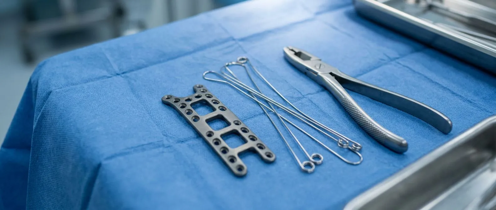
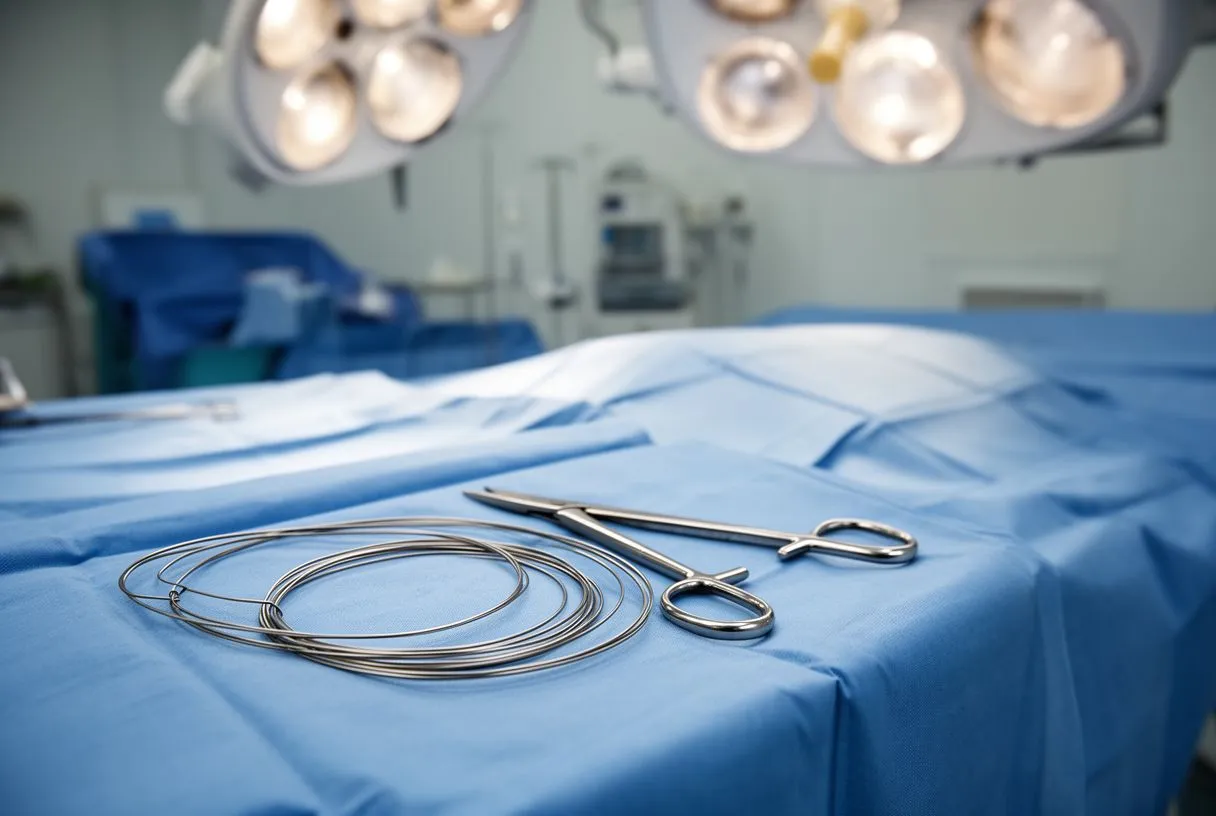
Deep sternal wound infections or complete bone separation require a much more aggressive approach. The patient will need to return to the operating room for an extensive debridement, where surgeons remove all infected, damaged, or dead tissue and bone. The old, broken sternal wires are extracted. The chest is then washed thoroughly with antibiotic solutions. Once the area is clean, the sternum is reapproximated using a more robust fixation method, sometimes utilizing specialized plates or a newer technique involving a rigid sternal plating system.
In severe cases where infection has destroyed too much bone or tissue, primary closure is impossible. Surgeons must then perform reconstructive surgery using muscle flaps, typically utilizing the pectoralis major muscles from the chest to cover the defect and provide healthy, well-perfused tissue. As highlighted in the document on Infective Endocarditis in Adults: Diagnosis, Antimicrobial Therapy, and Management of Complications, deep infections can sometimes spread to the heart valves, requiring prolonged courses of intravenous antibiotics alongside these structural repairs. When massive blood loss occurs during these revisions, the team will rely on Autologous Blood salvage techniques when possible to safely restore volume.
Recovering From a Reopened Chest Incision
Recovery from a wound revision is often more challenging than the initial postoperative period from the original heart surgery. Patients must be exceptionally gentle with their chest for a prolonged duration. Surgeons typically enforce strict sternal precautions—avoiding lifting anything heavier than a few pounds, refraining from pushing or pulling with the arms, and supporting the chest with a small pillow when coughing or sneezing. These precautions are not mere suggestions; they are vital to allow the newly repaired bone and tissues to fuse without mechanical interruption.
The healing timeline is extended. It can take several months for the revised sternum to achieve complete bony union, and during this time, patients will require frequent follow-up visits to monitor the incision. Physical therapy is usually delayed or heavily modified to focus on the lower body and gentle ambulation, avoiding any upper body resistance training. It is common for patients to feel frustrated or anxious during this prolonged recovery, especially if they felt they were just beginning to regain their strength before the complication occurred.
Managing pain and systemic health is crucial during this phase. The surgical team will carefully balance pain relief with the need to keep patients alert and breathing deeply to prevent pneumonia. Because postoperative inflammation can sometimes mimic other cardiac issues, doctors must carefully evaluate new chest pains. Differentiating between normal healing pain and more serious complications requires clinical expertise. For instance, if a patient develops a pericardial rub or effusion, the clinical team will consult resources like the 2015 ESC Guidelines for the diagnosis and management of pericardial diseases to rule out concurrent pericardial involvement.
Preventing Wound Dehiscence After Heart Surgery
Prevention begins long before the incision is made and continues well after you leave the hospital. Optimizing your health prior to surgery is the most effective preventative measure. If you are a smoker, quitting entirely before your operation dramatically improves the blood supply to your chest tissues. For patients with diabetes, maintaining strict glycemic control before and after surgery ensures that your white blood cells function properly and your body can synthesize the collagen necessary to heal the bone and skin.
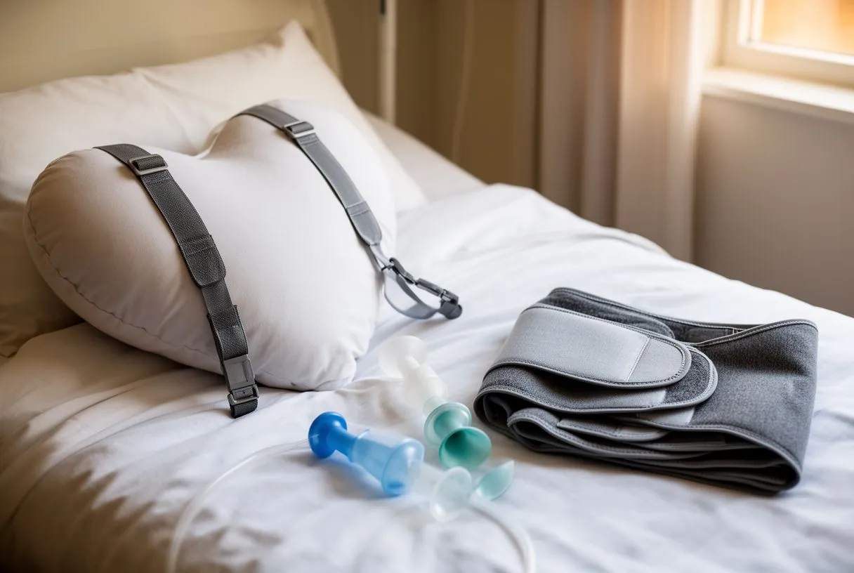
Postoperative adherence to sternal precautions is your primary defense against mechanical dehiscence. This means actively protecting your chest from sudden forces. You must avoid heavy lifting, pushing heavy doors, or performing exercises that engage the pectoral muscles too soon. Even simple daily activities, like getting out of a chair or rolling over in bed, must be done carefully without using your arms to bear your full body weight. Many patients find it helpful to sleep on their back in a slightly elevated position to minimize tension on the incision.
Nutrition plays a silent but powerful role in wound stability. Consuming a diet rich in protein, vitamin C, and zinc provides the essential building blocks for tissue repair. Staying hydrated keeps the skin supple and promotes healthy circulation. Proper wound care at home—keeping the incision clean and dry as instructed, and watching closely for any changes—remains your final line of defense. By combining strict physical precautions with excellent biological support, you significantly lower the risk of wound dehiscence after heart surgery.
- Smoking Cessation: Stop smoking before surgery to maximize vital blood flow to chest tissues.
- Sternal Precautions: Strictly avoid lifting, pushing, or pulling heavy objects to protect the healing bone.
- Protein-Rich Diet: Consume adequate protein and vitamins to fuel collagen production and tissue repair.
When to Seek Emergency Help for Your Incision
While some discomfort and mild bruising are normal after heart surgery, certain signs mandate an immediate trip to the emergency room. If you see a sudden, significant opening of the incision, especially if you can see deep tissue or bone, do not wait for an appointment. Similarly, if you experience a sudden gush of fluid, particularly blood or pus, from the wound, it requires immediate evaluation. These acute changes can signify a catastrophic failure of the chest closure or a rapidly progressing infection.
Systemic symptoms that develop alongside wound changes are also red flags. A fever over 101°F (38.3°C), shaking chills, or a rapid heart rate can indicate that a local infection has spread to the bloodstream. Furthermore, any sudden onset of severe chest pain that is different from your normal incisional pain, especially if accompanied by shortness of breath or unexplained arm pain, should be assessed immediately to rule out a concurrent cardiac event or severe sternal instability.
Recovering from heart surgery is a marathon, not a sprint, and complications like wound dehiscence require expert intervention. Never feel embarrassed to report a concern, no matter how small it may seem. If you have questions about your incision, the safest approach is always to contact your surgical team directly. If you or a family member are navigating the recovery from a cardiac procedure and have concerns about your healing, we encourage you to discuss your specific situation with a qualified cardiac team and book a consultation to ensure your recovery is as safe and successful as possible.






