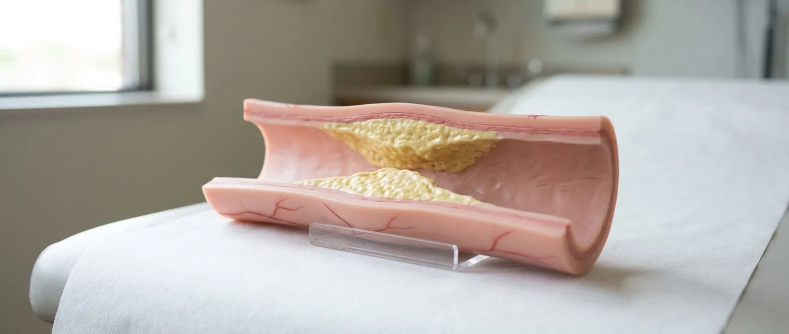
Atheromatous Plaque: How Arteries Become Blocked
Atheromatous plaque arteries cause narrowing that leads to heart attack and stroke.
Understanding how atheromatous plaque develops inside your arteries is one of the most empowering steps you can take toward protecting your heart. When fatty deposits accumulate along the inner walls of blood vessels, they narrow the passage that oxygen-rich blood travels through, and over time this process can lead to chest pain, heart attack, or stroke. This guide walks you through what atheromatous plaque is, why it forms, how doctors detect it, and what treatment options exist—so that when you sit down with your cardiac team, you arrive informed and ready to ask the right questions about your specific situation.
What Are Atheromatous Plaque Arteries?
Atheromatous plaque is a thick, waxy deposit that builds up along the inner lining of your arteries—the blood vessels that carry oxygen-rich blood away from your heart to every organ in your body. These deposits are not simple fat; they are a complex mixture of cholesterol, calcium, cellular waste, and inflammatory cells that gradually accumulate beneath the artery's surface. As this material collects, the artery wall thickens and stiffens, reducing the space available for blood flow. This narrowing is the fundamental process behind atherosclerosis, the most common form of arterial disease worldwide.
The process begins quietly, often decades before any symptom appears. When the innermost layer of an artery—the endothelium—becomes damaged by high blood pressure, smoking, or elevated blood sugar, cholesterol particles slip into the wall and trigger an immune response. White blood cells arrive to clean up the area, but many engulf so much cholesterol that they die in place, adding to the growing deposit. Over years, calcium crystals settle into this mixture, making the plaque hard and brittle. The result is an artery that is both narrower and less flexible than it should be.
For patients evaluating their cardiovascular risk, it helps to understand that not every atheromatous plaque causes immediate danger. Some plaques grow slowly and remain stable for decades, narrowing an artery only partially. Others develop thin caps that can rupture without warning, exposing the fatty core to the bloodstream and triggering a clot that blocks flow entirely. Knowing which type you carry—and whether your overall risk profile warrants intervention—is a conversation best had with a qualified cardiac specialist who can interpret your imaging and lab results in context.
What Causes Atheromatous Plaque Formation?
High cholesterol and Apolipoprotein B: The single most important driver of atheromatous plaque formation is elevated circulating cholesterol, particularly low-density lipoprotein (LDL). A protein called Apolipoprotein B wraps around each LDL particle and carries it through the bloodstream; the more Apolipoprotein B particles you have, the more raw material is available to infiltrate damaged artery walls. Genetics, diet, and metabolic health all influence these levels, which is why cholesterol testing remains a cornerstone of cardiovascular screening for adults of every age.
Hypertension and mechanical stress: High blood pressure does not just strain the heart; it physically hammers the delicate endothelial cells lining your arteries with every heartbeat. Over time, this repetitive force creates microscopic tears where cholesterol can lodge. The Ascending Aorta and other high-pressure vessels are especially vulnerable to this kind of wear. Controlling blood pressure through medication, diet, and regular monitoring reduces the mechanical injury that seeds plaque formation. Research summarized by Nature has helped clarify the molecular pathways through which arterial wall stress accelerates atherosclerosis and plaque progression.
Smoking, diabetes, and chronic inflammation: Tobacco smoke directly injures endothelial cells and makes blood more prone to clotting. Diabetes chronically elevates blood sugar, which damages proteins in vessel walls and accelerates plaque growth. Systemic inflammation—whether from autoimmune disease, chronic infection, or obesity—adds fuel to the fire by keeping immune cells in a constant state of activation inside artery walls. Each of these factors multiplies the effect of the others, which is why doctors assess them together rather than in isolation.
Atheromatous Plaque Symptoms: What Patients Notice
Atheromatous plaque is notoriously silent in its early stages. Most people with developing arterial plaque feel perfectly well until the narrowing reaches a critical threshold or a plaque ruptures. When symptoms do appear, they depend on which artery is affected. Plaque in the coronary arteries—the vessels that supply the heart muscle itself—often first announces itself as angina: a pressure, tightness, or heaviness in the chest that appears during exertion and eases with rest. Some patients describe it as a squeezing sensation rather than sharp pain.
When atheromatous plaque narrows arteries supplying the brain, the warning signs may include transient episodes of weakness, slurred speech, or visual disturbance—events sometimes called mini-strokes or TIAs. Plaque in the arteries of the legs produces claudication: cramping or aching in the calf or thigh that comes on with walking and improves after stopping. Atypical Infarction, a heart attack that presents without classic chest pain, is more common in women, older adults, and people with diabetes; it may show up as Arm Pain, jaw discomfort, profound fatigue, or nausea instead.
Because these symptoms can be subtle and intermittent, many patients delay seeking evaluation. It is important to recognise that the absence of symptoms does not mean the absence of disease. A person can have extensive atheromatous plaque and feel completely normal until the day a plaque ruptures. This is why guideline-based screening—cholesterol panels, blood pressure checks, and risk calculators such as EuroSCORE II—matters even for people who consider themselves healthy. If you have a family history of early heart disease or any of the symptoms above, discussing screening with a cardiologist is a reasonable and proactive step.
How Atherosclerosis Is Diagnosed and Screened
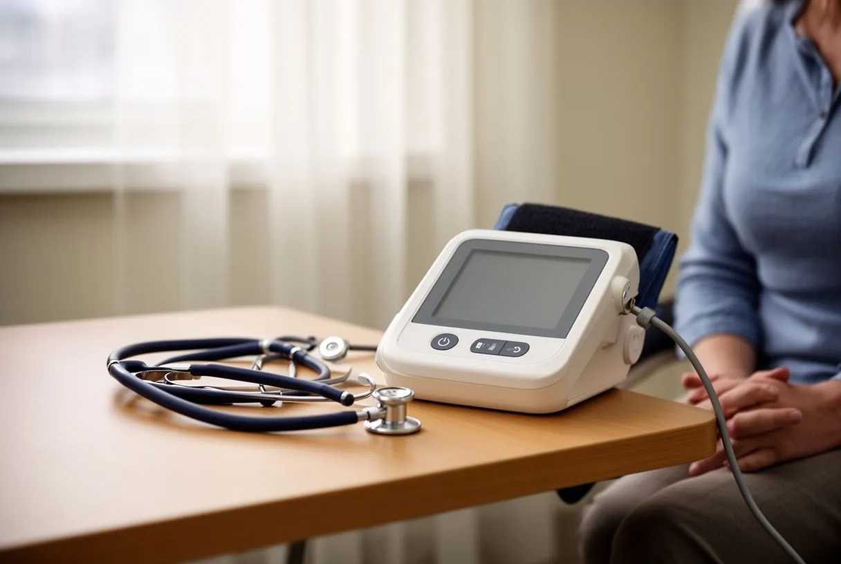
Diagnosis of atheromatous plaque begins with a thorough clinical assessment rather than a single test. Your cardiologist will review your personal and family history, measure blood pressure, listen to your heart and neck vessels using Auscultation (the clinical term for listening with a stethoscope), and order blood tests for cholesterol, blood sugar, and inflammatory markers. This initial evaluation helps estimate your overall cardiovascular risk and determines whether further imaging is warranted. The decision to proceed to advanced testing is always individualised—no two patients are identical.
Several imaging tools help visualise atheromatous plaque directly or measure its effects. A coronary calcium score uses a quick CT scan to detect calcium deposits in the heart's arteries, giving a numeric estimate of plaque burden. Carotid ultrasound measures the thickness of artery walls in the neck and can identify plaque there. Stress testing—either on a treadmill or with medication—evaluates whether the heart receives enough blood under exertion, which suggests functionally significant narrowing. More detailed imaging, such as CT coronary angiography, can show the exact location and severity of plaque within the coronary vessels. For research and clinical trial purposes, standardised reporting frameworks such as the CONSORT 2010 Explanation and Elaboration help ensure that diagnostic study results are reported transparently and can be compared across populations.
If screening reveals significant atheromatous plaque, the next step is a detailed conversation about what the findings mean for your individual risk. A calcium score of zero, for example, is reassuring but does not guarantee the absence of soft plaque. A high score does not automatically mean you need surgery; it means you and your doctor should discuss aggressive risk-factor management and possibly further testing. The goal of diagnosis is not to alarm but to inform—to give you and your cardiac team a clear picture of what is happening inside your arteries so that decisions about treatment are based on evidence, not guesswork.
Stable vs Unstable Plaque: Why It Matters
Not all atheromatous plaque behaves the same way, and the distinction between stable and unstable plaque is one of the most important concepts in modern cardiology. Stable plaques have a thick fibrous cap—a dense layer of tissue that holds the fatty core in place. These plaques grow slowly, and while they can eventually narrow an artery enough to cause symptoms during exertion, they rarely rupture suddenly. They are predictable, and doctors can often manage them with medication and lifestyle changes over many years.
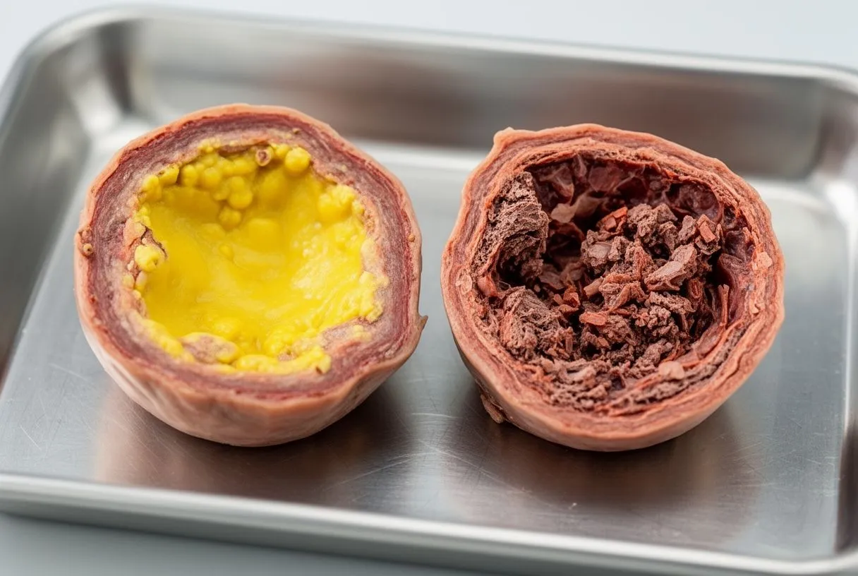
Unstable plaques, by contrast, have thin caps that are vulnerable to tearing. When the cap ruptures, the fatty interior spills into the bloodstream, and the body's clotting system reacts instantly. A clot forms at the rupture site, and if it is large enough, it can block the artery entirely—causing a heart attack if the artery feeds the heart, or a stroke if it feeds the brain. This is why some people with only moderate narrowing on imaging can still experience a sudden cardiac event: the dangerous plaque was not the one causing the most narrowing, but the one most likely to rupture.
Identifying unstable atheromatous plaque before it ruptures is an active area of research. Intravascular imaging techniques can sometimes characterise plaque composition, but these are not yet routine screening tools. What we do know is that aggressive cholesterol lowering, blood pressure control, and smoking cessation all help stabilise plaques by reducing inflammation within the artery wall. If you have been told you have atheromatous plaque, ask your cardiologist whether your imaging provides any information about plaque stability and what that means for your treatment plan.
Coronary Artery Blockage and Heart Attack Risk
When atheromatous plaque accumulates inside the coronary arteries—the vessels that wrap around the outside of the heart and supply the heart muscle with oxygen—the condition is called coronary artery disease. As plaque grows, the channel for blood flow narrows. At rest, the heart may still receive enough blood, but during exertion, stress, or even a heavy meal, the increased demand cannot be met through the narrowed passage, and the heart muscle signals distress through the sensation we call angina.
A heart attack, or myocardial infarction, occurs when blood flow through a coronary artery is abruptly cut off—most often because an atheromatous plaque has ruptured and a clot has formed on top of it. The heart muscle downstream of the blockage is starved of oxygen, and without rapid restoration of blood flow, that muscle begins to die. This is why time is critical: the sooner blood flow is restored, the more heart muscle is saved. Emergency departments are equipped to administer clot-busting medications or perform emergency Balloon Angioplasty, a procedure in which a small balloon is inflated inside the narrowed artery to compress the plaque and restore flow.
For patients with known coronary artery disease, certain arrhythmias raise additional concern. Atrial Flutter, a rapid and organised beating pattern in the upper chambers (the Atrium), can develop in hearts damaged by prior infarction or chronic plaque burden. Blood-thinning decisions in this context are complex; large studies such as ARISTOTLE have shaped how cardiologists balance stroke prevention against bleeding risk. If you carry both coronary disease and an arrhythmia, your treatment plan must be tailored to address both conditions in a coordinated way, which is why a single specialist or a coordinated team approach is preferable to fragmented care. For patients with clotting disorders, an International consensus statement on antiphospholipid syndrome provides classification criteria that may influence both diagnostic testing and anticoagulation choices.
Plaque in Arteries: Treatment Options
Treatment for atheromatous plaque falls into three broad categories: lifestyle modification, medication, and procedural intervention. The right combination depends on how much plaque is present, where it is located, whether it is causing symptoms, and your overall health. No single approach works for everyone, and the decision-making process should weigh the risks and benefits of each option against your individual circumstances.
- Medication: Statins lower LDL cholesterol and stabilise plaque; Aspirin at low dose reduces clotting risk; blood pressure medications protect the artery wall from mechanical stress.
- Balloon Angioplasty and stenting: A catheter is threaded through an Arterial Line into the blocked coronary artery; a balloon expands to compress plaque, and a metal scaffold (stent) holds the vessel open.
- Coronary artery bypass surgery: A surgeon uses an Arterial Graft or vein graft to reroute blood around a severely blocked artery, restoring flow to the heart muscle downstream.
Bypass surgery may be recommended when multiple coronary arteries are heavily blocked, when the left main coronary artery is narrowed, or when stenting is technically not feasible. During the operation, the surgeon may use an Arterial Cannula to connect the heart-lung machine, which maintains circulation while the heart is stopped. In selected cases, Autologous Blood (your own previously collected blood) may be used to reduce transfusion needs. Some centres are developing Awake ECMO techniques that allow certain procedures without general anaesthesia, though these remain specialised. Recovery from bypass surgery typically involves several days in hospital and a structured cardiac rehabilitation programme over weeks to months. Your surgical team should discuss the EuroSCORE II predicted risk for your specific case, including the trade-offs between surgery and stenting, so you can make an informed choice rather than a default one.

Can Atheromatous Plaque Be Reversed?
This is one of the questions patients ask most frequently, and the honest answer is nuanced. Complete reversal of established atheromatous plaque—returning an artery to its pristine, pre-disease state—is not something current medicine can reliably achieve. However, substantial evidence shows that aggressive risk-factor modification can shrink plaque volume modestly, halt its progression, and critically, stabilise it so it is far less likely to rupture. The distinction between reversal and stabilisation matters: stabilisation may be even more important for preventing heart attacks than shrinkage.
High-intensity statin therapy has been shown in imaging studies to reduce plaque volume over one to two years, particularly when LDL cholesterol is driven below 55 mg/dL (1.4 mmol/L). Combined with blood pressure control, smoking cessation, and regular exercise, these medications address the underlying biology of plaque growth. Some patients hope that supplements or extreme diets will dissolve atheromatous plaque without medication; while diet is a powerful preventive tool, there is no evidence that any supplement or food regimen can replace guideline-directed medical therapy once significant plaque is established.
For patients who have undergone stenting or bypass surgery, the question of reversal shifts toward prevention of new plaque. A stent opens one blockage but does nothing to stop plaque from forming elsewhere. This is why post-procedure care must include the same aggressive medical therapy and lifestyle changes used for patients who have never needed surgery. If you have been told you have atheromatous plaque, ask your cardiologist what your current LDL level is, what the target is, and what steps are needed to reach it. Those numbers, tracked over time, tell you more about your trajectory than any single scan.
Atherosclerosis Prevention: Daily Habits
Preventing atheromatous plaque is more achievable than reversing it, and the habits that help are well established. None of them are exotic, but consistency is what makes the difference. The goal is not perfection in any single category but a sustained pattern that lowers your cumulative cardiovascular risk over years and decades. Small, repeatable changes outperform dramatic short-term efforts that cannot be maintained.
- Diet: A Mediterranean-style eating pattern—rich in vegetables, legumes, whole grains, olive oil, and fish, low in processed meats and refined sugar—consistently associates with lower cardiovascular event rates.
- Exercise: At least 150 minutes per week of moderate aerobic activity (brisk walking, cycling, swimming) reduces blood pressure, improves insulin sensitivity, and raises protective HDL cholesterol.
- Smoking cessation: Stopping tobacco use is the single most impactful change a smoker can make; risk begins to decline within weeks and continues for years.
For patients with additional cardiac conditions, prevention strategies must be coordinated. If you have undergone valve procedures such as Balloon Aortic Valvuloplasty or received a Balloon-expandable Valve (a valve delivered via catheter and expanded within the diseased valve), managing cholesterol and blood pressure remains essential because the coronary arteries and the valves share the same neighborhood. An Automated External Defibrillator in the home may be recommended for patients with specific high-risk arrhythmias, but it does not substitute for plaque prevention. If you have had tissue replaced using an Autograft (your own tissue moved from one location to another), long-term arterial health still depends on the same fundamentals. The point is that prevention is not a one-time effort but a lifelong framework, refined at each follow-up visit based on how your body responds.

Living With Atheromatous Plaque: Next Steps
Being told you have atheromatous plaque can feel like a verdict, but it is better understood as a warning—one that gives you time to act. Many people live long, active, full lives with known arterial plaque, provided they engage with their medical team, adhere to prescribed therapy, and monitor their risk factors over time. The diagnosis is not an endpoint but a starting point for a more deliberate relationship with your cardiovascular health.
If you are reading this because a recent scan or blood test flagged concern, the most productive next step is a consultation with a cardiac specialist who can contextualise those results. A cholesterol number or calcium score in isolation means little without considering your age, family history, blood pressure, and overall health. A qualified cardiologist can translate your data into a personalised plan—whether that means reassurance and continued monitoring, medication initiation, or further testing. For patients in Mexico or travelling from abroad, Dr Alejandro Molina and the team at Cirugía de Corazón in Guadalajara, Querétaro, and Hermosillo are available to review your case and provide a clear, individualised assessment.
Atheromatous plaque is common, but its course is not predetermined. The combination of modern imaging, effective medications, and proven procedures means that most patients have multiple options at every stage. What matters most is that you understand those options, ask questions, and partner with a team you trust. Do not wait for symptoms to force the conversation—booking a consultation while you feel well is the most powerful step you can take. Your arteries have told you something; now is the time to respond with information, planning, and the right medical guidance for your situation.