Blood Pressure Difference Between Arms: A Warning Sign
Blood pressure difference between arms warning explained: discover risks, causes, and when to see a heart specialist in Guadalajara.
When a nurse takes your blood pressure in a clinic, they usually check only one arm. But is it normal for the readings to differ between your left and right limbs? Recognizing a blood pressure difference between arms warning is crucial for the early detection of underlying vascular and cardiac issues. While minor variations are completely common, a noticeable blood pressure asymmetry can indicate that blood flow is partially blocked to one arm, or that a broader cardiovascular risk requires specialized cardiology evaluation. Understanding this subtle warning sign empowers patients to seek timely treatment before severe complications arise.
What Is Blood Pressure Difference Between Arms Warning?
Understanding interarm blood pressure difference: A blood pressure difference between arms warning refers to a clinically significant gap in the systolic blood pressure readings when measured simultaneously or sequentially in both upper limbs. Systolic pressure is the top number in your reading, representing the force of blood against your artery walls when your heart beats. When one arm consistently registers a much lower pressure than the other, it suggests that the arterial pathway delivering blood to that arm is narrowed or blocked. This is not a disease itself, but rather a clinical sign pointing to an underlying structural or vascular problem.
Anatomical mechanisms behind the readings: Your heart pumps oxygen-rich blood into the aorta, the body's main artery. The arteries branching off the aorta supply both arms, but their anatomical origins differ slightly, which can lead to natural, minor discrepancies. However, when a severe blockage occurs—often due to plaque build-up—the blood struggles to reach the peripheral tissues. According to clinical guidelines from the American College of Cardiology, identifying these asymmetrical readings is a vital screening tool. It allows cardiologists to trace the blockage back to its source, which could be in the subclavian or brachial arteries.
Patient experience and initial recognition: For many patients, this warning sign is discovered by accident during a routine check-up or pre-operative assessment for heart surgery. You might not feel any symptoms initially, which is why measuring both arms is essential. In some cases, patients might notice a cold sensation in one hand, mild weakness, or numbness. By paying attention to these subtle cues and advocating for bilateral blood pressure measurements, patients take an active role in their cardiovascular health and ensure that silent arterial blockages do not go unnoticed.
Why Is Blood Pressure Higher in One Arm?
Natural anatomical variations in blood flow: It is extremely common for blood pressure to be slightly higher in the right arm compared to the left. This happens because the brachiocephalic artery, which supplies the right arm, branches directly off the aorta first, receiving blood at a marginally higher pressure. Conversely, the left subclavian artery branches off the aortic arch slightly further along the pathway. Understanding why is blood pressure higher in one arm helps patients realize that a discrepancy of a few millimeters of mercury (mmHg) is a normal physiological occurrence and not an immediate cause for panic.
Impact of arterial narrowing on pressure gradients: A significant elevation in one arm usually means the other arm is experiencing reduced blood flow due to an obstruction. If the left arm has a drastically lower reading, the left subclavian artery might be narrowed. This narrowing is most frequently caused by atherosclerosis, a condition where fatty deposits accumulate inside the artery walls. The resulting pressure gradient means the heart must pump harder, but the restricted vessel limits the volume of blood reaching the affected limb, causing the lower reading on that side.
Role of previous surgical interventions: If you have a history of cardiovascular procedures, the surgical approach itself might alter your blood pressure readings. For example, if a coronary artery bypass used an internal mammary artery, it could subtly change the hemodynamics of the chest and arm vasculature. Patients who have undergone major cardiac surgeries or have vascular grafts should always inform their care team, as these structural changes can perfectly explain why one arm consistently yields a different reading without necessarily indicating a new emergency.
Blood Pressure Asymmetry Causes and Risk Factors
Identifying the primary causes of unequal pressure: The most common blood pressure asymmetry causes revolve around structural changes in the blood vessels. Atheromatous plaque, a combination of cholesterol, fat, and calcium, builds up on the inner walls of the arteries. As this plaque accumulates, it restricts blood flow to the affected limb. Another potential cause is Takayasu arteritis, a rare inflammatory disease affecting the aorta and its main branches, which can cause severe narrowing and significantly lower blood pressure in one arm.
Modifiable and non-modifiable risk factors: Several factors increase the likelihood of developing these arterial blockages. Advanced age, a family history of heart disease, and smoking are primary drivers. Blood tests revealing high levels of Apolipoprotein B, a protein attached to bad cholesterol, can also indicate a higher risk for plaque accumulation. Patients facing international travel for cardiac care should be aware that unmanaged risk factors can complicate surgical outcomes and increase the necessity for thorough preoperative screening.
- Atherosclerosis: The buildup of fatty plaques in the arterial walls, restricting blood flow to one arm.
- Smoking history: Tobacco use severely damages the vascular endothelium, accelerating arterial blockages.
- Anatomical anomalies: Congenital differences in artery branching that alter natural blood flow patterns from birth.
- Inflammatory vascular disease: Conditions like Takayasu arteritis causing arterial wall thickening and narrowing.
The relationship between diabetes and vascular health: Patients with diabetes are at a particularly high risk for peripheral vascular issues. Chronically elevated blood sugar levels damage the delicate lining of the arteries, making them more susceptible to plaque rupture and clot formation. When a patient with diabetes presents with a blood pressure difference between arms warning, cardiologists prioritize aggressive screening for peripheral artery disease and coronary artery blockages, as the systemic damage often affects multiple vascular beds simultaneously.
How Much Blood Pressure Difference Is Normal?
Distinguishing between normal and abnormal readings: When assessing how much blood pressure difference is normal, cardiologists generally consider a systolic difference of less than 10 mmHg to be within the normal physiological range. Because our bodies are not perfectly symmetrical, slight variations in arterial structure and vessel elasticity naturally lead to minor differences. However, when the difference consistently exceeds 15 mmHg, it crosses the threshold into a clinical blood pressure difference between arms warning that necessitates further vascular investigation.
The significance of a 15 mmHg threshold: A difference of 15 mmHg or more is a critical marker because it strongly correlates with peripheral artery disease and cerebrovascular disease. Research detailing outcomes in structural heart issues, such as findings from Isolated tricuspid regurgitation: outcomes and therapeutic interventions, highlights how subtle hemodynamic markers are crucial for predicting severe systemic conditions. In the context of arm blood pressure, crossing this threshold means the blockage is significant enough to reduce distal blood flow, potentially leading to tissue ischemia if left untreated.
Avoiding common measurement errors: It is vital to ensure that a significant reading is not simply the result of improper technique. A cuff that is too small or too large can yield false readings. Furthermore, if a patient is anxious, in pain, or has recently consumed caffeine, their blood pressure may spike erratically. To confirm a true blood pressure discrepancy between arms, measurements should be taken in a calm environment, with the patient seated comfortably, and repeated over several different visits before a definitive diagnosis is made.
Blood Pressure Unequal Arms Peripheral Artery Disease Connection
Understanding peripheral artery disease (PAD): Peripheral artery disease occurs when the arteries outside the heart narrow, most commonly in the legs, but it can also affect the arms. The blood pressure unequal arms peripheral artery disease connection is direct: a blockage in the subclavian or brachial artery causes the lower reading in the affected arm. PAD is not just a localized problem; it indicates systemic atherosclerosis, meaning the same plaque building up in the arm is likely building up in the coronary arteries supplying the heart.
Recognizing physical symptoms of advanced PAD: When arterial blockage in the arm becomes severe, patients begin to experience noticeable physical symptoms. Arm pain during exertion, known as arm claudication, is a hallmark sign. The muscles demand more oxygen during activity, but the narrowed artery cannot deliver it, resulting in cramping or fatigue that resolves with rest. Patients might also notice that the affected hand feels colder than the other, or that the radial pulse at the wrist is significantly weaker or absent.
Implications for future surgical planning: For patients preparing for cardiac surgery, identifying PAD through an interarm blood pressure difference is essential for surgical planning. Surgeons must know which arteries are healthy enough to support an Arterial Cannula or an Arterial Line during heart-lung bypass. If a subclavian artery is severely blocked, using that vessel for monitoring or grafting could lead to catastrophic complications, making accurate preoperative diagnosis a matter of patient safety.
How to Measure Blood Pressure Left Arm vs Right Arm?
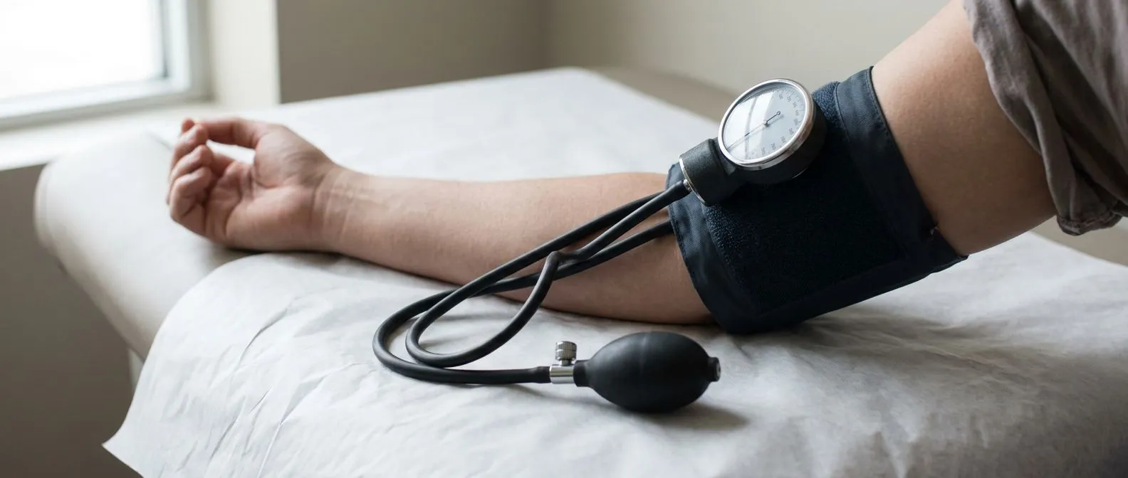
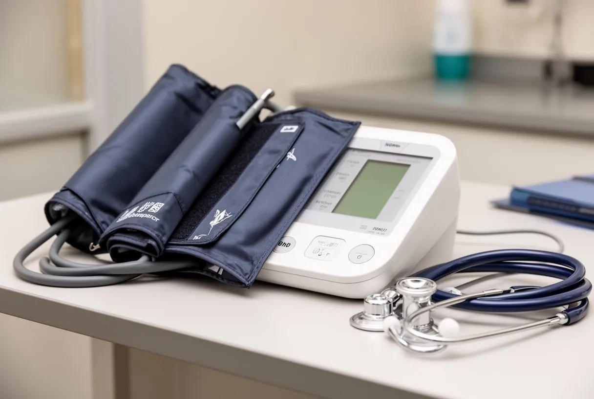
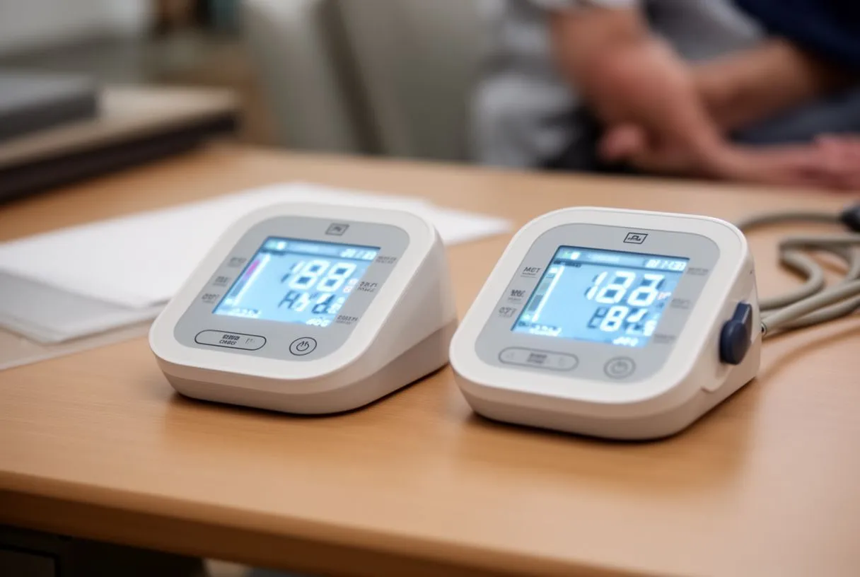
Proper technique for bilateral measurement: To accurately determine how to measure blood pressure left arm vs right arm, the ideal method involves using two separate blood pressure cuffs simultaneously. This eliminates the time delay between readings, which can cause fluctuations due to breathing, heart rate variations, or patient movement. If only one machine is available, measurements should be taken sequentially in the same arm first, and then immediately in the other, noting any consistent pattern over multiple readings.
Step-by-step guide for patients at home: Patients monitoring their blood pressure at home should follow a strict protocol to ensure accuracy. Rest for at least five minutes before taking a reading, sit with your back supported, and keep your feet flat on the floor. The cuff must be placed directly on the skin, at heart level.
- Rest: Sit quietly for five minutes before measuring to stabilize heart rate and vascular tone.
- Position: Keep the arm supported at heart level to prevent false high or low readings due to gravity.
- Auscultation: In clinical settings, doctors use a stethoscope for auscultation to listen to the Korotkoff sounds, ensuring maximum accuracy.
- Record: Document both the left and right arm readings immediately to track consistent discrepancies over time.
Understanding the limitations of automated cuffs: While home automated blood pressure monitors are highly convenient, they can sometimes fail to detect the true systolic pressure if the pulse is very weak, as is the case with severe peripheral artery disease. In these situations, a doctor may need to use a manual sphygmomanometer and a Doppler ultrasound device to detect blood flow. This ensures that a severe blockage is not mistakenly recorded as a normal or low blood pressure reading, which could delay critical intervention.
Blood Pressure Discrepancy Between Arms and Cardiovascular Risk
Linking interarm differences to systemic heart disease: A blood pressure discrepancy between arms is rarely an isolated vascular issue; it acts as a window into the health of the entire cardiovascular system. Patients with a significant interarm difference have a substantially higher risk of developing coronary artery disease, stroke, and transient ischemic attacks. The same atherosclerotic process blocking the arm artery is simultaneously threatening the delicate vessels supplying the brain and the heart muscle itself.
Connection to arrhythmias and heart function: Vascular resistance changes can also stress the heart muscle over time, potentially leading to electrical abnormalities. Patients with significant arterial stiffness or blockages are at a higher risk for developing Atrial Flutter, a rapid heart rhythm originating in the Atrium. This inefficient pumping can lead to blood pooling and clot formation. If a clot escapes into the circulation, it can cause a stroke or a severe peripheral blockage, underscoring the systemic danger of untreated vascular asymmetry.
Recognizing the signs of acute cardiac events: In some cases, vascular disease presents with silent or unusual symptoms. An Atypical Infarction, or silent heart attack, might not present with crushing chest pain but rather as fatigue, shortness of breath, or a sudden worsening of blood pressure asymmetry. Knowing your baseline interarm difference helps your cardiology team recognize when a sudden change might indicate an acute vascular event, prompting the immediate use of life-saving tools like an Automated External Defibrillator if a cardiac arrest occurs.

When to Worry About Blood Pressure Arms Difference?
Identifying red flag symptoms: Deciding when to worry about blood pressure arms difference depends on the severity of the discrepancy and the presence of accompanying symptoms. If your interarm difference is consistently greater than 15 mmHg, you should schedule a cardiology consultation. Immediate medical attention is required if the blood pressure drop is accompanied by sudden arm weakness, paralysis, profound coldness in the hand, or changes in skin color, as these may indicate an acute arterial occlusion.
Evaluating structural and aortic risks: Severe asymmetry can sometimes point to dangerous structural problems in the Ascending Aorta or aortic arch, such as an aneurysm or aortic dissection. These conditions are life-threatening and require immediate surgical intervention. Clinical trials, including those examining anticoagulation strategies like the ARISTOTLE trial, emphasize the importance of recognizing subtle hemodynamic signs to prevent catastrophic cardiovascular events. Prompt evaluation of significant interarm differences ensures that these critical structural issues are caught before they rupture.
Considering advanced cardiac support scenarios: In extreme cases where severe vascular disease leads to profound heart failure or cardiogenic shock, patients may require advanced life support. Interventions like Awake ECMO (Extracorporeal Membrane Oxygenation) might be utilized to rest the heart and lungs while the medical team addresses the underlying arterial blockages. Recognizing the early warning signs of vascular disease—like a significant blood pressure difference between arms—helps prevent the progression to these critical, highly invasive emergency interventions.
Diagnostic Tests and Cardiology Evaluation for Blood Pressure Asymmetry
Initial non-invasive vascular screening: When a patient presents with a blood pressure asymmetry, the first step in a cardiology evaluation is usually a vascular ultrasound. This painless test uses sound waves to visualize the arteries in the arms and neck, allowing the specialist to identify the exact location and severity of any plaque buildup. It provides a clear map of the blood flow without the need for radiation or invasive catheters, making it the safest initial diagnostic tool for patients.
Advanced imaging and hemodynamic monitoring: If ultrasound reveals significant disease, more advanced imaging like CT angiography or magnetic resonance angiography (MRA) may be ordered to get a more detailed view of the vascular anatomy. In intensive care or surgical settings, an Arterial Cannula or an Arterial Line might be inserted directly into the radial or femoral artery. This provides beat-to-beat, highly accurate blood pressure readings, which is crucial for hemodynamically unstable patients or those undergoing complex structural heart repairs.
Evaluating concurrent structural heart conditions: Because vascular disease often coexists with valve disease, the cardiology team will thoroughly evaluate the heart's structure and function. They will assess for conditions that might require intervention, ranging from valve repair to complex procedures utilizing a Balloon-expandable Valve or a Balloon Aortic Valvuloplasty. Comprehensive evaluations, like those discussed in studies regarding Transcatheter treatment for tricuspid valve disease, demonstrate the importance of treating the whole patient rather than just the localized arm symptom.

Treatment and Management for Upper Extremity Blood Pressure Variation
Medical management and risk factor modification: Treating a blood pressure difference between arms warning focuses primarily on managing the underlying systemic disease. Cardiologists usually prescribe medications to control blood pressure, lower cholesterol, and prevent blood clots. Daily low-dose Aspirin is commonly recommended to reduce the risk of thrombus formation in narrowed arteries. Lifestyle modifications, including smoking cessation, a heart-healthy diet, and controlled exercise, are essential to halt the progression of atherosclerosis and stabilize existing plaque.
Interventional procedures for severe blockages: If medication and lifestyle changes are insufficient, or if the arterial blockage is causing severe symptoms, interventional procedures may be required. Balloon Angioplasty is a common technique where a small balloon is inflated inside the narrowed artery to compress the plaque and widen the vessel. A stent is often placed afterward to keep the artery open. These minimally invasive procedures restore blood flow to the affected arm, effectively normalizing the interarm blood pressure readings and relieving symptoms like arm pain.
Surgical options and comprehensive cardiac care: In cases where minimally invasive techniques are not suitable, surgical bypass may be necessary to reroute blood around the blockage. This involves using an Arterial Graft or an Autograft—a blood vessel taken from another part of the patient's own body—to create a new pathway for blood flow. During complex surgeries, the medical team often utilizes Autologous Blood (the patient's own previously donated blood) to minimize transfusion risks. Ultimately, the most effective treatment plan is highly individualized, and we encourage you to discuss your specific situation with a qualified cardiac team and book a consultation rather than relying on self-diagnosis.