

Chordae Tendineae Rupture and Mitral Valve Repair
Chordae tendineae mitral repair restores normal heart valve function when cord rupture causes regurgitation.
If your cardiologist has told you the "strings" inside your heart are torn or stretched, you have likely encountered the term chordae tendineae. These thin, fibrous cords anchor the mitral valve leaflets so blood flows forward, not backward. When they rupture, the valve can leak severely — a condition called mitral regurgitation — and chordae tendineae mitral repair is often the treatment that restores normal valve function. This guide explains where these cords are, what happens when they break, how surgeons repair them, and what recovery looks like.
What Are Chordae Tendineae?
Thin cords with a heavy job. The chordae tendineae are delicate, tendon-like strands that connect the mitral valve leaflets to small muscles inside the left ventricle. Each strand is only a few millimetres wide, yet together they hold the valve shut against the full pressure of the heartbeat, millions of times over a lifetime.
Why they matter. Without the chordae, the valve leaflets — the thin flaps of the Mitral Valve — would flop backward into the upper heart chamber with every beat. Blood would then flow the wrong way, a leak called regurgitation. Surgeons studying the anatomy of these strands have shown that their arrangement is precise enough to allow targeted repair rather than replacement, which is why Mitral Valve Repair has become the preferred treatment for many patients (see Mitral Valve Repair: The Chordae Tendineae).
In our practice, we take the time to explain what each imaging study shows about these cords, in plain words, so patients understand exactly what is leaking before any decision is made.
Where Are the Chordae Tendineae Located?
Inside the left ventricle. The chordae tendineae sit in the left ventricle, the main pumping chamber of the heart. They run from the tips of the valve leaflets down to the papillary muscles, two finger-shaped muscles that rise from the ventricle wall.
Why they matter. The mitral valve sits between the left atrium and left ventricle. The chordae form a bridge across that space, holding the leaflets at the right tension so they meet cleanly in the middle of the valve opening when the heart contracts. This is what people mean when they ask "where do chordae tendineae?" — they are anchored at the bottom of the valve, not the top.
Their position matters in surgery: the posterior leaflet's cords are the most commonly ruptured, which is why posterior chordae tendineae rupture is one of the most frequent repair scenarios surgeons encounter.
What Is the Function of the Papillary Muscles?
Anchor points. The papillary muscles contract just before the ventricle does, pulling the chordae taut at exactly the moment the valve needs to close. Think of the valve leaflets as sails and the chordae as rigging lines — the papillary muscles are the sailors who hold the lines steady.
Timing is everything. If a papillary muscle contracts too late or too weakly, the leaflets billow backward and the valve leaks. When a heart attack damages the papillary muscle, it can tear away entirely, and the sudden loss of support can cause acute chordae tendineae rupture. This is one of the most dramatic leaks in cardiology, and off-pump surgical repair with artificial neochords has been described for exactly this situation (see Rescue transventricular off-pump mitral valve repair with artificial neochords).
In our practice, we review every case against international practice guidelines before recommending a treatment path, because the right approach depends on whether the muscle, the cords, or the leaflet itself is the problem.
What Happens if Chordae Tendineae Is Damaged?
The valve leaks. When a chord ruptures, the segment of leaflet it supports loses its tether and swings backward. Blood then flows backward into the left atrium with each beat — mitral regurgitation. The heart compensates by enlarging the atrium and working harder, and over time this can lead to heart rhythm problems and heart failure.
How badly depends on the cord. Rupture of a single minor strand may cause a mild leak that is simply monitored. Rupture of a major chord, or a set supporting a whole leaflet segment, can cause a sudden, severe leak with breathlessness, fatigue and even pulmonary congestion. Mitral Valve Prolapse is the commonest underlying condition where stretched or broken cords allow the leaflet to bulge back into the atrium.
- Mild damage: one small cord ruptured; leak is mild; doctors usually watch with yearly echo scans
- Severe damage: major cords broken; sudden breathlessness, rapid heart rhythms; surgery is usually needed promptly
We have seen in our practice how quickly a patient's condition can change from a stable, monitored leak to a severe one — which is why we treat new or worsening breathlessness as a reason to reassess promptly, not to wait.
Chordae Tendineae Rupture Symptoms
What patients feel first. A torn chordae tendineae treatment decision usually begins with symptoms. The most common is breathlessness, first on exertion, then at rest. Some patients notice a fluttering or pounding heartbeat, caused by the extra volume the atrium must handle. Fatigue and reduced exercise tolerance are also typical.
When it comes on suddenly. Acute chordae tendineae rupture — often after a heart attack or in connective tissue disorders — can cause sudden severe breathlessness and even coughing up pink, frothy fluid. That is a medical emergency. If you are unsure about your symptoms, the right move is not to self-diagnose but to be seen by a team that can evaluate you quickly.
In our practice, we get to know the patient first — a free video consultation lets us talk through the condition, review what the studies show, and order only the tests still needed, so no unnecessary ones are done.
Why Don't Semilunar Valves Have Chordae Tendineae?
A different design for different pressure. The semilunar valves — the aortic and pulmonary valves — have no chordae. Their leaflets, each a Valve Cusp shaped like a half-moon, close passively when the pressure on the far side of the valve exceeds the pressure behind it. The pressure gradient itself does the job that the chordae and papillary muscles do for the mitral valve.
Why the mitral valve needs help. The mitral valve faces far higher pressures than the semilunar valves and sits open during ventricular filling. Without the chordae, the leaflets would be forced backward. That is why the mitral valve, the Tricuspid Valve on the right side, and the mechanics of Tricuspid Valve Repair all involve chordal structures, while the aortic valve does not.
This is also why aortic valve disease and mitral valve disease are treated differently: aortic valve replacement is often done by catheter — Transcatheter Aortic Valve Implantation (commonly called TAVR) — while the mitral valve lends itself to repair.
How Chordae Tendineae Mitral Repair Works
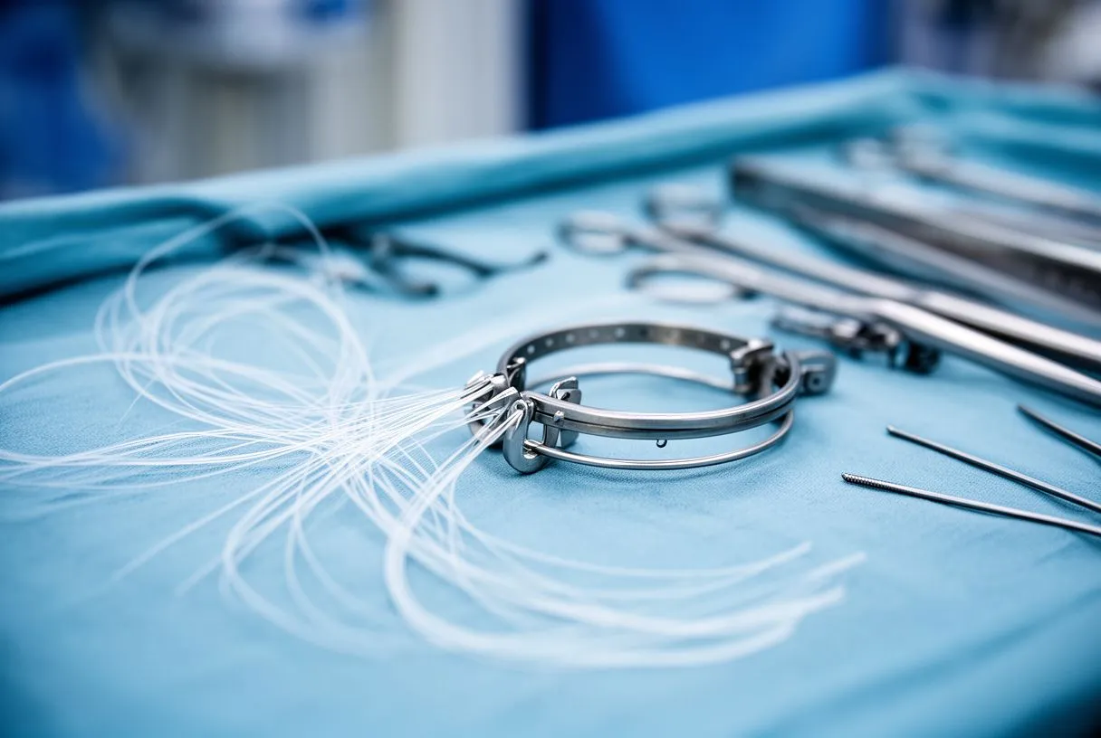
Rebuilding the tether. Chordae tendineae reconstruction restores the support the valve lost. Surgeons can transfer healthy chords from elsewhere on the leaflet — chordal transfer mitral repair — or implant artificial chordae tendineae made of fine Gore-Tex suture, a technique known as artificial chordae tendineae implantation. The new cords are cut to the exact length needed so the leaflet closes at the right level.
Why repair beats replacement. Keeping your own valve avoids the lifelong need for blood thinners that a mechanical Mitral Valve Replacement requires, and avoids the earlier wear-out associated with a Porcine Valve. Repair durability is excellent in experienced hands. Every case in our practice is reviewed by the multidisciplinary team against international practice guidelines before a recommendation is given; the patient receives a clear proposal with the alternatives, and the final decision is always theirs.
Other techniques. Depending on which leaflet is affected, repair may also involve removing excess leaflet tissue, folding the leaflet, or tightening the valve ring with an annuloplasty band. When the posterior chordae tendineae are the problem, repair is often straightforward; anterior chordae tendineae repair is more technically demanding and is best done by teams with high repair volumes.
Minimally Invasive Mitral Valve Repair Options
Small incisions, same repair. Minimally invasive mitral valve repair is done through a small cut between the ribs rather than opening the breastbone. The valve is repaired in exactly the same way — with artificial chordae or leaflet techniques — but the operation is gentler on the body, and recovery is typically faster.
Robotic and transcatheter options. Robotic mitral valve chordae repair uses the surgical robot to perform the same suturing through even smaller ports. For patients too frail for surgery, a catheter-based option called Transcatheter Edge-to-edge Repair — the same approach used for mitral regurgitation — clips the leaking leaflets together. Valve-in-valve procedures and Sutureless Valve options apply more to replacement scenarios than to primary chordal repair.
- Conventional open repair: breastbone incision; full access; standard choice in complex or urgent cases
- Minimally invasive / robotic: small side incision; less pain, faster discharge; requires suitable anatomy and an experienced team
- Catheter-based (edge-to-edge): no incision; for selected high-risk patients; less complete repair than surgery
In our practice, we perform conventional, minimally invasive, robotic and transcatheter procedures, and we recommend the less invasive route only where the evidence supports it for that specific patient.
Our work covers valve disease, coronary disease, the aorta and structural heart conditions — and Dr Molina performed the first robotic mitral valve repair in the Ángeles Health System and the first robotic heart surgery in Guadalajara.
Chordae Tendineae Repair Recovery Time
The usual timeline. Mitral valve chordae repair surgery generally involves a hospital stay of four to seven days for standard repair, with patients walking within a day or two of the operation. Most people are back to normal light activities around four to six weeks after a minimally invasive repair, and somewhat longer after conventional open surgery. Full recovery of energy can take a few months.
What helps. Following the cardiac rehabilitation plan, keeping the incision clean, and attending follow-up echo scans to confirm the valve stays competent all matter. Patients sometimes ask about restrictions — driving, lifting, returning to work — and these are agreed individually with the surgical team rather than by a fixed calendar.
In our practice, we take the time in consultation to explain what the studies actually show, in plain words, and to set out the alternatives, because the final decision is yours.

Are There Risks to Mitral Valve Repair?
Being honest about risk. Every open-heart operation carries risks: bleeding, infection, stroke, and rhythm disturbances. These are uncommon in planned mitral repair, and the risk profile depends heavily on the patient's age, other conditions, and how urgent the surgery is. A candid conversation — not reassurance without evidence — is what a good surgical team owes you.
Choosing the right team matters. Mitral repair outcomes correlate strongly with the surgeon's experience and the team's annual repair volume. Where the diagnosis is uncertain, or the anatomy is complex, seeking a second opinion is a normal part of deciding about heart surgery — and in our practice we encourage second opinions and treat them as such.
Costs and travel. Patients travelling for care in Guadalajara, Querétaro or Hermosillo should ask the team for a clear written proposal covering the operation, the expected stay, and follow-up arrangements, so there are no surprises.
In our practice, we encourage patients to bring their questions to the first consultation, and to weigh our proposal against any second opinion they receive. The final decision is always theirs.

In summary: the chordae tendineae are the small cords that keep your mitral valve working; when they rupture, the valve leaks and surgical repair — usually with artificial chords or leaflet techniques — can restore normal function. Chordae tendineae mitral repair is one of the most successful operations in modern heart surgery, and options now range from conventional open repair to minimally invasive, robotic and catheter-based approaches. If you have been told your valve is leaking, the next step is not to self-diagnose but to sit down with a qualified cardiac team, review your echo together, and decide the lowest-risk path for your situation. We invite you to book a consultation with our team in Guadalajara, Querétaro or Hermosillo to discuss your own case.






