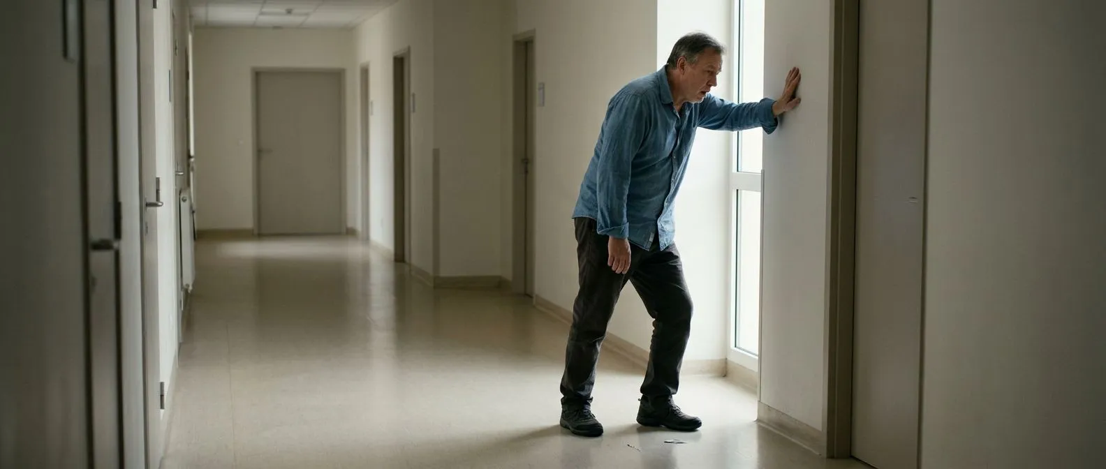
Dyspnoea on Exertion: Breathless When Active
Dyspnoea on exertion causes explained by heart specialists in Guadalajara.
What Are Dyspnoea on Exertion Causes?
Dyspnoea on exertion causes range from perfectly normal physiological responses to serious underlying heart or lung conditions. The term "dyspnoea" simply means an uncomfortable awareness of breathing — that sensation of breathlessness or "air hunger" you feel when climbing a steep flight of stairs or hurrying to catch a bus. For most healthy people, this settles within a minute or two of rest. When breathlessness appears at lower levels of effort than you would expect, or worsens over weeks and months, it becomes a symptom worth investigating rather than ignoring.
Understanding dyspnoea on exertion causes begins with recognising that your body demands more oxygen during physical activity. Your heart must pump faster and harder, your lungs must move more air, and your blood must carry that oxygen to working muscles. If any link in that chain — the pumping chambers, the valves, the airways, the blood vessels, or even the red blood cells themselves — is not functioning well, you will feel short of breath sooner than someone whose system is working efficiently. The question for clinicians is not just that you feel breathless, but where the bottleneck lies.
For patients and families in Mexico considering evaluation with a cardiac specialist, the key message is this: breathlessness on exertion is a symptom, not a diagnosis. It tells you something is limiting your exercise capacity, but it does not tell you what. That requires a structured assessment — listening to the heart through auscultation (the stethoscope examination), checking how the heart pumps on ultrasound, and sometimes performing stress testing. Many causes are manageable with medication or targeted procedures, and identifying the source early gives you the widest range of treatment options with the lowest risk for your situation.
How the Heart and Lungs Respond to Physical Effort
When you begin to walk uphill, climb stairs, or carry groceries, your body's demand for oxygen rises within seconds. Your nervous system signals the heart to beat faster and more forcefully. The left side of the heart — specifically the left atrium (the receiving chamber) and the left ventricle (the main pumping chamber) — must increase the volume of blood pushed out with each beat. At the same time, your breathing rate and depth increase so that more oxygen enters the lungs and more carbon dioxide is removed.
If the heart cannot keep up with this increased demand, oxygen-rich blood is diverted to essential organs and less reaches the exercising muscles. The muscles then switch to less efficient energy pathways that produce lactic acid, which stimulates breathing centres in the brain. You experience this as a mounting sense of air hunger that does not resolve until you stop and rest. In a healthy person, the system has enormous reserve capacity. In someone with a heart or lung condition, that reserve is narrowed or absent.
Cardiologists think of this reserve as your "exercise tolerance." A person with good exercise tolerance can briskly walk several blocks, climb two flights of stairs, and carry on a conversation without pausing for breath. When dyspnoea on exertion causes you to stop halfway up a single flight, or to avoid activities you once managed easily, your exercise tolerance has declined — and that decline is the clinical signal that investigation is needed.

Common Cardiac Causes of Breathlessness When Active
The heart-related dyspnoea on exertion causes fall into several broad categories, each with a different mechanism. Heart muscle weakness — often called heart failure or reduced ejection fraction — means the left ventricle cannot generate enough force to deliver blood to the body during effort. Valve problems, where doors inside the heart either narrow or leak, create backpressure or restrict forward flow. Coronary artery disease narrows the pipelines that supply the heart muscle itself, so the muscle starves during exertion. Arrhythmias — irregular heart rhythms such as atrial flutter (a fast, organised rhythm originating in the upper chambers) — can make the heartbeat too fast or too disorganised to pump effectively under stress.
Heart failure with reduced ejection fraction: When the pumping chamber weakens — from a previous heart attack, longstanding high blood pressure, or viral infection of the heart muscle — it cannot eject enough blood with each beat. During exertion, the shortfall becomes obvious. Fluid may also back up into the lungs, making breathing feel heavy and wet. This condition is often managed with medication that reduces the heart's workload and improves its efficiency over time.
Valvular heart disease: A narrowed aortic valve (aortic stenosis) forces the heart to push blood through a tiny opening, like pressing water through a pinched hose. A leaking mitral valve sends blood backwards into the left atrium with every beat, raising pressure in the lungs. Both situations limit exercise capacity and produce breathlessness. According to clinical research published through academic.oup.com, early valve evaluation significantly improves outcomes because it allows intervention before the heart muscle sustains lasting damage.
When Dyspnoea on Exertion Signals Heart Valve Disease
Valve disease is one of the most treatable dyspnoea on exertion causes, yet it is frequently overlooked because the symptoms creep in slowly. A patient may notice they no longer keep pace with their walking group, or that gardening leaves them winded for longer than it used to. The heart compensates for a diseased valve for months or even years before symptoms appear, which means that by the time you feel breathless, the valve problem may already be significant.
Aortic stenosis — narrowing of the valve between the left ventricle and the aorta, the large artery leaving the heart — classically produces three symptoms: breathlessness, chest tightness with effort, and fainting during exertion. The ascending aorta (the first segment of the body's largest artery, just above the aortic valve) carries the entire output of the heart; if the valve above it is stiff and calcified, the heart struggles to push blood through during activity. A thorough clinical evaluation including an echocardiogram (ultrasound of the heart) reveals the severity of narrowing and helps the cardiac team decide whether watchful monitoring, a balloon aortic valvuloplasty (a procedure that stretches the valve with a small balloon), or valve replacement is appropriate.
For patients whose surgical risk is high, transcatheter aortic valve replacement — known as TAVR — offers an alternative to open surgery. TAVR delivers a replacement valve through a catheter, usually entering through a blood vessel in the leg, and positions a new valve inside the old narrowed one. The decision between surgical replacement and TAVR depends on age, other medical conditions, and the anatomy of the valve and blood vessels — a conversation best had with a cardiac surgeon who can review your individual risk profile and explain which approach carries the lowest risk for your situation.
How Coronary Artery Disease Limits Exercise Tolerance
Coronary artery disease develops when fatty deposits called atheromatous plaque build up inside the arteries that supply the heart muscle with blood. During rest, the narrowed artery may deliver enough blood to meet the heart's modest needs. During exertion, the heart muscle demands more oxygen — and if the narrowed artery cannot deliver it, the muscle begins to starve. You may feel this as chest pressure, but many patients — particularly women, older adults, and people with diabetes — experience breathlessness as their main or only symptom, a pattern sometimes called atypical infarction when the blood supply becomes critically reduced.
Coronary disease is relevant to dyspnoea on exertion causes because it narrows the supply line precisely when demand rises. The heart, starved of oxygen, cannot pump as forcefully, and the lungs sense the mismatch. Sometimes the first sign is a decrease in exercise capacity over months; other times, a sudden worsening suggests an unstable plaque that needs urgent attention. Blood tests and markers such as apolipoprotein B (a protein carried on cholesterol particles that contributes to plaque formation) help clinicians assess your cardiovascular risk, but the definitive test for blocked arteries is a coronary angiogram — an X-ray study using contrast dye to map the coronary arteries directly.
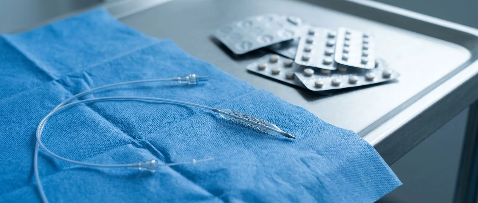
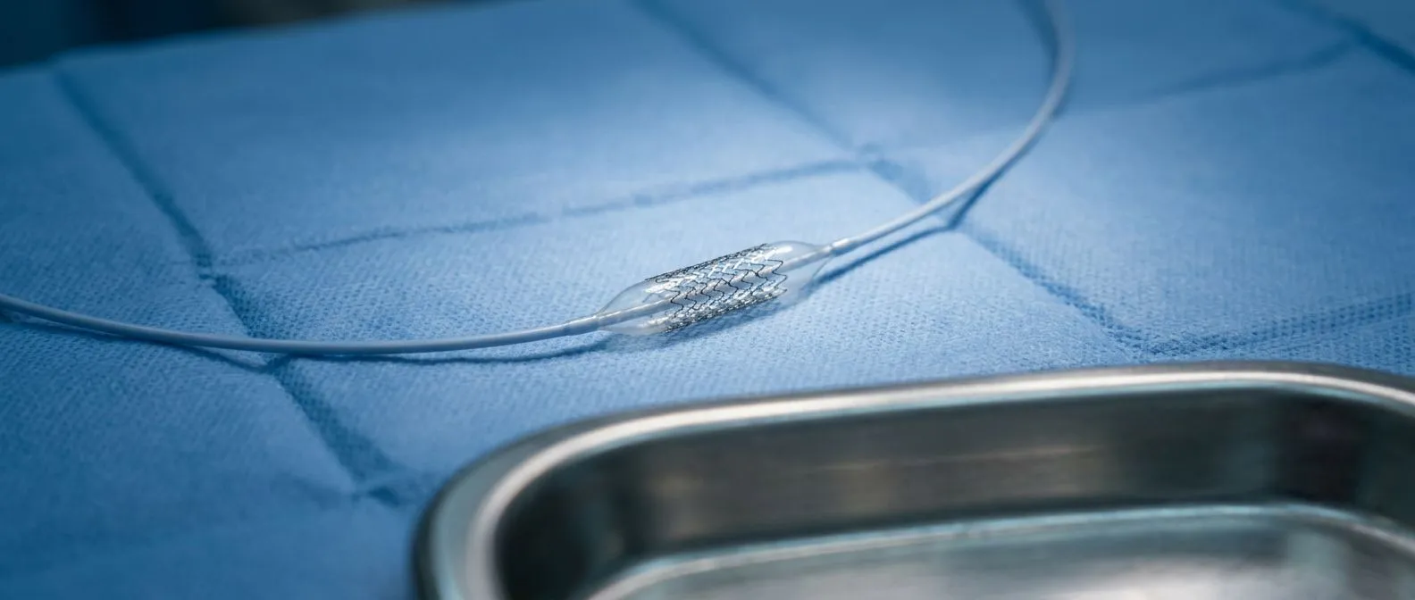
When significant blockages are found, treatment options include medication, balloon angioplasty (widening the narrowed artery with a small balloon, often followed by a stent to keep it open), or coronary artery bypass surgery using an arterial graft (a blood vessel taken from elsewhere in the body to reroute blood around the blockage). A study indexed at PubMed 33865571 underscores the importance of early identification of coronary disease in patients presenting with exertional symptoms, as timely revascularisation can prevent progression to heart muscle damage.
Are There Non-Cardiac Dyspnoea on Exertion Causes?
Not every case of breathlessness on exertion comes from the heart. The lungs are equally important in the oxygen chain, and conditions such as chronic obstructive pulmonary disease (COPD), asthma, pulmonary fibrosis, and pulmonary embolism can all produce exertional breathlessness. In these conditions, the problem lies in getting air into or out of the lungs, or in transferring oxygen from the air sacs into the blood. A careful history — including whether you smoked, your occupational exposures, and whether breathlessness comes with wheezing, cough, or chest tightness — helps distinguish lung causes from heart causes.
Anaemia is another frequently overlooked contributor. If your red blood cell count is low, your blood carries less oxygen regardless of how efficiently your heart and lungs work. Exertion demands oxygen delivery that the blood simply cannot meet, and the result is breathlessness and fatigue. This is particularly common in women with heavy menstrual cycles, older adults with poor nutrition, and patients with chronic kidney disease. A simple blood test reveals anaemia, and in many cases, it can be managed with medication, dietary changes, or treating the underlying cause.
Other possibilities include deconditioning from prolonged inactivity, thyroid disorders, obesity, and anxiety. None of these should be dismissed, but they should also not be assumed without testing — because attributing breathlessness to "just being out of shape" can delay the diagnosis of a serious heart or lung condition. A systematic approach, often coordinated between a cardiologist and a pulmonologist, ensures that no significant cause is missed.
- Heart causes: valve disease, heart failure, coronary artery disease, arrhythmias — each limits the heart's ability to meet exertional demand.
- Lung causes: COPD, asthma, pulmonary fibrosis, blood clots in the lungs — each limits oxygen uptake or airflow.
- Blood causes: anaemia reduces oxygen-carrying capacity, producing breathlessness even with a healthy heart and lungs.
- Systemic causes: thyroid imbalance, severe deconditioning, obesity — each reduces overall exercise tolerance.

What Tests Help Diagnose Breathlessness During Activity
The workup for dyspnoea on exertion causes follows a logical sequence. It usually begins with a thorough clinical interview and a physical examination, including auscultation — listening to the heart and lungs with a stethoscope to detect murmurs, irregular rhythms, or abnormal lung sounds. A resting electrocardiogram (ECG) records the heart's electrical activity and can reveal prior heart attacks, rhythm problems, or signs of heart strain. Blood tests check for anaemia, kidney function, thyroid levels, and cardiac biomarkers that suggest heart muscle stress.
An echocardiogram is often the single most informative test. This painless ultrasound study shows the heart's chambers and valves in real time, revealing how well the heart pumps, whether any valves are narrowed or leaking, and whether the heart muscle is thickened or weakened. For many patients, the echocardiogram alone clarifies whether the cause is cardiac and points to the specific problem. When the echocardiogram is normal but symptoms persist, the next step may be a stress test — exercising on a treadmill or stationary bike while monitored — to provoke the symptoms under controlled conditions and see how the heart responds to effort.
Additional testing might include a coronary CT angiogram to look for plaque in the coronary arteries, pulmonary function tests to assess lung capacity, or a six-minute walk test to objectively measure how far you can walk and whether your oxygen level drops during exertion. The goal is not to perform every test on every patient, but to use the clinical story to guide a focused, efficient investigation that identifies the cause and directs treatment.
Treatment Options for Exertional Breathlessness
Treatment for dyspnoea on exertion causes depends entirely on the underlying problem. For heart failure, guideline-directed medication — including drugs that reduce fluid overload, lower blood pressure, and help the heart remodel over time — forms the foundation. Many patients experience meaningful improvement in exercise capacity with optimised medical therapy alone, avoiding or delaying the need for procedures. Medications such as aspirin (a blood thinner that reduces clot risk in coronary disease) and anticoagulants — studied in large trials such as the ARISTOTLE trial for atrial fibrillation — address specific risks associated with particular conditions.
Valve interventions: When a severely narrowed or leaking valve is the cause, medication alone cannot fix the mechanical problem. Surgical valve replacement — using a mechanical valve, a biological tissue valve, or an autograft (a patient's own tissue, such as the pulmonary valve moved to replace the aortic valve in the Ross procedure) — directly addresses the obstruction or leak. For high-risk surgical patients, TAVR using a balloon-expandable valve (a new valve compressed onto a catheter and expanded into place with a balloon) offers a less invasive alternative. The choice depends on age, anatomy, and overall health.
Coronary revascularisation: When blocked arteries limit blood supply to the heart, balloon angioplasty with stenting opens specific narrowings, while bypass surgery creates new routes for blood flow. An ongoing clinical study — A Study in Patients with a Descending TAA or PAU Treated with the E-nya Thoracic Stent Graft System — illustrates how specialised stent-graft technology is being evaluated for complex thoracic vascular disease, reflecting the broader trend toward less invasive, catheter-based solutions. In complex surgery, techniques such as autologous blood (the patient's own blood, collected and returned during surgery) conservation and, in selected cases, awake ECMO (extracorporeal membrane oxygenation supporting heart and lung function while the patient remains awake) demonstrate how surgical practice continues to evolve to reduce risk.
Recovery and Quality of Life After Cardiac Intervention
Recovery from cardiac procedures varies widely depending on the intervention and the patient's baseline health. After TAVR, many patients leave the hospital within two to three days and notice improved exercise capacity within weeks. After open-heart surgery — whether valve replacement or bypass — the hospital stay is longer, typically five to seven days, followed by a structured recovery period at home over four to six weeks. Cardiac rehabilitation, a supervised exercise and education programme, plays a central role in rebuilding strength and confidence.
Most patients report significant improvement in their breathlessness after successful treatment of the underlying cause. A patient who could not walk a single block without stopping may, after valve replacement or coronary revascularisation, return to brisk walking, cycling, or swimming. The degree of improvement depends on how much the heart muscle was affected before treatment and whether other conditions — such as lung disease or kidney problems — coexist. Honest expectations matter: no procedure reverses all damage, and recovery requires commitment to medication, exercise, and follow-up.
Quality-of-life outcomes are closely tied to timeliness. Patients treated before the heart muscle has sustained permanent damage generally fare better than those treated late. This is why cardiac teams emphasise early evaluation when dyspnoea on exertion causes first appear, rather than waiting until symptoms become severe. For families weighing whether to travel for evaluation — from elsewhere in Mexico or from abroad — understanding that earlier assessment expands treatment options and typically lowers procedural risk can help frame the decision.
When to Seek Help for Worsening Breathlessness
Certain patterns of breathlessness warrant prompt medical attention rather than waiting for the next scheduled appointment. If you notice breathlessness that occurs at rest, that wakes you from sleep, or that forces you to prop yourself up on extra pillows to breathe comfortably at night, these may signal fluid accumulating in the lungs — a feature of worsening heart failure. Breathlessness accompanied by chest pain or pressure, sweating, nausea, or pain radiating to the arm, jaw, or back may indicate a heart attack and requires emergency evaluation.
A practical approach is to monitor your exercise tolerance over time. If you find yourself unable to complete activities that were easy a few months ago — walking the dog, climbing stairs, carrying groceries — schedule an evaluation. Keep a simple diary noting which activities trigger breathlessness, how long it takes to recover, and whether the pattern is worsening. This information is invaluable to the cardiac team because it reveals the trajectory, not just a single snapshot. Sudden changes are always more urgent than gradual ones, but even gradual decline deserves investigation.
- Seek emergency care if: breathlessness comes with chest pressure, fainting, sweating, or occurs at rest for the first time.
- Schedule a prompt appointment if: exercise tolerance has declined over weeks to months, or breathlessness now occurs with lighter activity than before.
- Keep a symptom diary: note triggering activities, recovery time, and whether symptoms are stable or worsening — this guides clinical decisions.
If you or a family member are experiencing breathlessness on exertion and would like a structured evaluation, the most important step is to speak with a qualified cardiac team. Every person's situation is unique, and decisions about testing, medication, or procedures should be made together with clinicians who understand your full medical picture. Dr Alejandro Molina and the team at Cirugía de Corazón see patients in Guadalajara, Querétaro, and Hermosillo, and offer consultation for those travelling from other regions or from abroad. Rather than self-diagnosing or waiting for symptoms to worsen, consider booking a consultation to understand what is causing your breathlessness and what options are available for your specific situation.